Automating Business Process via MuleSoft Composer | Bangalore MuleSoft Meetup...
AS Biology Lesson 2 - Measuring Cells
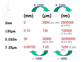
1. X 1000 X 1000
(mm) (µm) (nm)
2mm 2 2000 (2 x 10³) 2000000
(2 x 10⁶)
130µm 0.13 130 130000
(1.3 x 10⁵)
0.032m 32 32000 32000000
(3.2 x 10⁴) (3.2 x 10⁷)
7.25µm 0.00725 7.25 7250 (7.25 x
103)
÷ 1000 ÷ 1000
2. Learning Objectives
- [PA] use an eyepiece graticule and stage
micrometer scale to measure cells and be
familiar with units (millimetre, micrometre,
nanometre) used in cell studies;
-[PA] calculate linear magnification of
drawings and photographs;
-(h) [PA] calculate actual sizes of specimens
from drawings and photographs;
12. Eyepiece & stage graticules
Low magnification High magnification
13. Figure 4.3
Stage micrometer viewed at x100 magnification.
The total length of the micrometer is 1mm
total length = 1mm on this scale, 94
which is 1000μm divisions = 1000μm
Therefore, 1 division on the
eyepiece graticule represents
1000 ÷ 94 = 10.6 μm
at this magnification.
14. Figure 4.1
Cells of onion epidermis as viewed at x100 magnification
with a graticule in the eyepiece of the microscope
We know that at this
In the two columns covered
magnification, 1 division
by the graticule there is an
on the eyepiece graticule
average of five cells in the
represents 10.6 μm
length of the graticule
1060μm
Therefore the average
Therefore the total length
oflength of one cell is
the eyepiece graticule
represents = 212μm = 1060μm
1060 ÷ 5 10.6 x 100
at this magnification
15. Figure 4.4
Part of the stage micrometer viewed at x400
magnification
remember thatshown
so the length each on this scale, 90
division here is 10μm
by the bracket is
240μm divisions = 240μm
Therefore, 1 division on the
eyepiece graticule represents
240 ÷ 90 = 2.67 μm
at this magnification.
16. Figure 4.2
Cells of onion epidermis as viewed at x400 magnification with
the same graticule in the eyepiece
We know that at this magnification,
each division of the eyepiece graticule
represents 2.67μm
The length of the cell covered
by the graticule is 98 divisions,
therefore the length of this cell
is 2.67 x 98 = 262μm
17. We now have two measurements for the length of an onion cell;
212μm and 262 μm.
Which of these is the more accurate estimate of the length of onion
epidermal cells?
• The answer from Q. 2 [212 μm]
• because this is a mean of several cells.
• Only one cell was measured in Q.3, and this one
may not be representative.
18. Estimating cell width. Figure 4.5.
Cells of the onion epidermis as viewed at x100 magnification
with a graticule in the eyepiece of the microscope
Remember the total length
of the eyepiece graticule
represents 1060μm
at this magnification
There are approximately
thirteen cells in the
length of the graticule
Therefore the average
width of one cell is
1060 ÷ 13 = 81.5μm
19. Figure 4.6.
Cells of the onion epidermis as viewed at x400 magnification
with the same graticule in the eyepiece of the microscope
Remember, we know that at this
magnification, each division of
the eyepiece graticule
represents 2.67μm 62 divisions
Here, two cells span 62 divisions
on the eyepiece graticule. This
represents 2.67 x 62 = 165.5 μm
Therefore the average
width of one cell is
165.5 ÷ 2 = 82.8μm
![Learning Objectives
- [PA] use an eyepiece graticule and stage
micrometer scale to measure cells and be
familiar with units (millimetre, micrometre,
nanometre) used in cell studies;
-[PA] calculate linear magnification of
drawings and photographs;
-(h) [PA] calculate actual sizes of specimens
from drawings and photographs;](data:image/gif;base64,R0lGODlhAQABAIAAAAAAAP///yH5BAEAAAAALAAAAAABAAEAAAIBRAA7)