
Recommended
More Related Content
What's hot
What's hot (18)
Viewers also liked
Similar to Anatomy and Physiology Times Sensory Receptors
Similar to Anatomy and Physiology Times Sensory Receptors (20)
More from KatKatKatGarGar
Anatomy and Physiology Times Sensory Receptors
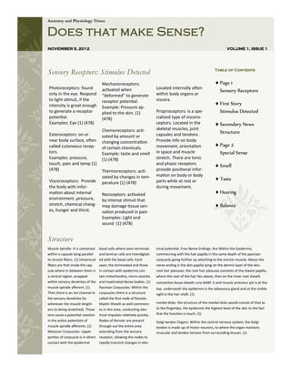
- 1. Anatomy and Physiology Times Does that make Sense? NOVEMBER 5, 2012 VOLUME 1, ISSUE 1 Sensory Receptors: Stimulus Detected Table of Contents Mechanoreceptors: Page 1 Photoreceptors: found activated when Located internally often Sensory Receptors only in the eye. Respond “deformed” to generate within body organs or to light stimuli, if the receptor potential. viscera. intensity is great enough First Story Example: Pressure ap- to generate a receptor plied to the skin. (1) Proprioceptors: is a spe- Stimulus Detected potential. (478) cialized type of viscero- Examples: Eye (1) (478) ceptors. Located in the Secondary News Chemoreceptors: acti- skeletal muscles, joint Exteroceptors: on or capsules and tendons . Structure vated by amount or near body surface, often changing concentration Provide info on body called cutaneous recep- of certain chemicals. movement, orientation Page 2 tors. Example: taste and smell in space and muscle Special Sense Examples: pressure, (1) (478) stretch. There are tonic touch, pain and temp (1) and phasic receptors Smell (478) Thermoreceptors: acti- provide positional infor- mation on body or body Visceroceptors: Provide vated by changes in tem- parts while at rest or Taste perature (1) (478) the body with infor- during movement. mation about internal Nociceptors: activated Hearing environment ,pressure, by intense stimuli that stretch, chemical chang- may damage tissue sen- Balance es, hunger and thirst. sation produced in pain Examples: Light and sound (1) (478) Structure Muscle Spindle- It is contained basal cells where axon terminals trical potential. Free Nerve Endings: Are Within the Epidermis, within a capsule lying parallel and laminar cells are interdigitat- commecning with the hair papilla in the same depth of the pacinian to muscle fibers. (1) Intramural ed with the basal cells. Each corpuscle going further up attaching to the erector muscle. Above the fibers are that inside the cap- axon; the terminated and those nerve ending is the skin papilla lying on the dermis layer of the skin. sule where in-between them is in contact with epidermis con- root hair plexuses: the root hair plexuses constists of the lowest papilla, a central region, wrapped tain mitochondria, micro vesicles where the root of the hair lies above, then on the inner root sheath within sensory dendrites of the and myelinated dense bodies. (1) connective tissue sheath runs over it and muscle arrecteur pili is at the muscle spindle afferent. (1) Pacinian Corpuscles: Within the top. underneath the epidermis is the sebaceous gland and at the visible Then there is an ion channel in corpuscles there is a structure sight is the hair shaft. (1) the sensory dendrites for called the first node of Ranvier. whenever the muscle length- Myelin Sheath as well commenc- merkel disks: the structure of the merkel disks would consist of that as ens (is being stretched). Those es in this area, conducting elec- to the fingertips, the epidermis the highest level of the skin to the fact ions cause a potential reaction trical impulses relatively quickly. that the function is touch. (1) in the action potentials of Nodes of Ranvier are present Golgi tendon Organs: Within the central nervous system, the Golgi muscle spindle afferents. (1) through out the entire area tendon is made up of motor neurons, to where the organ monitors Meissner Corpuscles- Upper extending from the sensory muscular and tendon tension from surrounding tissues. (1) portion of corpuscle is in direct receptor, allowing the nodes to contact with the epidermal rapidly transmit changes in elec-
- 2. DOES THAT MAKE SENSE? Page 2 Structure Continued... of Ranvier. Withing the Epidermis, then on the inner root sheath commecning with the hair Myelin Sheath well com- connective tissue sheath runs papilla in the same depth of mences in this area, con- over it and muscle arrecteur the pacinian corpuscle ducting electrical impulses pili is at the top. underneath relatively quickly. going further up attaching to the epidermis is the seba- the erector muscle. Above ceous gland and at the visible Nodes of Ranvier are present the nerve ending is the skin sight is the hair shaft through out the entire area papilla lying on the dermis extending from the sensory layer of the skin. receptor, allowing the nodes merkel disks: the structure of to rapidly transmit changes in the merkel disks would con- electrical potential. root hair plexuses: the root sist of that as to the finger- hair plexuses constists of the tips, the epidermis the high- lowest papilla, where the est level of the skin to the Free Nerve Endings: Are root of the hair lies above, fact that the function is touch EXTRA! EXTRA! READ ALL ABOUT THE SPECIAL SENCES What are Special Senses? Special Senses are characterized by receptors grouped closely together or grouped in spe- cialized organs; sense of smell, taste, hearing, equilibrium, and vision. SMELL: Olfactory Receptors epithelium stimulate the olfactory cell. Olfactory sense organs consist of Olfactory Epithelium- located in most superior portion of the epithelial support cells and nasal cavity specialized olfactory receptor Olfactory Receptors- extremely sensitive and easily fatigued neurons Olfactory Cilia- located on the Olfactory Pathways olfactory receptor neu- When the level of SMELL odor- producing chemicals reaches a rons that touch the olfac- threshold level, the following occurs tory epithelium lining the Receptor potential, and then action potential, is generated and upper surface of the nasal passed to the olfactory nerves in the olfactory bulb cavity. The impulse then passes through the olfactory tract and into Olfactory Cells- chemorecep- the thalamic and olfactory centers of the brain for inter- tors; gas molecules or pretation, integration, and memory storage chemicals dissolved in the mucus covering the nasal
- 3. Page 3 VOLUME 1, ISSUE 1 Taste: Taste Buds- sense organs that (1) chemicals in the saliva Adaptation and sensitivity thresh- Nerve impulses from the respond to gustatory, or taste, Taste buds are similar in struc- olds are different for each of anterior two thirds of the stimuli; associated with papillae ture; functionally, each taste the primary taste sensations tongue travel dover the bud responds most effec- (1) Chemoreceptors that are facial nerve; those from stimulated by chem- Neural the posterior one-third of icals dissolved in the Pathway- the tongue travel over saliva (1) taste the glossopharyngeal Gustatory cells special- sensation nerve; vagus nerve plays ized cells in taste begins a minor role in taste. buds; gustatory with a Nerve impulses are hairs extend from receptor carried to the medulla each other into the potential oblongata, relayed into taste pores (1) in the the thalamus and then Sense of taste depends on the gustatory cells of a taste bud; into the gustatory area of tively to one of four primary creation of a receptor po- generation and propagation of an the cerebral cortex in the taste sensations: sour, sweet, tential in gustatory cells action potential then transmits parietal lobe of the brain. bitter, and salty(1) because of taste-producing the sensory input to the brain.(1) (1) Hearing: Neural Pathway of hearing– a move- External ear is divided into two divi- The inner ear has a variety of struc- ment of hair cells against the tectorial sions. Auricle or pinna which is the tures that consist of the a bony laby- membrane stimulates the dendrites visible portion. The external auditory rinth, a membrane labyrinth, the vesti- that terminate around the base of the meatus is the tube leading from the bule and semicircular canals which are hair cells and initiates impulse conduc- auricle into the temporal bone and involved with balance, the cochlea, the tion by the cocniear nerve to the brain- ending at the tympanic membrane. (1) end lymph and the perilymph (1) stem. The impulses pass through “relay stations” in the nuclei in the medulla, Middle Ear has tiny epithelium which pons, midbrain and thalamus before is lined cavity hollowed out of the reaching the auditory are of the tem- temporal lobe. It also contain 3 audito- poral lobe.(1) ry ossicles the malleolus (hammer), incus (anvil), and stapes (stirrup). (1) Mechanisms of Hearing Balance: Sense of balance- related to head posi- Static equilibrium is the abil- tion and deceleration ity to sense the position Otoliths are located with- of the head relative to in the matrix of the gravity or to sense accel- macula eration or deceleration Dynamic Equilibrium- needed Movements of the macu- to maintain to balance when la, located in both the head or body is rotated or the utricle and sac- suddenly moved; able to de- cule almost right tect changes in direction and angles to each other, rate at which movement oc- provide information curs (1)