2. Amyloid-Induced Activity in Follicles
Jorge Parodi et al.
gregate. This approach will potentially speed up the screening Table 1. Decay constants (τ) and amplitude variance of blips in the
2+
for drugs against amyloid aggregation and its action on mem- absence or presence of amyloid aggregate in normal or in Ca -free
branes. Ringer’s, amphotericin B, and gramicidin
Condition τ (ms) Variance (nA)
MATERIALS AND METHODS
Control 1,900 ± 1,000 0.7 ± 0.01
Amyloid aggregation Gramicidin 0.004 ± 0.0002 7.2 ± 0.12
Human synthetic Aβ1-40 peptides were dissolved in dimethyl Amphotericin B 00.01 ± 0.0004 1.6 ± 0.03
sulfoxide (DMSO) at a concentration of 10 mg/ml and immedi- 2+
ately stored in aliquots at -20°C. Twenty-five μl of this Aβ- Amyloid aggregate Ca free 0.100 ± 10 0.1 ± 0.01
peptide stock solution (10 mg/ml) was diluted to a final concen- Amyloid aggregate 00.03 ± 0.0005 7.3 ± 0.1
tration of 80 μM in 725 μl of PBS (Tocris Bioscience, USA) and
stirred continuously at 200 rpm for 150 min at 37°C (Parodi et
al., 2009).
ted using pClampfit 9; the membrane conductance was deter-
Electrophysiological recordings and transmission electron mined as previously described (Reiser and Miledi, 1989), and
microscopy the variance of the events and average decay constants (τ)
All the animals were handled in accordance with the guidelines were derived and are shown in Table 1. To determine what ion
of the National Institutes of Health Guide for Care and Use of is responsible for generating the blips, the current-voltage rela-
Laboratory Animals and with the approval of the Institutional tion of blips was constructed by applying a pulse protocol from
Animal Care and Use Committee of the National University of -150 mV to 60 mV.
Mexico. X. laevis frogs were anesthetized with 0.1% 3-amino- Recordings were acquired using a Warner amplifier (OC-
benzoic acid methylester (MS-222) for 10 min, and follicles 765C, Warner Instruments, LLC., USA) and an AC/DC con-
were isolated manually using fine-tipped forceps. To determine verter (Digidata 1322A, Axon, Molecular Devices, USA) and
the effect of amyloid aggregate on endogenous oocyte currents, stored in winWCP. Files were analyzed and plotted using
the cells were treated with 0.3 μg/μl collagenase type I at room pClampfit 9 (Axon, Molecular Devices, USA) and Origin 8.0.
temperature for 35 min to remove the follicular cell layer, and ANOVA (confidence interval of 95%) was used to analyze mul-
then kept at 16°C in Barth’s medium: 88 mM NaCl, 1 mM KCl, tiple means and was followed by Bonferroni’s test to correct for
0.33 mM Ca2(NO)3, 0.41 mM CaCl2, 0.82 mM MgSO4, 2.4 mM type I errors. Student’s t-test with confidence interval of 95%
NaHCO3, 5 mM HEPES, pH 7.4, containing 0.1 mg/ml genta- was used to compare the means of controls versus experimen-
mycin sulfate. The electrophysiological recordings were ob- tal conditions.
tained 24 h after isolation.
For electrophysiological recordings, two electrodes were in- RESULTS
serted into the oocyte, and membrane currents were recorded
in the voltage-clamp configuration at -60 mV (Miledi, 1982). The Blips of oocytes exposed to membrane-perforating
cells were superfused with frog Ringer’s solution: 115 mM NaCl, molecules and amyloid aggregate
2 mM KCl, 1.8 mM CaCl2, 5 mM HEPES, pH 7.4, at room tem- The stability of the oocyte plasma membrane was assessed by
perature (20-25°C). Gramicidin (100 μg/μl), amphotericin-B (250 recording the basal electric activity of cells held at -60 mV for
μg/μl), or amyloid aggregate (Aβ) (1 μM or 22 μg/μl) were per- periods of 15 min before adding any substances. During these
fused while recording the resting membrane potential for 15 recordings the spontaneous activity was minimal in control cells,
min. To test the role of extracellular cations on the effect of and the amplitude of the blips fluctuated from 3.8 ± 0.9 nA at
amyloid aggregate, we substituted the Na+, K+, or Ca2+ in the the beginning to 4.5 ± 0.8 nA at the end of the recording. In
Ringer’s solution. Na+ was partially replaced by N-Methyl-D- contrast, the spontaneous activity increased greatly when
glucamine (NMDG+ 50%), K+ by an equimolar concentration of gramicidin (100 μg/μl) or amphotericin B (250 μg/μl) were su-
CsCl (100%), and Ca2+ by Mg2+ (5 mM). High Ca2+ Ringer’s perfused onto the oocytes, and at the end of the recording time-
was prepared by increasing the concentration of Ca2+ to 10 mM. lapse we found that the amplitude of blips increased to an av-
To determine the effect of the amyloid aggregate on the elec- erage of 7.8 ± 1.2 nA for gramicidin and 6.3 ± 1.1 nA for am-
trical coupling between oocytes and surrounding follicular cells, photericin B (Fig. 1A and plotted in 1B). Furthermore, when
follicles were exposed to 100 μM ATP to induce the oscillatory, oocytes were bathed in a 1 μM solution of amyloid aggregate
Ca2+-dependent Cl- current (Arellano et al., 1996; 1998) before for 15 min, the amplitude of blips increased to an average of 6.8
and after a 15-min exposure to amyloid aggregate. After elec- ± 1.7 nA, i.e., a value very similar to that generated by the other
trophysiological recordings, the oocytes were processed for molecules.
electron microscopy (Saldana et al., 2009) by fixation in 4% The amplitude of blips from the control cells was concen-
glutaraldehyde, and embedded in the plastic resin “EPOL”; trated near -2.5 nA; in contrast, amyloid aggregate, gramicidin,
slices of 2 μm were obtained, contrasted with 2% uranyl ace- or amphotericin induced blips with a broad range of ampltitudes,
tate, placed on lead grids, rinsed, and observed under the elec- ranging from -6 to +0.5 nA (Fig. 2A). The membrane conduc-
tron microscope (JEOL JEM 1010, JEOL Ltd., Japan). tance increased when cells were exposed to each of the three
molecules tested, from an average of -141 ± 14 nS in control
Data analysis samples to -647 ± 78, -522 ± 51, and -577 ± 25 nS for grami-
Spontaneous electrical events of the oocyte plasma membrane cidin, amphotericin B, and amyloid aggregate, respectively (Fig.
were recorded for 15 min in the presence or not of amyloid 2B). Current-voltage relations were constructed to gain some
aggregate (22 μM), amphotericin B (200 μM), or gramicidin understanding of the ion(s) responsible for the currents ob-
(100 μM). The frequency of blips was analyzed using the Root served. The plot in Fig. 2C shows an almost linear relation for
Mean Square (RMS) tool of Mini Analysis 6 Software and plot- all the conditions and voltages tested. Gramicidin and amyloid
350 Mol. Cells http://molcells.org
3. Amyloid-Induced Activity in Follicles
Jorge Parodi et al.
A B Fig. 1. Spontaneous electric blip activity
of X. laevis oocytes. (A) Sample recor-
dings from oocytes exposed to grami-
cidin (100 µg/µl), amphotericin-B (250
µg/µl), or amyloid aggregate (1 µM). (B)
Average amplitude of blip currents from
sample traces (% of control). Each bar
(mean ± SEM) was obtained from at
least six different cells. Amph B, am-
photericin B. The asterisk indicates P <
0.05 (ANOVA).
A B C
Fig. 2. Distribution of blip amplitudes and current-voltage relation. (A) Histogram distribution of frequencies of blip amplitudes from oocytes
exposed to amyloid aggregate, gramicidin, or amphotericin. (B) Bar graph of average membrane conductance in different conditions. Each bar
(mean ± SEM) represents measurements of at least six different oocytes. The asterisks indicate P < 0.05 (ANOVA). (C) Current-voltage rela-
tions for blip amplitude at different voltages in the presence of amyloid aggregate, gramicidin, or amphotericin.
aggregate showed reversal potentials of -51 ± 5 mV and -69 ± Ca2+-free Ringer’s the amyloid aggregate did not increase the
4 mV, respectively (Fig. 2C), suggesting a cationic conductance amplitude of blips, which remained at the same levels as in
near the equilibrium potential for Ca2+ in oocytes bathed in control cells (12.6 ± 1.7 nA, Fig. 3B, Student’s t-test, p < 0.05;
Ringer’s, although other conductances cannot be excluded. control vs. Ca2+ free Ringer’s). This observation suggests that
The observations above suggested that the amyloid aggre- extracellular Ca2+ generates, at least in part, the blips induced
gate could be inducing pores in the plasma membrane of the by amyloid aggregate.
oocyte; thus, we examined the effect of removing extracellular We explored the effect of reducing Na+ or K+ in the Ringer’s
Ca2+ on the generation of spontaneous activity. Ca2+-free Ringer’s solution on blip amplitude in the presence of amyloid aggregate.
was superfused onto the oocytes either in the presence or ab- In both cases we observed slight increases in the amplitudes of
sence of 1 μM amyloid aggregate. When Ca2+ was removed the blips, also these increases were statistically significant
from the extracellular medium of the oocytes, the amyloid ag- when compared to the effect of the amyloid aggregate in nor-
gregate did not elicit evident changes in the amplitude of blips mal Ringer’s (indicated as ρ’s in Fig. 3C), however all groups
as compared to control oocytes bathed in normal Ringer’s solu- showed differences when compared to the untreated control
tion (11.5 ± 1.7 vs. 12.3 ± 1.1 nA, respectively; Fig. 3A, Stu- (indicated as * in Fig. 3C ANOVA, p < 0.05). Oocytes exhibit
dent’s t-test, p < 0.05; control vs. normal Ringer’s). Perfusion of endogenous conductances generated by entry of extracellular
1 μM amyloid aggregate for 15 min in normal Ringer’s in- Ca2+ as well as by Ca2+ released from intracellular stores; for
creased the amplitude to 21.8 ± 1.3 nA; in sharp contrast, in example the depolarization-induced Tout current (Miledi, 1982)
http://molcells.org Mol. Cells 351
4. Amyloid-Induced Activity in Follicles
Jorge Parodi et al.
A B 2+
Fig. 3. Ca dependence. (A)
Sample recordings from an
oocyte exposed to amyloid
aggregate in the presence or
2+
absence of extracellular Ca
at different times, from 0 to
15 min. (B) Bar graph of aver-
2+
age blip amplitudes in Ca -
free medium. (C) Bar graph
for average blip amplitudes
and effect of amyloid aggre-
+ +
gate with or without Na or K
solution. Each bar (mean ±
C SEM) represents measure-
ments of at least six different
oocytes. The asterisks (*)
indicate significant difference
compared to control and ρ’s
indicate significant difference
compared to normal Ringer’s
solution (P < 0.05, ANOVA).
A B
C D
2+
Fig. 4. Effect of amyloid aggregate and extracellular Ca on endogenous oocyte currents. (A, B) Tout currents in the absence or presence of
2+
amyloid aggregate. The high Ca media (10 mM) (arrowhead) was used to demonstrate the response of the Tout current generated at +20 mV.
(B) Bar graph of the current at +20 mV in control and in presence of amyloid aggregate. Observe that amyloid aggregate induces current am-
plitudes larger than control oocytes. (C) Responses to rabbit serum, in the absence or presence amyloid aggregate. (D) Bar graph for the
maximum serum-induced current. Note that the second application (15 min) of serum elicited a smaller response due to receptor desensitization,
whereas in cells exposed to amyloid aggregate the response is enhanced. In all these experiments we used oocytes without follicular cells (see
“Materials and Methods”). Each bar (mean ± SEM) represents measurements of at least six different oocytes. The asterisks indicate P < 0.05 (ANOVA).
352 Mol. Cells http://molcells.org
5. Amyloid-Induced Activity in Follicles
Jorge Parodi et al.
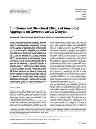
A B Fig. 5. Amyloid aggregate uncouples
the follicular cells. (A) Electron mi-
croscopy of a control follicle (A) con-
trasted with a follicle exposed for 15
min to 1 µM amyloid aggregate (B).
Follicular cells (FC) are collapsed
and detached from the vitelline enve-
lope (double-headed arrow). The
arrow in (B) points to the multiple
granules generated after exposing
the follicle to the amyloid aggregate.
In (C), sample traces of 100 µM
ATP-induced currents of follicles at 0
and 15 min under control conditions
and after a 15-min incubation with 1
µM amyloid aggregate. The inset plot
shows the drop in the ATP-induced
currents of follicles exposed to amy-
loid aggregate. Each bar or dot
(mean ± SEM) was measured in a
least 6 oocytes. The asterisk indi-
cates P < 0.05 (ANOVA).
C
is a Ca2+-dependent Cl- current mediated by TMEM16A (Schro- Morphological changes and uncoupling of follicular cells
eder et al., 2008; Scudieri et al., 2012); this same channel is The amyloid aggregate induced several morphological changes
also responsible for the oscillatory currents generated by serum in the follicles after 15 min of exposure (Fig. 5). First, the colla-
activation of lysophosphatidic acid (LPA) receptors (Tigyi et al., gen fibers and vitelline envelope were found to be disorganized
1991). We tested the effect of exposing the oocytes to amyloid as compared with the control; second, follicular cells were com-
aggregate for 15 min and observed larger Tout currents. This pacted and frequently detached from the oocyte proper; third,
effect was even more evident when high Ca2+ Ringer’s medium cortical granules were smaller and more numerous in the
was used in the recording bath (Figs. 4A and 4B), in which the treated oocytes. Physical detachment of the follicular cells was
Tout current reached up to 200 ± 0.12 nA when oocytes were clearly evident in the EM images, suggesting that the follicular
exposed to the amyloid, i.e., 150% higher than control Tout cur- cells had become uncoupled from the oocyte. To explore this
rents. Thus, the high-Ca2+ Ringer’s consistently increased the idea, we measured the currents induced by ATP in follicles
Tout current by 75 ± 2% compared to the currents reached by before and after exposure to amyloid aggregate. If the follicular
oocytes in normal Ringer’s, whereas the increases due to the cells were detached, smaller ATP-induced currents would be
presence of amyloid aggregate were 48 ± 1.9% and 149 ± expected due to a reduction in the number of gap junctions that
3.1%, in normal and high-Ca2+ Ringer’s, respectively. allow the passage of second messengers from the follicular
To test the effect of amyloid aggregate on the oscillatory Cl- cells to the oocyte (Miledi and Woodward, 1989). Figure 5C
current elicited by serum activation, we applied rabbit serum shows sample currents elicited by ATP (100 µM) in control
(1:100) onto oocytes held at -60 mV, which induced oscillatory follicles and after 15 min exposure to 1 µM amyloid aggregate.
currents (Fig. 4C) due to the release of Ca2+ from intracellular In control oocytes, consecutive applications of 100 µM ATP
stores. Two consecutive applications of serum, separated by 15 reduced the currents to 60 ± 12% of the initial response (Fig.
min, illustrate that there is some receptor desensitization, to 85 5C, inset), a typical response due to desensitization of the ATP
± 3.1% the control; in contrast, when amyloid aggregate are receptors. In contrast, when the follicle was exposed to the
included in the bath, the response to serum increases to about amyloid aggregate, the second response to ATP was reduced
50 ± 1.8% relative to the control (Figs. 4C and 4D). to 20 ± 4.3% of the first application. This reduction of the ATP-
induced current indicates that amyloid aggregate induces the
http://molcells.org Mol. Cells 353
6. Amyloid-Induced Activity in Follicles
Jorge Parodi et al.
physical detachment of the follicular cells from the oocyte and gregate on the plasma membrane, and may therefore be useful
thereby disrupts their electrical coupling, as indicated by the against Alzheimer’s disease. This model will enable the study of
reduction of the ATP currents and is consistent with the ultra- basic molecular mechanisms associated with amyloid pore
structural images. formation and its effect on cell to cell communication.
DISCUSSION ACKNOWLEDGMENTS
This work was partially supported by grants from CONACYT
The observations described in this report derive from our finding 101851 and UNAM-PAPIIT 204806 (to AM-T and RM). J.P. is a
that X. laevis follicles exposed to amyloid aggregate increased postdoctoral fellow from CTIC-UNAM and was supported by a
their electrical activity, observed as changes in the amplitude of travel grant from MECESUP - PUC/0708 of Pontificia Universi-
spontaneous activity (blips). One of the fundamental problems dad Católica de Chile. The authors thank Dr. D. Pless (INB-
to solve in the field of Alzheimer’s disease is to elucidate the UNAM) for editing the manuscript. We are also in debt to E.
modes of action of the Aβ peptide; here, we demonstrate the Ruiz Alcíbar, E. Espino, L. Palma, and E. Pérez for technical
ability of the amyloid aggregate to disrupt the contacts between assistance. AM-T thanks support from PASPA (Programa de
oocyte and follicular cells, thus uncoupling their electro-che- Apoyo para la Superación del Personal Académico-UNAM) for
mical communication. This paves the way to use the Xenopus sabatical fellowship. WinWCP was kindly provided by Dr. J.
follicle as a tool to screen for molecules that could block the Dempster, University of Strathclyde in Glasgow, U.K.
action of amyloid aggregate on the plasma membrane.
Amyloid aggregate induces “channel activity” in neurons REFERENCES
grown in vitro as well as in immortalized cell lines (Kawahara et
al., 2000), and more recently, this activity was probed in hippo- Arellano, R.O., Woodward, R.M., and Miledi, R. (1996). Ion chan-
campal neurons (Sepulveda et al., 2010). In neurons in culture, nels and membrane receptors in follicle-enclosed Xenopus oo-
cytes. Ion Channels 4, 203-259.
β-amyloid acts like a pore-forming neurotoxin, increasing intra- -
Arellano, R.O., Garay, E., and Miledi, R. (1998). Cl currents acti-
cellular Ca2+ and leading to depletion of synaptic vesicles. This vated via purinergic receptors in Xenopus follicles. Am. J. Phy-
observation led us to compare the membrane electric behavior siol. 274, C333-340.
of oocytes exposed to known membrane-perforating agents Arispe, N. (2004). Architecture of the Alzheimer’s A beta P ion
with that induced by amyloid aggregate. The blips recorded in channel pore. J. Membr. Biol. 197, 33-48.
oocytes exposed to gramicidin, amphotericin B, or amyloid Arispe, N., Pollard, H.B., and Rojas, E. (1994). The ability of amy-
2+
loid beta-protein [A beta P (1-40)] to form Ca channels pro-
aggregate had similar amplitudes: 7.8, 6.3, and 6.8 nA, respec- vides a mechanism for neuronal death in Alzheimer’s disease.
tively, and were clearly larger than those recorded in control Ann. N Y Acad. Sci. 747, 256-266.
cells (4.5 nA). Several studies suggest that amyloid aggregate Avila, M.E., Sepulveda, F.J., Burgos, C.F., Moraga-Cid, G., Parodi,
permit Ca2+ entry into the cells and increase membrane con- J., Moon, R.T., Aguayo, L.G., Opazo, C., and De Ferrari, G.V.
ductance in artificial lipid bilayers, clonal cell lines, and neurons (2010). Canonical Wnt3a modulates intracellular calcium and
in culture (Arispe, 2004; Sepulveda et al., 2010). Furthermore, enhances excitatory neurotransmission in hippocampal neurons.
J. Biol. Chem. 285, 18939-18947.
modifications in the Ca2+ concentration are important for devel- Bourin, M., Ripoll, N., and Dailly, E. (2003). Nicotinic receptors and
oping AD. In the oocyte, the amyloid aggregate induces blips Alzheimer’s disease. Curr. Med. Res. Opin. 19, 169-177.
whose reverse equilibrium potential is near to that of Ca2+ and Daniels, W.M., Hendricks, J., Salie, R., and Taljaard, J.J. (2001). The
whose amplitude is significantly affected by removal of Na+ or role of the MAP-kinase superfamily in beta-amyloid toxicity. Me-
K+, suggesting that Ca2+ is the main ion responsible for these tab. Brain Dis. 16, 175-185.
Haass, C., and Selkoe, D.J. (2007). Soluble protein oligomers in
spontaneous electric events, nevertheless other cationic con-
neurodegeneration: lessons from the Alzheimer’s amyloid β-
ductances could not be excluded. These observations are con- peptide. Nat. Rev. Mol. Cell Biol. 8, 101-112.
sistent with the results showing that reducing extracellular Ca2+ Hsieh, H., Boehm, J., Sato, C., Iwatsubo, T., Tomita, T., Sisodia, S.,
decreased the blips generated by amyloid aggregate and have and Malinow, R. (2006). AMPAR removal underlies Abeta-in-
an impact on the oocyte’s endogenous, Ca2+-dependent cur- duced synaptic depression and dendritic spine loss. Neuron 52,
rents, such as the Tout and Ca2+- dependent oscillatory Cl- cur- 831-843.
Jang, H., Zheng, J., and Nussinov, R. (2007). Models of beta-amy-
rents generated by serum. In addition, membrane blips are loid ion channels in the membrane suggest that channel forma-
practically absent when Ca2+- free Ringer´s was used to bathe tion in the bilayer is a dynamic process. Biophys. J. 93, 1938-1949.
the oocytes. Kawahara, M., Kuroda, Y., Arispe, N., and Rojas, E. (2000). Alz-
Although our results do not show direct evidence for the gen- heimer’s beta-amyloid, human islet amylin, and prion protein
eration of an Aβ amyloid ion channel, they clearly show the fragment evoke intracellular free calcium elevations by a com-
induction of new spontaneous conductances in the plasma mon mechanism in a hypothalamic GnRH neuronal cell line. J.
Biol. Chem. 275, 14077-14083.
membrane of the oocyte and the eventual detachment of fol- Maccioni, R.B., Otth, C., Concha, II., and Munoz, J.P. (2001). The
licular cells from the oocyte. Follicular cells were collapsed and protein kinase Cdk5. Structural aspects, roles in neurogenesis
separated from the oocyte after only 15 min of contact with and involvement in Alzheimer’s pathology. Eur. J. Biochem. 268,
amyloid aggregate. Other major structural changes included 1518-1527.
degeneration of the basal lamina and collapse of intracellular Mattson, M.P., and Chan, S.L. (2001). Dysregulation of cellular cal-
cium homeostasis in Alzheimer's disease: bad genes and bad
granules within the oocyte. Thus, amyloid aggregate impacted
habits. J. Mol. Neurosci. 17, 205-224.
directly the ultrastructure of X. laevis follicles, leading to changes Mezler, M., Barghorn, S., Schoemaker, H., Gross, G., and Nim-mrich,
in the chemical communication among these cells. This may V. (2012). Abeta oligomer directly modulates P/Q-type calcium
help to understand how the amyloid aggregate modify, in the currents in Xenopus oocytes. Br. J. Pharmacol. 165, 1572-1583
short term, the neuronal structure and neurotransmission prop- Miledi, R. (1982). A calcium-dependent transient outward current in
erties in patients affected by Alzheimer’s disease. Xenopus laevis oocytes. Proc. R Soc. Lond. B. Biol. Sci. 215, 491-
497.
Xenopus frog oocytes could be a suitable experimental Miledi, R., and Woodward, R.M. (1989). Effects of defolliculation on
model for screening drugs that block the effect of amyloid ag- membrane current responses of Xenopus oocytes. J. Physiol.
354 Mol. Cells http://molcells.org
7. Amyloid-Induced Activity in Follicles
Jorge Parodi et al.
416, 601-621. pression cloning of TMEM16A as a calcium-activated chloride
Pandya, A., and Yakel, J.L. (2011). Allosteric modulator Desfor- channel subunit. Cell 134, 1019-1029.
mylflustrabromine relieves the inhibition of alpha2beta2 and al- Scudieri, P., Sondo, E., Ferrera, L., and Galietta, L.J. (2012). The
pha4beta2 nicotinic acetylcholine receptors by beta-amyloid(1- anoctamin family: TMEM16A and TMEM16B as calcium-activa-
42) peptide. J. Mol. Neurosci. 45, 42-47. ted chloride channels. Exp. Physiol. 97, 177-183.
Parodi, J., Sepulveda, F.J., Roa, J., Opazo, C., Inestrosa, N.C., and Selkoe, D.J. (2002). Alzheimer’s disease is a synaptic failure. Sci-
Aguayo, L.G. (2009). Beta-amyloid causes depletion of synaptic ence 298, 789-791.
vesicles leading to neurotransmission failure. J. Biol. Chem. 285, Sepulveda, F.J., Parodi, J., Peoples, R.W., Opazo, C., and Aguayo,
2506-2514. L.G. (2010). Synaptotoxicity of Alzheimer beta amyloid can be
Quist, A., Doudevski, I., Lin, H., Azimova, R., Ng, D., Frangione, B., explained by its membrane perforating property. PLoS One 5,
Kagan, B., Ghiso, J., and Lal, R. (2005). Amyloid ion channels: a e11820.
common structural link for protein-misfolding disease. Proc. Natl. Shankar, G.M., Bloodgood, B.L., Townsend, M., Walsh, D.M.,
Acad. Sci. USA 102, 10427-10432. Selkoe, D.J., and Sabatini, B.L. (2007). Natural oligomers of the
Reiser, G., and Miledi, R. (1989). Changes in the properties of syn- Alzheimer amyloid-beta protein induce reversible synapse loss
aptic channels opened by acetylcholine in denervated frog mus- by modulating an NMDA-type glutamate receptor-dependent
cle. Brain Res. 479, 83-97. signaling pathway. J. Neurosci. 27, 2866-2875.
Roberson, E.D., and Mucke, L. (2006). 100 years and counting: Texido, L., Martin-Satue, M., Alberdi, E., Solsona, C., and Matute, C.
prospects for defeating Alzheimer’s disease. Science 314, 781- (2011). Amyloid beta peptide oligomers directly activate NMDA
784. receptors. Cell Calcium 49, 184-190.
Saldana, C., Garay, E., Rangel, G.E., Reyes, L.M., and Arellano, Tigyi, G., Henschen, A., and Miledi, R. (1991). A factor that acti-
R.O. (2009). Native ion current coupled to purinergic activation vates oscillatory chloride currents in Xenopus oocytes copurifies
via basal and mechanically induced ATP release in Xenopus fol- with a subfraction of serum albumin. J. Biol. Chem. 266, 20602-
licles. J. Cell. Physiol. 218, 355-365. 20609.
Schroeder, B.C., Cheng, T., Jan, Y.N., and Jan, L.Y. (2008). Ex-
http://molcells.org Mol. Cells 355