Premium Bangalore Call Girls Jigani Dail 6378878445 Escort Service For Hot Ma...
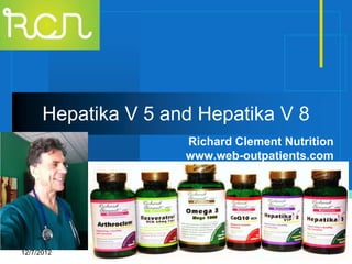
Hepatika V 5 and V 8
1. Hepatika V 5 and Hepatika V 8
Richard Clement Nutrition
www.web-outpatients.com
Company
LOGO
12/7/2012 1
2. Hepatika V 5 or Hepatika V 8
HEPATIKA V 5 (90 Capsules) HEPATIKA 8 VIP (90 Capsules)
Helps Maintain a Healthy Liver Helps Maintain a Healthy Liver
One before each meal with ½ glass of water One before each meal with ½ glass of water
Ingredients: Ingredients:
Milk Thistle (Std. Extract 80% silymarin) 150 mg Milk Thistle (Std. Extract 80% silymarin) 150 mg
Curcuma longa (Std. Extract 95% L,Taurine 150 mg
curcuminoids) 125 mg (Anti-inflammatory, Curcuma longa (Std. Extract 95%
hypocholesterolemic, seems to be a strong curcuminoids) 125 mg (Anti-inflammatory,
anti-cancer substance in many digestive hypocholesterolemic, seems to be a strong
cancer (colon, pancreas, liver) and a cancer anti-cancer substance in many digestive
preventative. cancer (colon, pancreas, liver) and a cancer
Artichoke (Std. Extract cynarine) 125 mg preventative.
(Kidney Drainage Antibacterial, Antifungal) Artichoke (Std. Extract cynarine) 125 mg
Boldo (Std. Extract boldine) 100 mg (Kidney Drainage Antibacterial, Antifungal)
(Vermifuge, Anti-oxidant, Anti-inflammatory, Boldo (Std. Extract boldine) 100 mg
Anti-colitis) (Vermifuge, Anti-oxidant, Anti-inflammatory,
Alpha lipoic acid 50 mg (is Anti-poison and Anti-colitis)
Hepato -protective, it decrease levels of toxicants in Alpha lipoic acid 100 mg (is Anti-poison and
the brain and increase Glutathion levels) Hepato -protective, it decrease levels of toxicants in
the brain and increase Glutathion levels)
N Acetyl L Cysteine 80 mg
Zinc 3 mg
12/7/2012 2
5. Milk Thistle
PLoS One. 2012;7(4):e34630. Epub 2012 Apr 13.
Angiopreventive efficacy of pure flavonolignans from milk thistle extract against prostate cancer:
targeting VEGF-VEGFR signaling.
Deep G, Gangar SC, Rajamanickam S, Raina K, Gu M, Agarwal C, Oberlies NH, Agarwal R.
Source
Skaggs School of Pharmacy and Pharmaceutical Sciences, University of Colorado Denver, Aurora, Colorado,
United States of America.
Abstract
The role of neo-angiogenesis in prostate cancer (PCA) growth and metastasis is well established, but the
development of effective and non-toxic pharmacological inhibitors of angiogenesis remains an unaccomplished
goal. In this regard, targeting aberrant angiogenesis through non-toxic phytochemicals could be an attractive
angiopreventive strategy against PCA. The rationale of the present study was to compare the anti-angiogenic
potential of four pure diastereoisomeric flavonolignans, namely silybin A, silybin B, isosilybin A and isosilybin B,
which we established previously as biologically active constituents in Milk Thistle extract. Results showed that oral
feeding of these flavonolignans (50 and 100 mg/kg body weight) effectively inhibit the growth of advanced human
PCA DU145 xenografts. Immunohistochemical analyses revealed that these flavonolignans inhibit tumor
angiogenesis biomarkers (CD31 and nestin) and signaling molecules regulating angiogenesis (VEGF, VEGFR1,
VEGFR2, phospho-Akt and HIF-1α) without adversely affecting the vessel-count in normal tissues (liver, lung, and
kidney) of tumor bearing mice. These flavonolignans also inhibited the microvessel sprouting from mouse dorsal
aortas ex vivo, and the VEGF-induced cell proliferation, capillary-like tube formation and invasiveness of human
umbilical vein endothelial cells (HUVEC) in vitro. Further studies in HUVEC showed that these diastereoisomers
target cell cycle, apoptosis and VEGF-induced signaling cascade. Three dimensional growth assay as well as co-
culture invasion and in vitro angiogenesis studies (with HUVEC and DU145 cells) suggested the differential
effectiveness of the diastereoisomers toward PCA and endothelial cells. Overall, these studies elucidated the
comparative anti-angiogenic efficacy of pure flavonolignans from Milk Thistle and suggest their usefulness in PCA
12/7/2012 5
6. Milk Thistle
Bratisl Lek Listy. 2012;113(3):145-51.
The comparison of the effects of hepatic regeneration after partial hepatectomy, silybum marinaum,
propofol, N-acetylcysteine and vitamin E on liver.
Yormaz S, Bulbuloglu E, Kurutas EB, Ciralik H, Yuzbasioglu MF, Yildiz H, Coskuner I, Silay E, Kantarceken B,
Goksu M, Senoglu N, Kale IT.
Source
Department of General Surgery, KSU, Faculty of Medicine, Kahramanmaras, Turkey.
Abstract
AIM:
We investigated the comparison of the effects of N-acetylcysteine, silybum marinaum, propofol, and vitamin E on
liver hepatic regeneration after partial hepatectomy.
RESULTS:
Blood samples were used for biochemical parameters (AST, ALT). Ki-67 proliferation index was used for
histopathologic parameters. A statistically meaningful difference was detected in silybum, vitamin E, N-
acetylcysteine, and propofol groups for AST, ALT levels when compared to control and sham groups (p<0.05). Ki-
67 regeneration proliferation index of all groups, which were given agents on the third and seventh days were
statistically higher than the control and sham groups (p<0.05). During the evaluation, AST, ALT, Ki-67, Ro
(regeneration value) levels of silybum group displayed a statistically significant difference according to other
groups (p<0.05).
CONCLUSION:
Our experimental study indicates that hepatic regeneration after partial hepatectomy was meaningful and
significant in groups with intraperitoneal administration of silybum marinaum,vitamin E, N-acetylcysteine and
propofol. Hepatic regeneration rate was particularly higher in silybum group compared to other groups (Fig. 16,
Ref. 26).
12/7/2012 6
7. Milk Thistle
J Oncol Pharm Pract. 2012 Sep;18(3):360-5. doi: 10.1177/1078155212438252. Epub 2012 Feb 29.
Silybum marianum (milk thistle) in the management and prevention of hepatotoxicity in a patient
undergoing reinduction therapy for acute myelogenous leukemia.
McBride A, Augustin KM, Nobbe J, Westervelt P.
Source
Arthur G. James Cancer Hospital, The Ohio State University Department of Pharmacy, Columbus, OH 43210,
USA. alimcbride@gmail.com
Abstract
Hepatotoxicity has been observed with several chemotherapy agents and combination regimens. Conventional
treatment methods often include supportive care or observation. We report a case of a patient with noted
transaminitis presumed secondary to chemotherapy, which did not resolve with supportive care but was shown to
respond to milk thistle. The patient had an immediate decrease in liver function tests and showed decreased
elevation in levels upon treatment with subsequent chemotherapy regimens. This case demonstrates the potential
efficacy of milk thistle as a unique hepatoprotective agent
12/7/2012 7
8. Milk Thistle
Parasit Vectors. 2012 Jan 11;5:9.
Anti-inflammatory/anti-fibrotic effects of the hepatoprotective silymarin and the schistosomicide
praziquantel against Schistosoma mansoni-induced liver fibrosis.
El-Lakkany NM, Hammam OA, El-Maadawy WH, Badawy AA, Ain-Shoka AA, Ebeid FA.
Source
Department of Pharmacology, Theodor Bilharz Research Institute, Warrak El-Hadar, Imbaba, P,O Box 30, Giza
12411, Egypt. naglaaellakkany@yahoo.com
Abstract
BACKGROUND:
Praziquantel (PZQ) is an isoquinoline derivative (2-cyclohexylcarbonyl-1, 2, 3, 6, 7, 11b-hexahydro-4H-
pyrazino{2,1-a}-isoquinoline-4-one), and is currently the drug of choice for all forms of schistosomiasis. Silymarin,
a standardized milk thistle extract, of which silibinin is the main component, is known for its hepatoprotective, anti-
inflammatory, antioxidant activities, and hepatocyte regeneration. This study investigates the anti-
inflammatory/anti-fibrotic effects of silymarin and/or PZQ on schistosomal hepatic fibrosis.
, biochemical and histological parameters that reflect disease severity and morbidity were examined.
RESULTS:
Silymarin caused a partial decrease in worm burden; hepatic tissue egg load, with an increase in percentage of
dead eggs; modulation of granuloma size, with significant reduction of hepatic HYP content; tissue expression of
MMP-2, TGF-β1; number of mast cells, with conservation of hepatic reduced glutathione (GSH). PZQ produced
complete eradication of worms, eggs and alleviated liver inflammation and fibrosis. The best results were obtained,
in most parameters studied, in groups of mice treated with silymarin in addition to PZQ.
CONCLUSIONS:
Our results point to silymarin as a promising anti-inflammatory and anti-fibrotic agent; it could be introduced as a
therapeutic tool with PZQ in the treatment of schistosomal liver fibrosis, but further studies on mechanisms of
silymarin and PZQ in chronic liver diseases may shed light on developing therapeutic methods in clinical practice.
12/7/2012 8
9. Milk Thistle
J Res Med Sci. 2011 Mar;16(3):287-90.
Effects of silybum marianum on patients with chronic hepatitis C.
Kalantari H, Shahshahan Z, Hejazi SM, Ghafghazi T, Sebghatolahi V.
Source
Associate Professor of Gastroenterology, School of Medicine, Isfahan University of Medical Sciences, Isfahan,
Iran.
Abstract
BACKGROUND:
Silymarin derived from silybum marianum (milk thistle), a flowering member of the daisy family, may benefit liver
function in people infected with the hepatitis C virus. The aims of this pilot study were to assess the efficacy and
safety of silymarin on serum hepatitis C virus (HCV) RNA, serum aminotransferases (ALT, AST) levels, liver
fibrosis and well-being in patients with chronic hepatitis C (CHC).
RESULTS:
There was statistically difference in mean of ALT (108.7 ± 86.6 vs 70.3 ± 57.7) before and after the treatment (p <
0.001). The means of AST were 99.4 ± 139.7 and 59.7 ± 64.32 before and after the treatment with statistically
differences (p = 0.004). After the treatment, nine patients were found with negative HCV-RNA (p = 0.004) and
statistically significant improvement in results of liver fibrosis markers were found only in fibrosis group (p = 0.015).
Quality of life was improved significantly (p < 0.001).
CONCLUSIONS:
This study indicated that in patients with CHC performing silymarin (650 mg/day) for 6 months, improved serum
HCV-RNA titer, serum aminotransferases (ALT, AST), hepatic fibrosis and patient's quality of life. More future
studies are warranted.
12/7/2012 9
10. Milk Thistle
Phytother Res. 2012 May;26(5):709-15. doi: 10.1002/ptr.3618. Epub 2011 Oct 20.
Silymarin inhibits cervical cancer cell through an increase of phosphatase and tensin homolog.
Yu HC, Chen LJ, Cheng KC, Li YX, Yeh CH, Cheng JT.
Source
Department of Obstetrics and Gynecology, Zhudong Veterans Hospital, Zhudong City, Taiwan.
Abstract
Silymarin is an active constituent contained in the seeds of the milk thistle plant and is widely used as a hepatic
protection agent due to its antioxidant-like activity. In the present study we evaluated the potential action of
silymarin against cervical cancer and investigated its mechanism of action. Treatment of cervical cancer cells (C-
33A) with silymarin resulted in a significant decrease in cell viability. Silymarin induced apoptosis through the
modulation of Bcl-2 family proteins and activation of caspase 3. Silymarin also inhibited the phosphorylation of Akt
with an increase in expression of phosphatase and tensin homolog (PTEN). We also observed that silymarin
suppressed C-33A cell invasion and wound-healing migration in a concentration-dependent manner. Western-blot
analysis showed that silymarin significantly inhibited the expression of matrix metalloproteinase-9 (MMP-9) in C-
33A cells. Furthermore, we applied siRNA to lower the PTEN gene, which diminished the anticancer actions of
silymarin. Taken together, these results show that silymarin has the potential to suppress the survival, migration
and invasion of C-33A cancer cells; thus, it could be developed as a promising agent for the treatment of cervical
cancer in the future.
12/7/2012 10
11. Milk Thistle
Molecules. 2011 Oct 12;16(10):8601-13.
The protective effects of silymarin against doxorubicin-induced cardiotoxicity and hepatotoxicity in rats.
Rašković A, Stilinović N, Kolarović J, Vasović V, Vukmirović S, Mikov M.
Source
Department of Pharmacology, Toxicology and Clinical Pharmacology, School of Medicine, University of Novi Sad,
21000 Novi Sad, Serbia.
Abstract
Silymarin is a complex of five major compounds, and silibinin is the most biologically active component of the
complex. The aim of this study was to investigate, evaluate and confirm the potential cardioprotective and
hepatoprotective effects of administration of silymarin, rich in silibinin, at a dose of 60 mg/kg orally for a time-span
of 12 days on doxorubicin induced toxicity in male Wistar rats. The in vivo model was used to explore whether
silymarin could prevent damage of liver and heart tissue induced by doxorubicin administered every other day at
dose of 1.66 mg/kg intraperitoneally for twelve days. In the study the change of body weight, ECG changes,
biochemical parameters of oxidative stress, serum activity of alanine and aspartate transaminase, lactate
dehydrogenase, creatine kinase and histological preparations of heart and liver samples of treated animals were
examined. According to physiological, pharmacological, microscopic and biochemical results, we confirmed that at
the examined dose, silymarin exhibits a protective influence on the heart and liver tissue against toxicity induced
by doxorubicin.
12/7/2012 11
13. Alpha Lipoic Acid
Food Chem Toxicol. 2012 Nov 28. pii: S0278-6915(12)00833-2. doi: 10.1016/j.fct.2012.11.026. [Epub ahead of
print]
The effects of alpha lipoic acid on liver cells damages and apoptosis induced by polyunsaturated fatty
acids.
Kaya-Dagistanli F, Tanriverdi G, Altinok A, Ozyazgan S, Ozturk M.
Source
Istanbul University Cerrahpaşa Medical Faculty Medical Biology Department 34098-Cerrahpasa, Istanbul, Turkey.
Electronic address: fkaya@istanbul.edu.tr.
Abstract
We studied the effect of alpha-lipoic acid (ALA) on the liver cell damages and apoptosis by n-6 polyunsaturated
fatty acids (PUFA) rich diet in young rats. 24 Wistar rats were divided into four groups. During the study, 12 of
them (control) were fed with standard chow and other 12 (n-6) were fed with the food containing high-fat n-6 for 8
weeks. At the end of the fourth week, control and n-6 groups were randomly divided into two groups and then, 4
weeks, 35mg/kg ALA are injected. Groups; control, control+ALA, n-6, n-6+ALA. The liver tissue glutathione (GSH)
activity was determined. Immunohistochemistry for caspase-3 and TUNEL method for apoptosis were performed.
The GSH levels have significantly decreased (p<0,001), and vacuolization in the hepatocytes, infiltration and the
collagen accumulation around the central vein, hepatic stellate cells in the sinusoids have increased in n-6 group
compared with the other groups. TUNEL (p<0,001) and caspase-3 (p<0,001) positive cells increased in n-6 group
whereas all degenerative observations decreased in n-6+ALA group. Our results demonstrate that the feeding with
n-6 PUFA causes fatty liver, fibrosis development, inflammations and apoptosis in the liver of young rats. ALA has
a beneficial effects on these degenerative effects.
12/7/2012 13
14. Alpha Lipoic Acid
Biochim Biophys Acta. 2013 Jan;1830(1):2226-32. doi: 10.1016/j.bbagen.2012.10.010. Epub 2012 Oct 17.
Lipoic acid prevents liver metabolic changes induced by administration of a fructose-rich diet.
Castro MC, Massa ML, Schinella G, Gagliardino JJ, Francini F.
Source
CENEXA, Centro de Endocrinología Experimental y Aplicada, UNLP-CONICET LA PLATA, Centro Colaborador
OPS/OMS, 1900 La Plata, Argentina.
Abstract
BACKGROUND:
To evaluate whether co-administration of R/S-α-lipoic acid can prevent the development of oxidative stress and
metabolic changes induced by a fructose-rich diet (F).
RESULTS:
R/S-α-lipoic acid co-administration to F-fed rats a) prevented hyperinsulinemia, hypertriglyceridemia and insulin
resistance, b) improved hepatic insulin sensitivity and glucose tolerance, c) decreased liver oxidative stress and
increased antioxidant capacity and antioxidant enzymes expression, d) decreased uncoupling protein 2 and
PPARδ protein expression and increased PPARγ levels, e) restored the basal gene expression of PPARδ,
SREBP-1c and the lipogenic genes fatty acid synthase and glycerol-3-phosphate acyltransferase, and f)
decreased the fructose-mediated enhancement of glucokinase activity.
CONCLUSIONS:
Our results suggest that fructose-induced oxidative stress is an early phenomenon associated with compensatory
hepatic metabolic mechanisms, and that treatment with an antioxidant prevented the development of such
changes.
GENERAL SIGNIFICANCE:
This knowledge would help to better understand the mechanisms involved in liver adaptation to fructose-induced
oxidative stress and to develop effective strategies to prevent and treat, at early stages, obesity and type 2
diabetes mellitus.
12/7/2012 14
15. Alpha Lipoic Acid
Eur J Nutr. 2012 Oct 12. [Epub ahead of print]
Alpha-lipoic acid upregulates antioxidant enzyme gene expression and enzymatic activity in diabetic rat
kidneys through an O-GlcNAc-dependent mechanism.
Arambašić J, Mihailović M, Uskoković A, Dinić S, Grdović N, Marković J, Poznanović G, Bajec D, Vidaković M.
Source
Department of Molecular Biology, Institute for Biological Research, University of Belgrade, Bulevar despota
Stefana 142, 10060, Belgrade, Serbia.
Abstract
PURPOSE:
The combined hyperglycemia lowering and antioxidant actions of α-lipoic acid (LA) contribute to its usefulness in
preventing renal injury and other diabetic complications. The precise mechanisms by which LA alters diabetic
oxidative renal injury are not known. We hypothesized that LA through its hypoglycemic effect lowers O-
GlcNAcylation which influences the expression and activities of antioxidant enzymes which assume important
roles in preventing diabetes-induced oxidative renal injury.
RESULTS:
An improved glycemic status of LA-treated diabetic rats was accompanied by a significant suppression of oxidative
stress and a reduction of oxidative damage of lipids, proteins and DNA. LA treatment normalized CuZn-superoxide
dismutase (SOD) and catalase activities in renal tissue of diabetic rats. These changes were allied with
upregulated gene expression and lower levels of O-GlcNA glycosylation. The accompanying increase in MnSOD
activity was only linked with upregulated gene expression. The observed antioxidant enzyme gene regulation was
accompanied by nuclear translocation of Nuclear factor-erythroid-2-related factor 2 (Nrf2), enhanced expression of
heat shock proteins (HSPs) and by reduction in O-GlcNAcylation of HSP90, HSP70, and extracellular regulated
kinase and p38.
CONCLUSION:
α-Lipoic acid administration activates a coordinated cytoprotective response against diabetes-induced oxidative
injury in kidney tissue through an O-GlcNAc-dependent mechanism
12/7/2012 15
16. Alpha Lipoic Acid
ScientificWorldJournal. 2012;2012:509838. Epub 2012 May 1.
Alfa-lipoic acid controls tumor growth and modulates hepatic redox state in ehrlich-ascites-carcinoma-
bearing mice.
Al Abdan M.
Source
Zoology Department, Faculty of Science, Princess Nora Bint AbdulRahman University, Riyadh 11481, Saudi
Arabia. moalabdan@yahoo.com
Abstract
The effect of oral supplementation of α-lipoic (LA) on growth of Ehrlich ascites carcinoma cells (EACs) and hepatic
antioxidant state in mice was investigated. The results revealed that α-lipoic (LA) acid at 50 mg/kg body wt
reduced the viability and volume of EAC cells and increased the survival of the treated animals. In addition, LA
normalized oxidative stress in liver of mice-bearing EAC cells evidenced by increasing the levels of total thiols,
glutathione, glutathione-S-transferase, superoxide dismutase, and catalyse. On the other hand, significant
decreases in the levels of malondialdehyde and protein carbonyl were demonstrated in liver indicating controlled
oxidative stress in these animals. As a consequence, LA regulated liver enzymes, alkaline phosphatase, aspartate
aminotransferase, alanine aminotransferase, and gamma-glutamyl transferase. The data also indicated the
efficiency of LA as cancer inhibitor and therapeutic influence. In conclusion, the present data suggest LA as a
potential therapeutic complement in the treatment or prevention of different pathologies that may be related to an
imbalance of the cellular oxidoreductive status associated with malignancy
12/7/2012 16
17. Alpha Lipoic Acid
Neurochem Int. 2012 Dec;61(7):1231-41. doi: 10.1016/j.neuint.2012.09.003. Epub 2012 Sep 17.
The oxidative damage and inflammation caused by pesticides are reverted by lipoic acid in rat brain.
Astiz M, de Alaniz MJ, Marra CA.
Source
INIBIOLP (Instituto de Investigaciones Bioquímicas de La Plata), CCT La Plata, CONICET-UNLP, Cátedra de
Bioquímica y Biología Molecular, Facultad de Ciencias Médicas, Universidad Nacional de La Plata, Calles 60 y
120, 1900 La Plata, Argentina.
Abstract
We have previously demonstrated that the administration of low doses of dimethoate, glyphosate and zineb to rats
(i.p. 1/250 LD50, three times a week for 5weeks) provokes severe oxidative stress (OS) in specific brain regions:
substantia nigra, cortex and hippocampus. These effects were also observed in plasma. Lipoic acid (LA) is
considered an "ideal antioxidant" due to its ability to scavenge reactive species, reset antioxidant levels and cross
the blood-brain barrier. To investigate its protective effect we administered LA (i.p. 25, 50 and 100mg/kg)
simultaneously with the pesticide mixture (PM) for 5weeks. After suppression of PM administration, we evaluated
the restorative effect of LA for a further 5weeks. LA prevented OS and the production of nitrites+nitrates [NOx]
caused by PM in a dose-dependent manner. The PM-induced decrease in reduced glutathione and α-tocopherol
levels in all brain regions was completely restored by LA at both high doses. PM administration also caused an
increase in prostaglandins E(2) and F(2α) in brain that was reduced by LA in a dose-dependent fashion. Taking
into account the relationship between OS, inflammation and apoptosis, we measured caspase and calpain activity.
Only milli- and micro-calpain isoforms were increased in the PM-treated group and LA reduced the activities to
basal levels. We also demonstrated that interrupting PM administration is not enough to restore the levels of all the
parameters measured and that LA is necessary to achieve basal status. In our experimental model LA displayed a
protective role against pesticide-induced damage, suggesting that LA administration is a promising therapeutic
strategy to cope with disorders suspected to be caused by OS generators, especially in brain.
12/7/2012 17
18. Alpha Lipoic Acid
Cancer Biol Ther. 2012 Sep 6;13(14). [Epub ahead of print]
Lipoic acid inhibits cell proliferation of tumor cells in vitro and in vivo.
Feuerecker B, Pirsig S, Seidl C, Aichler M, Feuchtinger A, Bruchelt G, Senekowitsch-Schmidtke R.
Abstract
Cancer cells convert glucose preferentially to lactate even in the presence of oxygen (aerobic glycolysis-Warburg
effect). New concepts in cancer treatment aim at inhibition of aerobic glycolysis. Pyruvate dehydrogenase converts
pyruvate to acetylCoA thus preventing lactate formation. Therefore, the aim of this study was to evaluate
compounds that could activate pyruvate dehydrogenase in cancer cells. We investigated the effects of (R)-(+)-
alpha-lipoic acid (LPA) and dichloroacetate (DCA), possible activators of pyruvate dehydrogenase, on suppression
of aerobic glycolysis and induction of cell death. The neuroblastoma cell lines Kelly, SK-N-SH, Neuro-2a and the
breast cancer cell line SkBr3 were incubated with different concentrations (0.1-0 mM) of LPA and DCA. The
effects of both compounds on cell viability/proliferation (WST-1 assay), [18F]-FDG uptake, lactate production and
induction of apoptosis (flow cytometric detection of caspase-3) were evaluated. Furthermore, NMRI nu/nu mice
that had been inoculated s.c. with SkBr3 cells were treated daily for four weeks with LPA (i.p, 18.5 mg/kg) starting
at day 7 p.i.. Tumour development was measured with a sliding calliper and monitored via [18F]-FDG-PET.
Residual tumours after therapy were examined histopathologically. These data suggests that LPA can reduce (i)
cell viability/proliferation, (ii) uptake of [18F]-FDG and (iii) lactate production and increase apoptosis in all
investigated cell lines. In contrast, DCA was almost ineffective. In the mouse xenograft model with s.c. SkBr3
cells, daily treatment with LPA retarded tumor progression. Therefore, LPA seems to be a promising compound for
cancer treatment.
12/7/2012 18
19. Alpha Lipoic Acid
Biogerontology. 2012 Oct;13(5):479-88. doi: 10.1007/s10522-012-9392-5. Epub 2012 Aug 1.
Dietary supplementation with N-acetyl cysteine, α-tocopherol and α-lipoic acid reduces the extent of
oxidative stress and proinflammatory state in aged rat brain.
Thakurta IG, Chattopadhyay M, Ghosh A, Chakrabarti S.
Source
Department of Biochemistry, Institute of Postgraduate Medical Education and Research, 244, Acharya J.C. Bose
Road, Kolkata, 700020, India.
Abstract
The present study has attempted to understand how oxidative stress contributes to the development of
proinflammatory state in the brain during aging. Three groups of rats have been used in this study: young (4-
6 months, Group I), aged (22-24 months, Group II) and aged with dietary antioxidant supplementation (Group III).
The antioxidants were given daily from 18 months onwards in the form of a combination of N-acetyl cysteine
(50 mg/100 g body weight), α-lipoic acid (3 mg/100 g body weight), and α-tocopherol (1.5 mg/100 g body weight)
till the animals were used for the experiments between 22 and 24 months. Several measurements have been
made to evaluate the ROS (reactive oxygen species) production rate, the levels of proinflammatory cytokines (IL-
1β, IL-6 and TNF-α) and the activation status of NF-κβ (p65 subunit) in brain of the three groups of rats under the
study. Our results reveal that brain aging is accompanied with a significant increase in NADPH oxidase activity
and mitochondrial ROS production, a distinct elevation of IL-1β, IL-6 and TNF-α levels along with increased
nuclear translocation of NF-κβ (p65 subunit) and all these phenomena are partially but significantly prevented by
the long-term dietary antioxidant treatment. The results imply that chronic dietary antioxidants by preventing
oxidative stress and proinflammatory state may produce beneficial effects against multiple age-related deficits of
the brain.
12/7/2012 19
20. Curcuma Longa
AAPS J. 2012 Nov 10. [Epub ahead of print]
Therapeutic Roles of Curcumin: Lessons Learned from Clinical Trials.
Gupta SC, Patchva S, Aggarwal BB.
Source
Cytokine Research Laboratory, Department of Experimental Therapeutics, The University of Texas MD Anderson
Cancer Center, 1901 East Road, Unit # 1950, Houston, Texas, 77054, USA.
Abstract
Extensive research over the past half century has shown that curcumin (diferuloylmethane), a component of the
golden spice turmeric (Curcuma longa), can modulate multiple cell signaling pathways. Extensive clinical trials
over the past quarter century have addressed the pharmacokinetics, safety, and efficacy of this nutraceutical
against numerous diseases in humans. Some promising effects have been observed in patients with various pro-
inflammatory diseases including cancer, cardiovascular disease, arthritis, uveitis, ulcerative proctitis, Crohn's
disease, ulcerative colitis, irritable bowel disease, tropical pancreatitis, peptic ulcer, gastric ulcer, idiopathic orbital
inflammatory pseudotumor, oral lichen planus, gastric inflammation, vitiligo, psoriasis, acute coronary syndrome,
atherosclerosis, diabetes, diabetic nephropathy, diabetic microangiopathy, lupus nephritis, renal conditions,
acquired immunodeficiency syndrome, β-thalassemia, biliary dyskinesia, Dejerine-Sottas disease, cholecystitis,
and chronic bacterial prostatitis. Curcumin has also shown protection against hepatic conditions, chronic arsenic
exposure, and alcohol intoxication. Dose-escalating studies have indicated the safety of curcumin at doses as high
as 12 g/day over 3 months. Curcumin's pleiotropic activities emanate from its ability to modulate numerous
signaling molecules such as pro-inflammatory cytokines, apoptotic proteins, NF-κB, cyclooxygenase-2, 5-LOX,
STAT3, C-reactive protein, prostaglandin E(2), prostate-specific antigen, adhesion molecules, phosphorylase
kinase, transforming growth factor-β, triglyceride, ET-1, creatinine, HO-1, AST, and ALT in human participants. In
clinical trials, curcumin has been used either alone or in combination with other agents. Various formulations of
curcumin, including nanoparticles, liposomal encapsulation, emulsions, capsules, tablets, and powder, have been
examined. In this review, we discuss in detail the various human diseases in which the effect of curcumin has
been investigated.
12/7/2012 20
21. Curcuma Longa
Curr Pharm Des. 2012 Oct 23. [Epub ahead of print]
The Clinical Applications of Curcumin: Current State and the Future.
Fan X, Zhang C, Liu DB, Yan J, Liang HP.
Source
State Key Laboratory of Trauma, Burns and Combined Injury, Research Institute of Surgery, Daping Hospital, The
Third Military Medical University, Chongqing 400042, China. huaping_liang@yahoo.com.cn.
Abstract
Curcumin is a natural polyphenol product derived from the rhizome of the Curcuma longa. In vivo and in vitro
studies have uncovered many important bioactivities of curcumin, such as antioxidant activity, inducing cell
apoptosis, inhibiting cell proliferation, anti-cell adhesion and motility, anti-angiogenesis and anti-microbe
properties. Based on these functions, curcumin has been used in clinical trials on various inflammatory diseases
and cancers. In the future, it will be necessary to focus attention partly on the clinical application of curcumin in
neurodegenerative diseases, cardiovascular diseases and diabetes, because many experiments have clarified the
potential value of curcumin in these areas. As a diet-derived agent, curcumin has no severe toxicity except for
minor gastrointestinal side effects even up to the dosage of 8 grams for 3 months. However, curcumin has a low
systemic bioavailability, so it is imperative to improve the bioavailability of curcumin in its clinical application. Many
methods, such as adjuvant drug delivery system and structural modification have been demonstrated to have a
potential effect.
12/7/2012 21
22. Curcuma Longa
Biofactors. 2012 Sep 20. doi: 10.1002/biof.1041. [Epub ahead of print]
Molecular mechanisms of curcumin action: Gene expression.
Shishodia S.
Source
Department of Biology, Texas Southern University, Houston, TX, USA. shishodias@tsu.edu,
shishodia@gmail.com.
Abstract
Curcumin derived from the tropical plant Curcuma longa has a long history of use as a dietary agent, food
preservative, and in traditional Asian medicine. It has been used for centuries to treat biliary disorders, anorexia,
cough, diabetic wounds, hepatic disorders, rheumatism, and sinusitis. The preventive and therapeutic properties of
curcumin are associated with its antioxidant, anti-inflammatory, and anticancer properties. Extensive research over
several decades has attempted to identify the molecular mechanisms of curcumin action. Curcumin modulates
numerous molecular targets by altering their gene expression, signaling pathways, or through direct interaction.
Curcumin regulates the expression of inflammatory cytokines (e.g., TNF, IL-1), growth factors (e.g., VEGF, EGF,
FGF), growth factor receptors (e.g., EGFR, HER-2, AR), enzymes (e.g., COX-2, LOX, MMP9, MAPK, mTOR,
Akt), adhesion molecules (e.g., ELAM-1, ICAM-1, VCAM-1), apoptosis related proteins (e.g., Bcl-2, caspases, DR,
Fas), and cell cycle proteins (e.g., cyclin D1). Curcumin modulates the activity of several transcription factors (e.g.,
NF-κB, AP-1, STAT) and their signaling pathways. Based on its ability to affect multiple targets, curcumin has the
potential for the prevention and treatment of various diseases including cancers, arthritis, allergies,
atherosclerosis, aging, neurodegenerative disease, hepatic disorders, obesity, diabetes, psoriasis, and
autoimmune diseases. This review summarizes the molecular mechanisms of modulation of gene expression by
curcumin.
12/7/2012 22
23. Artichoke
Int J Food Sci Nutr. 2012 Jun 29. [Epub ahead of print]
Beneficial effects of artichoke leaf extract supplementation on increasing HDL-cholesterol in subjects
with primary mild hypercholesterolaemia: a double-blind, randomized, placebo-controlled trial.
Rondanelli M, Giacosa A, Opizzi A, Faliva MA, Sala P, Perna S, Riva A, Morazzoni P, Bombardelli E.
Source
Section of Human Nutrition, Health Sciences Department, Azienda di Servizi alla Persona, University of Pavia ,
Pavia , Italy.
Abstract
The aim of this study was to evaluate the effects of artichoke leaf extract (ALE) supplementation (250 mg, 2 b.i.d.)
on the lipid pattern. A randomized, double-blind, placebo-controlled clinical trial was performed on 92 overweight
subjects with primary mild hypercholesterolaemia for 8 weeks. Forty-six subjects were randomized to
supplementation (age: 54.2 ± 6.6 years, body mass index (BMI): 25.8 ± 3.9 kg/m(2), male/female: 20/26) and 46
subjects to placebo (age: 53.8 ± 9.0 years, BMI: 24.8 ± 1.6 kg/m(2), male/female: 21/25). Verum supplementation
was associated with a significant increase in mean high-density lipoprotein (HDL)-cholesterol (p < 0.001) and in
mean change in HDL-cholesterol (HDL-C) (p = 0.004). A significantly decreased difference was also found for the
mean change in total cholesterol (p = 0.033), low-density lipoprotein (LDL)-cholesterol (p < 0.001), total
cholesterol/HDL ratio (p < 0.001) and LDL/HDL ratio (p < 0.001), when verum and placebo treatment were
compared. These results indicate that ALE could play a relevant role in the management of mild
hypercholesterolaemia, favouring in particular the increase in HDL-C, besides decreasing total cholesterol and
LDL-cholesterol.
12/7/2012 23
24. Artichoke
J Cell Physiol. 2012 Sep;227(9):3301-9. doi: 10.1002/jcp.24029.
Artichoke polyphenols induce apoptosis and decrease the invasive potential of the human breast cancer
cell line MDA-MB231.
Mileo AM, Di Venere D, Linsalata V, Fraioli R, Miccadei S.
Source
Department of Development of Therapeutic Programs, Regina Elena National Cancer Institute, Rome, Italy.
Abstract
The human breast cancer cell line, estrogen receptor negative, MDA-MB231, was used to evaluate the antitumor
effect of polyphenolic extracts from the edible part of artichokes (AEs). Treatment of cancer cells reduced cell
viability and inhibited cell growth in a dose-dependent manner. Importantly, AEs did not have any effect on normal
breast epithelial cell line, MCF10A. Chlorogenic acid (ChA), the most representative component of the
polyphenolic fraction of artichoke, had no prominent effects on the cell death rate of MDA-MB231 cells. The
addition of AEs to the cells, rather than ChA, triggered apoptosis via a mitochondrial and a death-receptor
pathway, as shown by the activation of caspase-9 and caspase-8, respectively. Furthermore, an increase of the
Bax:Bcl2 ratio and up-regulation of cyclin-dependent kinase inhibitor, p21(WAF1), crucial apoptotic players, were
documented. According to our data on activation of caspase-9, a loss of mitochondrial transmembrane potential
(Ψ(m)) was shown. Cell motility and invasion capabilities were remarkably inhibited by AEs-treatment in highly
invasive MDA-MB231 cells. In addition, a significant decrease of proteolytic activity of metalloproteinase-2 protein
(MMP-2), involved in degrading components of the extracellular matrix, was detected. Our findings indicate that
AEs reduced cell viability, inhibited cell growth, triggered apoptotic mechanisms, and showed inhibitory properties
against the invasive behavior of MDA-MB231 cancer cell line. Altogether, these data indicate the potential
chemopreventive activity of artichoke polyphenolic extracts.
12/7/2012 24
25. Artichoke
Phytother Res. 2010 Jun;24 Suppl 2:S123-8.
The effect of artichoke (Cynara scolymus L.) extract on respiratory chain system activity in rat liver
mitochondria.
Juzyszyn Z, Czerny B, Myśliwiec Z, Pawlik A, Droździk M.
Source
Department of Medical Chemistry, Pomeranian Medical University, Szczecin, Poland. zjuzyszyn@wp.pl
Erratum in
Phytother Res. 2010 Jun;24 Suppl 2:S233-4.
Abstract
The effect of artichoke extract on mitochondrial respiratory chain (MRC) activity in isolated rat liver mitochondria
(including reaction kinetics) was studied. The effect of the extract on the activity of isolated cytochrome oxidase
was also studied. Extract in the range of 0.68-2.72 microg/ml demonstrated potent and concentration-dependent
inhibitory activity. Concentrations > or =5.4 microg/ml entirely inhibited MRC activity. The succinate oxidase
system (MRC complexes II-IV) was the most potently inhibited, its activity at an extract concentration of 1.36
microg/ml being reduced by 63.3% compared with the control (p < 0.05). The results suggest a complex inhibitory
mechanism of the extract. Inhibition of the succinate oxidase system was competitive (K(i) = 0.23 microg/ml),
whereas isolated cytochrome oxidase was inhibited noncompetitively (K(i) = 126 microg/ml). The results of this
study suggest that the salubrious effects of artichoke extracts may rely in part on the effects of their active
compounds on the activity of the mitochondrial respiratory chain system.
12/7/2012 25
26. Artichoke
Biol Trace Elem Res. 2010 Jun;135(1-3):264-74. Epub 2009 Aug 4.
Effect of artichoke leaf extract on hepatic and cardiac oxidative stress in rats fed on high cholesterol diet.
Küçükgergin C, Aydin AF, Ozdemirler-Erata G, Mehmetçik G, Koçak-Toker N, Uysal M.
Source
Department of Biochemistry, Istanbul Medical Faculty, Istanbul University, Capa, 34093 Istanbul, Turkey.
Abstract
Hypercholesterolemia and lipid peroxidation play complementary roles in atherosclerosis. Artichoke (Cynara
scolymus L., Asteraceae) leaf extract (ALE), rich in antioxidants, has cholesterol-reducing effect. We investigated
the effect of ALE on serum and hepatic lipid levels and pro-oxidant-antioxidant balance in the liver and heart of
hypercholesterolemic rats. Rats were fed on 4% (w/w) cholesterol and 1% cholic acid (w/w) supplemented diet for
1 month. ALE (1.5 g/kg/day) was given by gavage during the last 2 weeks. High cholesterol (HC) diet caused
significant increases in serum and liver cholesterol and triglyceride levels. It increased malondialdehyde (MDA)
and diene conjugate (DC) levels in both tissues. Hepatic vitamin E levels and hepatic and cardiac glutathione
peroxidase (GSH-Px) activities decreased, but superoxide dismutase and glutathione transferase activities,
glutathione, and vitamin C levels remained unchanged due to HC diet. Serum cholesterol and triglyceride levels
and ratio of cholesterol to high-density lipoprotein (HDL)-cholesterol decreased in ALE plus HC-treated rats, but
liver cholesterol and triglyceride levels remained unchanged. Significant decreases in hepatic and cardiac MDA
and DC levels and increases in hepatic vitamin E and GSH-Px activities were observed in ALE-treated
hypercholesterolemic rats. Our results indicate that ALE decreases serum lipids and hypercholesterolemia-
induced pro-oxidant state in both tissues
12/7/2012 26
27. Boldo
Biochem Pharmacol. 2012 Nov 22. pii: S0006-2952(12)00751-4. doi: 10.1016/j.bcp.2012.11.010. [Epub ahead of
print]
Boldine protects endothelial function in hyperglycemia-induced oxidative stress through an antioxidant
mechanism.
Lau YS, Tian XY, Huang Y, Murugan D, Achike FI, Mustafa MR.
Source
Department of Pharmacology, Faculty of Medicine, University of Malaya, Kuala Lumpur 50603, Malaysia.
Abstract
Increased oxidative stress is involved in the pathogenesis and progression of diabetes. Antioxidants are
therapeutically beneficial for oxidative stress-associated diseases. Boldine ([s]-2,9-dihydroxy-1,10-
dimethoxyaporphine) is a major alkaloid present in the leaves and bark of the boldo tree (Peumus boldus Molina),
with known an antioxidant activity. This study examined the protective effects of boldine against high glucose-
induced oxidative stress in rat aortic endothelial cells (RAEC) and its mechanisms of vasoprotection related to
diabetic endothelial dysfunction. In RAEC exposed to high glucose (30mM) for 48hours, pre-treatment with boldine
reduced the elevated ROS and nitrotyrosine formation, and preserved nitric oxide (NO) production. Pre-incubation
with β-NAPDH reduced the acetylcholine-induced endothelium-dependent relaxation; this attenuation was
reversed by boldine. Compared with control, endothelium-dependent relaxation in the aortas of streptozotocin
(STZ)-treated diabetic rats was significantly improved by both acute (1μM, 30min) and chronic (20mg/kg/daily, i.p.,
7 days) treatment with boldine. Intracellular superoxide and peroxynitrite formation measured by DHE
fluorescence or chemiluminescence assay were higher in sections of aortic rings from diabetic rats compared with
control. Chronic boldine treatment normalized ROS over-production in the diabetic group and this correlated with
reduction of NAD(P)H oxidase subunits, NOX2 and p47(phox). The present study shows that boldine reversed the
increased ROS formation in high glucose-treated endothelial cells and restored endothelial function in STZ-
induced diabetes by inhibiting oxidative stress and thus increasing NO bioavailability.
12/7/2012 27
28. Boldo
J Pharm Pharmacol. 2011 Sep;63(9):1219-29. doi: 10.1111/j.2042-7158.2011.01320.x. Epub 2011 Jun 27.
Boldo prevents UV light and nitric oxide-mediated plasmid DNA damage and reduces the expression of
Hsp70 protein in melanoma cancer cells.
Russo A, Cardile V, Caggia S, Gunther G, Troncoso N, Garbarino J.
Source
Department of Biological Chemistry, Medical Chemistry and Molecular Biology, University of Catania, Catania,
Italy. alrusso@unict.it
Abstract
OBJECTIVES:
This study was designed to investigate the potential protective effect of a methanolic extract of Peumus boldus
leaves on UV light and nitric oxide (NO)-mediated DNA damage. In addition, we investigated the growth inhibitory
activity of this natural product against human melanoma cells (M14).
KEY FINDINGS:
The extract (demonstrating the synergistic effect of the constituents boldine and flavonoids), showed a protective
effect on plasmid DNA and selectively inhibited the growth of melanoma cells. But a novel finding was that
apoptosis evoked by this natural product in M14 cells, appears to be mediated, at least in part, via the inhibition of
Hsp70 expression, which may be correlated with a modulation of redox-sensitive mechanisms.
CONCLUSIONS:
These results confirm the promising biological properties of Peumus boldus and encourage in-vivo investigations
into its potential anti-cancer activity.
12/7/2012 28
29. Boldo
Phytother Res. 2009 Jul;23(7):1024-7.
Effect of boldo (Peumus boldus Molina) infusion on lipoperoxidation induced by cisplatin in mice liver.
Fernández J, Lagos P, Rivera P, Zamorano-Ponce E.
Source
Universidad del Bío-Bío, Casilla 447, Chillán, Chile.
Abstract
Peumus boldus Molina (Monimiaceae), commonly referred to as 'boldo', is used in traditional Chilean medicine to
treat hepatic and gastrointestinal diseases. Its leaves are rich in antioxidant compounds, principally alkaloids and
flavonoids. This study evaluates the protective effect of a complete boldo leaf infusion on lipoperoxidation (MDA
determination at 532 nm) induced by cisplatin in mice liver. To determine if the observed effect can be explained
by the action of boldine or catechin, each compound was studied separately. The mice were divided into 8 groups
(n = 6): (I) not treated; (II) treated with cisplatin 6 mg/Kg b.w.; (III) treated with boldo leaf infusion 5%; (IV)
pretreated with boldo leaf infusion 5% and treated with cisplatin 6 mg/Kg b.w.; (V) treated with boldine 50 mg/Kg
b.w.; (VI) pretreated with boldine 50 mg/Kg b.w. and treated with cisplatin 6 mg/kg.b.w.; (VII) treated with catechin;
and (VIII) pretreated with catechin 50 mg/Kg b.w. and treated with cisplatin 6 mg/Kg b.w. As expected, the
treatment with cisplatin significantly increased (p < 0.01) lipoperoxidation in comparison with the non-treated
group. Pretreatment with boldo leaf infusion significantly diminished (p < 0.05) the lipoperoxidation induced by
cisplatin with respect to the animals not pretreated with the infusion. The pretreatments with boldine and catechin
significantly diminished (p < 0.05) the lipoperoxidation induced by cisplatin with respect to the group treated only
with cisplatin. The results suggest that the boldo infusion is acting as a protector with respect to the oxidative
hepatic damage caused by cisplatin, and that this protective ability would be due to the presence in the infusion of
the natural antioxidants boldine and principally catechin. These findings suggest the potential use of the infusion
as a chemoprotector.
12/7/2012 29
30. Boldo
Phytother Res. 2009 Jul;23(7):1024-7.
Effect of boldo (Peumus boldus Molina) infusion on lipoperoxidation induced by cisplatin in mice liver.
Fernández J, Lagos P, Rivera P, Zamorano-Ponce E.
Source Invest New Drugs. 2009 Dec;27(6):517-25. doi: 10.1007/s10637-008-9203-7. Epub 2008 Dec 3.
Boldine: a potential new antiproliferative drug against glioma cell lines.
Gerhardt D, Horn AP, Gaelzer MM, Frozza RL, Delgado-Cañedo A, Pelegrini AL, Henriques AT, Lenz G, Salbego
C.
Source
Programa de Pós graduação em Ciências Biológicas: Bioquímica, Departamento de Bioquímica, Instituto de
Ciências Básicas da Saúde, UFRGS, Rua Ramiro Barcelos 2600, 90035.003, Porto Alegre, RS, Brazil.
Abstract
Malignant gliomas are the most common and devastating primary tumors of the central nervous system. Currently
no efficient treatment is available. This study evaluated the effect and underlying mechanisms of boldine, an
aporphine alkaloid of Peumus boldus, on glioma proliferation and cell death. Boldine decreased the cell number of
U138-MG, U87-MG and C6 glioma lines at concentrations of 80, 250 and 500 muM. We observed that cell death
caused by boldine was cell-type specific and dose-dependent. Exposure to boldine for 24 h did not activate key
mediators of apoptosis. However, it induced alterations in the cell cycle suggesting a G(2)/M arrest in U138-MG
cells. Boldine had no toxic effect on non-tumor cells when used at the same concentrations as those used on
tumor cells. Based on these results, we speculate that boldine may be a promising compound for evaluation as an
anti-cancer agent.
12/7/2012 30
31. L Taurine
Metabolism. 2012 Nov 12. pii: S0026-0495(12)00387-3. doi: 10.1016/j.metabol.2012.10.005. [Epub ahead of print]
Effect of taurine and N-acetylcysteine on methionine restriction-mediated adiposity resistance.
Elshorbagy AK, Valdivia-Garcia M, Mattocks DA, Plummer JD, Orentreich DS, Orentreich N, Refsum H, Perrone
CE.
Source
Department of Pharmacology, University of Oxford, UK; Department of Physiology, Faculty of Medicine, University
of Alexandria, Egypt. Electronic address: amany.elshorbagy@pharm.ox.ac.uk.
Abstract
OBJECTIVES:
Methionine-restricted (MR) rats, which are lean and insulin sensitive, have low serum total cysteine (tCys) and
taurine and decreased hepatic expression and activity indices of stearoyl-coenzyme A desaturase-1 (SCD1).
These effects are partly or completely reversed by cysteine supplementation. We investigated whether reversal of
MR phenotypes can be achieved by other sulfur compounds, namely taurine or N-acetylcysteine (NAC).
RESULTS:
Taurine supplementation of MR rats did not restore weight gain or hepatic Scd1 expression or indices to CF levels,
but further decreased adiposity. Taurine supplementation of CF rats did not affect adiposity, but lowered
triglyceridemia. NAC supplementation in MR rats raised tCys and partly or completely reversed MR effects on
weight, fat %, Scd1 expression in liver and white adipose tissue, and estimated SCD1 activity. In CF rats, NAC
decreased body fat % and lowered SCD1-18 activity index (P<0.001). Serum triglycerides and leptin were over
40% lower in CF+NAC relative to CF rats (P≤0.003 for both). In all groups, change in tCys correlated with change
in SCD1-16 index (partial r=0.60, P<0.001) independent of other SAA.
CONCLUSION:
The results rule out taurine as a mediator of increased adiposity produced by cysteine in MR, and show that NAC,
similar to L-cysteine, blocks anti-obesity effects of MR. Our data show that dietary SAA can influence adiposity in
part through mechanisms that converge on SCD1 function. This may have implications for understanding and
preventing human obesity.
12/7/2012 31
32. L Taurine
Blood Coagul Fibrinolysis. 2012 Dec;23(8):677-9. doi: 10.1097/MBC.0b013e3283597586.
Lowering homocysteine levels may prevent cardiovascular impairments? Possible therapeutic behaviors.
Cacciapuoti F.
Source
Department of Internal Medicine, Second University of Naples, Naples, Italy.
Abstract
Homocysteine (Hcy) is metabolized through two pathways, requiring folates and B6-12 vitamins as cofactors.
Increased Hcy concentration is responsible for early atherosclerosis with possible acute cardiovascular events.
Ample evidence has demonstrated that Hcy lowering with folic acid and B vitamin supplementation, even if
reduces Hcy serum levels, is unable to lower cardiovascular risk. On the contrary, omega-3 fatty acids and some
nutraceuticals, such as N-acetyl cysteine, taurine, or S-adenosyl-methionine, reduce both Hcy serum
concentration and cardiovascular risk. Instead, antiplatelet drugs, such as aspirin and clopidogrel or ticlopidine and
statins only antagonize vascular derangements. Finally, metformin, some lipid-lowering drugs, and some diuretics
should be avoided because they can increase Hcy levels.
12/7/2012 32
33. L Taurine
Exp Mol Med. 2012 Sep 21. [Epub ahead of print]
Taurine ameliorates hyperglycemia and dyslipidemia by reducing insulin resistance and leptin level in
Otsuka Long-Evans Tokushima fatty (OLETF) rats with long-term diabetes.
Kim KS, Oh DH, Kim JY, Lee BG, You JS, Chang KJ, Chung HJ, Yoo MC, Yang HI, Kang JH, Hwang YC, Ahn KJ,
Chung HY, Jeong IK.
Abstract
This study aimed to determine whether taurine supplementation improves metabolic disturbances and diabetic
complications in an animal model for type 2 diabetes. We investigated whether taurine has therapeutic effects on
glucose metabolism, lipid metabolism, and diabetic complications in Otsuka Long- Evans Tokushima fatty
(OLETF) rats with long-term duration of diabetes. Fourteen 50week-old OLETF rats with chronic diabetes were fed
a diet supplemented with taurine (2%) or a non-supplemented control diet for 12 weeks. Taurine reduced blood
glucose levels over 12 weeks, and improved OGTT outcomes at 6 weeks after taurine supplementation, in OLETF
rats. Taurine significantly reduced insulin resistance (HOMA-IR) but did not improve β-cell function (HOMA-β) or
islet mass. After 12 weeks, taurine significantly decreased serum levels of lipids such as triglyceride, cholesterol,
high density lipoprotein cholesterol (HDL-C), and low density lipoprotein cholesterol (LDL-C). Taurine significantly
reduced serum leptin, but not adiponectin levels. However, taurine had no therapeutic effect on damaged tissues.
Taurine ameliorated hyperglycemia and dyslipidemia, at least in part, by improving insulin sensitivity and leptin
modulation in OLETF rats with long-term diabetes. Additional study is needed to investigate whether taurine has
the same beneficial effects in human diabetic patients.
12/7/2012 33
34. L Taurine
Am J Cardiovasc Dis. 2011;1(3):293-311. Epub 2011 Sep 10.
Role of taurine in the vasculature: an overview of experimental and human studies.
Abebe W, Mozaffari MS.
Source
Department of Oral Biology, College of Dental Medicine, Georgia Health Sciences University Augusta, GA, USA.
Abstract
Taurine is a sulfur-containing amino acid-like endogenous compound found in substantial amounts in mammalian
tissues. It exerts a diverse array of biological effects, including cardiovascular regulation, antioxidation, modulation
of ion transport, membrane stabilization, osmoregulation, modulation of neurotransmission, bile acid conjugation,
hypolipidemia, antiplatelet activity and modulation of fetal development. This brief review summarizes the role of
taurine in the vasculature and modulation of blood pressure, based on experimental and human studies. Oral
supplementation of taurine induces antihypertensive effects in various animal models of hypertension. These
effects of taurine have been shown to be both centrally and peripherally mediated. Consistent with this, taurine
produces endothelium-dependent and independent relaxant effects in isolated vascular tissue preparations. Oral
administration of taurine also ameliorates impairment of vascular reactivity, intimal thickening, arteriosclerosis,
endothelial apoptosis, oxidative stress and inflammation, associated primarily with diabetes and, to a lesser extent
with obesity, hypertension and nicotine-induced vascular adverse events. In rat aortic vascular smooth muscle
cells (VSMCs), taurine acts as an antiproliferative and antioxidant agent. In endothelial cells, taurine inhibits
apoptosis, inflammation, oxidative stress and cell death while increasing NO generation. Oral taurine in
hypertensive human patients alleviates the symptoms of hypertension and also reverses arterial stiffness and
brachial artery reactivity in type 1 diabetic patients. However, despite these favorable findings, there is a need to
further establish certain aspects of the reported results and also consider addressing unresolved related issues. In
addition, the molecular mechanism (s) involved in the vascular effects of taurine is largely unknown and requires
further investigations. Elucidation of the mechanisms through which taurine affects the vasculature could facilitate
the development of therapeutic and/or diet-based strategies to reduce the burdens of vascular diseases.
12/7/2012 34
35. L Taurine
Am J Cardiovasc Dis. 2011;1(3):293-311. Epub 2011 Sep 10.
Role of taurine in the vasculature: an overview of experimental and human studies.
Abebe W, Mozaffari MS.
Source
Department of Oral Biology, College of Dental Medicine, Georgia Health Sciences University Augusta, GA, USA.
Abstract
Taurine is a sulfur-containing amino acid-like endogenous compound found in substantial amounts in mammalian
tissues. It exerts a diverse array of biological effects, including cardiovascular regulation, antioxidation, modulation
of ion transport, membrane stabilization, osmoregulation, modulation of neurotransmission, bile acid conjugation,
hypolipidemia, antiplatelet activity and modulation of fetal development. This brief review summarizes the role of
taurine in the vasculature and modulation of blood pressure, based on experimental and human studies. Oral
supplementation of taurine induces antihypertensive effects in various animal models of hypertension. These
effects of taurine have been shown to be both centrally and peripherally mediated. Consistent with this, taurine
produces endothelium-dependent and independent relaxant effects in isolated vascular tissue preparations. Oral
administration of taurine also ameliorates impairment of vascular reactivity, intimal thickening, arteriosclerosis,
endothelial apoptosis, oxidative stress and inflammation, associated primarily with diabetes and, to a lesser extent
with obesity, hypertension and nicotine-induced vascular adverse events. In rat aortic vascular smooth muscle
cells (VSMCs), taurine acts as an antiproliferative and antioxidant agent. In endothelial cells, taurine inhibits
apoptosis, inflammation, oxidative stress and cell death while increasing NO generation. Oral taurine in
hypertensive human patients alleviates the symptoms of hypertension and also reverses arterial stiffness and
brachial artery reactivity in type 1 diabetic patients. However, despite these favorable findings, there is a need to
further establish certain aspects of the reported results and also consider addressing unresolved related issues. In
addition, the molecular mechanism (s) involved in the vascular effects of taurine is largely unknown and requires
further investigations. Elucidation of the mechanisms through which taurine affects the vasculature could facilitate
the development of therapeutic and/or diet-based strategies to reduce the burdens of vascular diseases.
12/7/2012 35
36. L Taurine
Am J Physiol Regul Integr Comp Physiol. 2011 Dec;301(6):R1710-22. Epub 2011 Sep 28.
Experimental evidence for therapeutic potential of taurine in the treatment of nonalcoholic fatty liver
disease.
Gentile CL, Nivala AM, Gonzales JC, Pfaffenbach KT, Wang D, Wei Y, Jiang H, Orlicky DJ, Petersen DR,
Pagliassotti MJ, Maclean KN.
Source
Department of Food Science and Human Nutrition, Colorado State University, Fort Collins, Colorado 80523, USA.
Abstract
The incidence of obesity is now at epidemic proportions and has resulted in the emergence of nonalcoholic fatty
liver disease (NAFLD) as a common metabolic disorder that can lead to liver injury and cirrhosis. Excess sucrose
and long-chain saturated fatty acids in the diet may play a role in the development and progression of NAFLD.
One factor linking sucrose and saturated fatty acids to liver damage is dysfunction of the endoplasmic reticulum
(ER). Although there is currently no proven, effective therapy for NAFLD, the amino sulfonic acid taurine is
protective against various metabolic disturbances, including alcohol-induced liver damage. The present study was
undertaken to evaluate the therapeutic potential of taurine to serve as a preventative treatment for diet-induced
NAFLD. We report that taurine significantly mitigated palmitate-mediated caspase-3 activity, cell death, ER stress,
and oxidative stress in H4IIE liver cells and primary hepatocytes. In rats fed a high-sucrose diet, dietary taurine
supplementation significantly reduced hepatic lipid accumulation, liver injury, inflammation, plasma triglycerides,
and insulin levels. The high-sucrose diet resulted in an induction of multiple components of the unfolded protein
response in the liver consistent with ER stress, which was ameliorated by taurine supplementation. Treatment of
mice with the ER stress-inducing agent tunicamycin resulted in liver injury, unfolded protein response induction,
and hepatic lipid accumulation that was significantly ameliorated by dietary supplementation with taurine. Our
results indicate that dietary supplementation with taurine offers significant potential as a preventative treatment for
NAFLD.
12/7/2012 36
37. L Taurine
Nutr Hosp. 2010 Nov-Dec;25(6):910-9.
Taurine and glucose metabolism: a review.
De la Puerta C, Arrieta FJ, Balsa JA, Botella-Carretero JI, Zamarrón I, Vázquez C.
Source
Unidad de Nutrición Clínica y Dietética, Servicio de Endocrinología y Nutrición, Hospital Universitario Ramón y
Cajal, Madrid, España. cpuerta@us.es
Abstract
INTRODUCTION:
Taurine has probed to be involved in a wide range of biological processes and to provide several different
important health benefits. Its effects have been revealed to be exerted mainly through its antioxidant and anti-
inflammatory effects, among other mechanisms.
OBJECTIVES AND METHODS:
The present review is aimed to provide a solid body of evidence regarding the beneficial effects of taurine in the
context of diabetes and its complications, with an special focus on the cardiovascular health impairments so
frequently associated to this disease, so that data from this updated systematic review of the literature, may
constitute a base to back up future clinical and epidemiological studies, on the possibilities of taurine
supplementation as a useful tool for both prevention and treatment of diabetes complications.
CONCLUSIONS:
We consider results from the different experimental, in vitro studies as well as some clinical ones reviewed, to
provide sufficient evidence as to constitute a solid base to back up future clinical and epidemiological studies on
the usefulness of taurine supplementation both in the prevention and treatment of diabetes and its complications.
12/7/2012 37
38. N-acetylcysteine
Comp Hepatol. 2012 Dec 4;11(1):4. [Epub ahead of print]
N-acetylcysteine improves antitumoural response of Interferon alpha by NF-kB downregulation in liver
cancer cells.
Kretzmann NA, Chiela E, Matte U, Marroni N, Marroni CA.
Abstract
ABSTRACT:
BACKGROUND: Liver cancer is one of the most common malignancies in the world and at the moment, there is
no drug intervention effective for the treatment of liver tumours. Investigate the effect of N-acetylcysteine (NAC),
which has been studied for its antitumoural properties, on the toxicity of hepatocarcinoma (HCC) cells in vitro
when used with the drug interferon alpha-2A (IFN), which is used clinically to treat HCC.
RESULTS:
NAC, IFN and NAC plus IFN reduced cell viability, as determined by MTT assay. More importantly, NAC
potentiates the cytotoxic effect of IFN, with the best response achieved with 10 mM of NAC and 2.5 x 10⁴ of IFN.
These results were confirmed by Annexin/PI staining through flow cytometry and morphologic analyses. Co-
treatment reduced the expression of the nuclear transcription factor kappa-B (NF-kB). In a similar way to NAC,
RNAi against p65 potentiated the toxic effect of IFN, suggesting that, indeed, NAC may be enhancing the effect of
IFN through inhibition of NF-kB.
CONCLUSIONS:
Our results support the notion that NAC may be an important drug for the treatment of liver tumours as primary or
adjuvant therapy. IFN has a limited clinical response, and therefore, the anti-proliferative properties of NAC in the
liver should be explored further as an alternative for non-responders to IFN treatment.
12/7/2012 38
39. N-acetylcysteine
Chem Biol Interact. 2012 Nov 16. pii: S0009-2797(12)00234-7. doi: 10.1016/j.cbi.2012.10.027. [Epub ahead of
print]
Biochemical and molecular mechanisms of N-acetyl cysteine and silymarin-mediated protection against
maneb- and paraquat-induced hepatotoxicity in rats.
Ahmad I, Shukla S, Kumar A, Singh BK, Kumar V, Chauhan AK, Singh D, Pandey HP, Singh C.
Source
CSIR-Indian Institute of Toxicology Research (CSIR-IITR), Lucknow 226 001, India.
Abstract
Oxidative stress is one of the major players in the pathogenesis of maneb (MB) and paraquat (PQ)-induced
disorders. N-acetyl cysteine (NAC), a glutathione (GSH) precursor and silymarin (SIL), a naturally occurring
antioxidant, encounter oxidative stress-mediated cellular damage. The present study was aimed to investigate the
effects of NAC and SIL against MB and/or PQ-induced hepatotoxicity in rats. The levels of hepatotoxicity markers
- alanine aminotransaminase (ALT), aspartate aminotransaminase (AST) and total bilirubin, histological changes,
oxidative stress indices, phase I and phase II xenobiotic metabolizing enzymes - cytochrome P450 (CYP) and
glutathione S-transferase (GST) and pro-inflammatory molecules - inducible nitric oxide synthase (iNOS), tumor
necrosis factor-α/TNF-α and interleukin-1β/IL-1β) were measured in animals treated with MB and/or PQ in the
presence or absence of NAC and SIL. MB and/or PQ augmented ALT, AST, total bilirubin, lipid peroxidation and
nitrite contents and catalytic activities of superoxide dismutase and glutathione peroxidase however, the GSH
content was attenuated. NAC and SIL restored the above-mentioned alterations towards basal levels but the
restorations were more pronounced in SIL treated groups. Similarly, MB and/or PQ-mediated histopathological
symptoms and changes in the catalytic activities/expressions of CYP1A2, CYP2E1, iNOS, TNF-α, and IL-1β were
alleviated by NAC and SIL. Conversely, MB and/or PQ-induced GSTA4-4 expression/activity was further
increased by NAC/SIL and glutathione reductase activity was also increased. The results obtained thus suggest
that NAC and SIL protect MB and/or PQ-induced hepatotoxicity by reducing oxidative stress, inflammation and by
modulating xenobiotic metabolizing machinery and SIL seems to be more effectiv
12/7/2012 39
40. N-acetylcysteine
Scand J Clin Lab Invest. 2012 Oct 30. [Epub ahead of print]
N-acetylcysteine expresses powerful anti-inflammatory and antioxidant activities resulting in complete
improvement of acetic acid-induced colitis in rats.
Uraz S, Tahan G, Aytekin H, Tahan V.
Source
Florence Nightingale Hospital Department of Gastroenterology , Istanbul , Turkey.
Abstract
High free radical production, low antioxidant capacity and excessive inflammation are well known features in the
pathogenesis of inflammatory bowel disease. N-acetylcysteine (NAC) is a powerful antioxidant and a scavenger of
hydroxyl radicals. Recently, NAC has also been shown to have anti-inflammatory activities in tissues. Our study
objective was to investigate the effects of NAC on tissue inflammatory activities using an ulcerative colitis model
induced by acetic acid (AA) in rats. Wistar rats (n = 32) were divided into four groups. AA-induced colitis was
performed in two of the groups while the other two groups were injected with saline intrarectally. One of the AA-
induced colitis groups and one of the control groups were administered NAC (500 mg/kg/day) intrarectally, and the
other control groups were given saline. After 4 days, colonic changes were evaluated biochemically by measuring
proinflammatory cytokines [tumor necrosis factor (TNF)-α, interleukin (IL)-1β and IL-6], myeloperoxidase (MPO),
malondialdehyde (MDA), glutathione (GSH) and superoxide dismutase (SOD) levels in tissue homogenates and
by histopathological examination. AA caused colonic mucosal injury, whereas NAC administration suppressed
these changes in the AA-induced colitis group (p < 0.001). AA-administration resulted in increased TNF-α, IL-1β,
IL-6, MPO and MDA levels, and decreased GSH and SOD levels, whereas NAC reversed these effects (all p <
0.001). In conclusion, the present study proposes that intrarectal NAC therapy has a dual action as an effective
anti-inflammatory and an antioxidant, and may be a promising therapeutic option for ulcerative colitis.
12/7/2012 40
41. N-acetylcysteine
Diabetologia. 2012 Nov;55(11):2920-8. doi: 10.1007/s00125-012-2685-z. Epub 2012 Aug 31.
N-Acetylcysteine inhibits platelet-monocyte conjugation in patients with type 2 diabetes with depleted
intraplatelet glutathione: a randomised controlled trial.
Treweeke AT, Winterburn TJ, Mackenzie I, Barrett F, Barr C, Rushworth GF, Dransfield I, Macrury SM, Megson
IL.
Source
Free Radical Research Facility, Department of Diabetes & Cardiovascular Science, University of the Highlands &
Islands, Centre for Health Science, Inverness, IV2 3JH, UK.
Abstract
AIMS/HYPOTHESIS:
The aim of this study was to determine whether oral dosing with N-acetylcysteine (NAC) increases intraplatelet
levels of the antioxidant, glutathione (GSH), and reduces platelet-monocyte conjugation in blood from patients with
type 2 diabetes.
RESULTS:
Oral NAC reduced platelet-monocyte conjugation (from 53.1 ± 4.5% to 42.5 ± 3.9%) at 2 h after administration and
the effect was maintained after 7 days of dosing. Intraplatelet GSH was raised in individuals with depleted GSH
and there was a negative correlation between baseline intraplatelet GSH and platelet-monocyte conjugation.
There were no adverse events.
CONCLUSIONS/INTERPRETATION:
The NAC-induced normalisation of intraplatelet GSH, coupled with a reduction in platelet-monocyte conjugation,
suggests that NAC might help to reduce atherothrombotic risk in type 2 diabetes.
12/7/2012 41
42. N-acetylcysteine
Zinc and human health: an update.
Chasapis CT, Loutsidou AC, Spiliopoulou CA, Stefanidou ME.
Source
Department of Pharmacy, School of Health Sciences, University of Patras, Patras, Greece.
Abstract
The importance of micronutrients in health and nutrition is undisputable, and among them, zinc is an essential
element whose significance to health is increasingly appreciated and whose deficiency may play an important role
in the appearance of diseases. Zinc is one of the most important trace elements in the organism, with three major
biological roles, as catalyst, structural, and regulatory ion. Zinc-binding motifs are found in many proteins encoded
by the human genome physiologically, and free zinc is mainly regulated at the single-cell level. Zinc has critical
effect in homeostasis, in immune function, in oxidative stress, in apoptosis, and in aging, and significant disorders
of great public health interest are associated with zinc deficiency. In many chronic diseases, including
atherosclerosis, several malignancies, neurological disorders, autoimmune diseases, aging, age-related
degenerative diseases, and Wilson's disease, the concurrent zinc deficiency may complicate the clinical features,
affect adversely immunological status, increase oxidative stress, and lead to the generation of inflammatory
cytokines. In these diseases, oxidative stress and chronic inflammation may play important causative roles. It is
therefore important that status of zinc is assessed in any case and zinc deficiency is corrected, since the unique
properties of zinc may have significant therapeutic benefits in these diseases. In the present paper, we review the
zinc as a multipurpose trace element, its biological role in homeostasis, proliferation and apoptosis and its role in
immunity and in chronic diseases, such as cancer, diabetes, depression, Wilson's disease, Alzheimer's disease,
and other age-related diseases.
12/7/2012 42