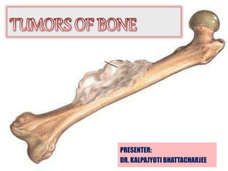
SlideShare a Scribd company logo
1 of 121
PRESENTER:
DR. KALPAJYOTI BHATTACHARJEE
CONTENTS
• Introduction
• Classification
• Benign tumors of bone
• Malignant tumors of bone
• Conclusion
• References
Introduction
• Primary bone tumors are rare;
• Non-neoplastic conditions, metastatic disease, and
lymphohematologic malignancies, which may simulate
primary bone tumors, by far outnumber genuine bone tumors.
• The classification of bone tumor is based on histologic criteria,
particularly on the type of differentiation shown by tumor cells
and the type of intercellular material they produce, as seen by
conventional light microscopy.
• However, it is recognized that electron microscopy and especially
immunohistochemical techniques may be relevant for precise
classification and diagnosis in specific instances.
• The final diagnosis of bone tumors should be based on a
synthesis of histopathologic findings, clinical presentation,
and imaging characteristics.
Predominant tissue Benign Malignant
Bone forming Osteoma
Osteoid osteoma and
osteoblastoma
Osteosarcoma
-central
-peripheral
-parosteal
Cartilage forming Chondroma
Osteochondroma
Chondroblastoma
Chondromyxoid
fibroma
Chondrosarcoma
-Juxtacortical chondrosarcoma
-Mesenchymal chondrosarcoma
-Dedifferentiated chondrosarcoma
-Clear cell chondrosarcoma
-Malignant chondroblastoma
Marrow tumors -Ewing sarcoma
-Primitive neuroectodermal tumor of
bone (PNET)
-Malignant lymphoma of bone
-Myeloma
WHO Histologic Classification of Bone Tumors 1993
Chondroma
• A benign central tumor composed of mature cartilage, is a well-
recognized entity in certain areas of the bony skeleton
• Considerable clinical importance because of the propensity of the
tumor to undergo malignant degeneration in some instances, even
after remaining quiescent for long periods of time.
• Types: A) Central chondroma / Enchondroma- develop within
medullary cavity
• B) Periosteal chondroma / Ecchondroma - develops on the surface
• C) Soft tissue chondroma.
Clinical feature
• Develop at any age and
• Shows no apparent gender predilection
• Site: maxilla: anterior portion of the maxilla
mandible: posterior to the cuspid tooth, involving the
body of the mandible, the coronoid or condylar processes.
• c/p: arises as a painless, slowly progressive swelling of the
jaw, may cause loosening of the teeth
Radiographic Features
• Destructive lesion
• Irregular radiolucent or mottled area in the bone
• Root resorption of teeth adjacent to it
Histological Features
• Chondroma is made up of a mass of hyaline cartilage which
may exhibit areas of calcification or of necrosis.
• The cartilage cells appear small, contain only single nuclei and
do not exhibit great variation in size, shape or staining reaction.
• vary considerably in appearance from area to area.
A, Chondroma. Strands of epithelium-like cells with abundant eosinophilic cytoplasm
reside in a blue-gray mucinous stroma. B, Area of chondroma with tumor cells showing
cytoplasmic vacuolation with the formation of multivacuolated physaliferous cells.
A, Enchondroma shows small, uniform chondrocytes whose nuclei are densely
hyperchromatic (ink dot) without a visible chromatin pattern. Cells are well separated from
each other. B, An island of hyaline cartilage in an enchondroma is separated from the
adjacent bone trabeculae by a zone of normal marrow tissue
Osteoma
• Osteoma is a benign neoplasm characterized by a
proliferation of either compact or cancellous bone,
usually in an endosteal or periosteal location.
• Described as a specific entity by Jaffe in 1935
• Occurs almost exclusively in the head and neck region
• Not a common oral lesion.
Types:
Compact osteoma: consists of compact bone, which has a dense
lamellae of bone
Cancellous osteoma: consisting of trabeculae of bone
Periosteal, peripheral or exophytic osteoma: arise on the
surface of bone as sessile mass
Endosteal or central osteoma: located in the medullary bone
Osteoma cutis: extraskeletal lesion of soft tissue seen in the
dermis of the skin
Clinical features
• Age: second to fourth decades of life,
• males> female
• c/p: slow growing tumor.
• Periosteal origin → circumscribed swelling → obvious asymmetry.
• Endosteal origin → expansion of the cortical plates.
• Multiple osteomas of the jaws, as well as of long bones and skull,
are a characteristic manifestation of Gardner syndrome.
GARDNER SYNDROME
• It is an autosomal dominant disorder.
• Mutation in APC gene
• characterized by the triad of colonic polyposis, multiple
osteomas and mesenchymal tumors of the skin and soft tissues
including epidermal inclusion cyst, lipoma, fibroma, and
fibromatosis
Oral manifestations
• Multiple odontomas, Compound odontomas
• Supernumerary teeth
• Impacted permanent teeth
• One or more osteomas of the jaws
• Occult radiopaque lesions of the jaws are common
• Various incidental findings include hypercementosis, root
resorption, ankylosis and persistent primary teeth.
Histologic Features.
• Composed either of extremely dense, compact bone or of
coarse cancellous bone.
• Bone formed appears normal
• Well circumscribed.
• In some tumors foci of cartilage may be found, in which
case the term ‘osteochondroma’ is often used.
• Myxomatous tissue also may be intermingled on rare
occasions.
Compact and trabecular bone is present beneath intact mucosa at the left of the field.
B, Compact cortical-type bone of the osteoma shown in A contains haversian systems
Treatment and Prognosis
• Symptomatic lesions→ local excision.
• No recurrence after surgical removal.
Osteoid osteoma
• Benign tumor of bone, seldom been described in the jaws.
Etiology:
• True nature → unknown.
• Jaffe and Lichtenstein have suggested “A true neoplasm of
osteoblastic derivation”.
• Trauma
• Inflammation
Classification
Cortical
Cancellous
Intra- articular
Subperiosteal
Clinical Features.
• Age: young persons, under the age of 10 years
• Sex: males:female - 2:1.
• Site: Frequently in the femur or in the tibia.
• In head and neck→ Cervical spine > mandible and maxilla.
• Chief symptoms → severe pain → unrelenting and sharp, worse at
night.
• Classically, the pain is relieved by aspirin.
Pathologic Features
• In its active growth phase
considerable vascularity.
• Grossly appears as a discrete, round to oval lesion marked by a
cherry-red or reddish-brown color. Quite granular and friable and
easily displaced from the adjacent bone.
• In its mature phase → more calcification and bone production, the
lesion is hard and gritty and blends with the bone around it.
Histologic Features
• Characteristic and consists of a central nidus composed of compact
osteoid tissue, varying in degree of calcification, interspersed by a
vascular connective tissue.
• Formation of definite trabeculae occurs, particularly in older
lesions, outlined by active osteoblasts.
• Osteoclasts and foci of bone resorption are also usually evident.
• Overlying periosteum exhibits new bone formation, and in this
interstitial tissue collections of lymphocytes seen.
A, Nidus of osteoid osteoma abuts thickened mature bone. B, Osteoid trabeculae, some
partially calcified, within the nidus of an osteoid osteoma. The trabeculae are rimmed with
plump osteoblasts with occasional osteoclasts. The stroma is hemorrhagic.
Ultrastructural investigation by Steiner,
• The morphology of the osteoblasts to be similar to that of
normal osteoblasts.
• Although atypical mitochondria could be seen.
• Neural staining → many axons throughout an osteoid osteoma,
which probably accounts for the pain (the nidus).
• ↑Levels of prostaglandin E2 in the nidus; this is presumably
the cause of pain and vasodilatation.
Treatment
• NSAIDS- Aspirin relieves the pain
• Surgical curettage
• Lasers
Benign Osteoblastoma
(Giant osteoid osteoma)
• Osteoblastoma accounts →1% of primary bone tumors.
• It is typically a slow-growing, benign bone tumor.
• Incidence in the head and neck → 13% to 26%.
• Osteoblastoma frequently lacks the characteristic pain and the
halo of sclerotic bone associated with osteoid osteoma.
• Benign osteoblastoma → Jaffe and by Lichtenstein in 1956.
Etiopathogenesis
• Jaffe and Lichtenstein stated this lesion to be “a true neoplasm
of osteoblastic derivation”.
• Trauma,
• Inflammation,
• Abnormal local response of the tissues to injury, and
• Local alteration in bone physiology
Clinical Features
• Age: in young persons, 20-30 years.
• Sex: Males>Females.
• C/P: characterized clinically by pain and swelling.
pain → more generalized and less likely relieved by salicylates.
• Most common site → vertebral column.
• Mandible > Maxilla
• Occurs in Periosteal, cortical, or medullary location
Pathologic Features.
On gross examination,
• Well delimited within either the cortex
or cancellous bone.
• Hemorrhagic
• Gritty or granular consistency with
cystic regions.
Histologic Features
The hallmark of the benign osteoblastoma consists of:
The vascularity of the lesion with many dilated capillaries
scattered throughout the tissue
The moderate numbers of multinucleated giant cells scattered
throughout the tissue, and
The actively proliferating osteoblasts which pave the
irregular trabeculae of new bone
It may or may not have a central sclerotic nidus
• In the less mature lesion → abundance of connective tissue
stroma in which osteoclast-type giant cells and small foci of
osteoid are present, some in a lacelike pattern.
• With maturation → progressive mineralization of the osteoid
with conversion to trabeculae of coarse woven bone, rimmed by
plump osteoblasts. The trabeculae may fuse to form an
anastomosing, netlike pattern.
• The osteoblasts usually lack any significant atypia, having
round to oval regular nuclei, often with prominent nucleoli.
Mitotic activity is infrequent.
• The combination of bone production and resorption → pagetoid
- appearing bone with prominent cement lines
A, Nidus of osteoblastoma shows active production of osteoid trabeculae, some in the early stage
of bone formation. The trabeculae are lined with enlarged osteoblasts with occasional osteoclasts.
Numerous dilated capillaries are present in the stroma. B, Large epithelioid osteoblasts, in
osteoblastoma, have abundant cytoplasm and large nuclei containing prominent nucleoli.
Formation of lacelike osteoid is seen.
Osteoblastoma comprises interanastomosing
trabeculae of woven bone lined by a single
layer of osteoblasts within a loosely textured
fibrovascular stroma
The nidus is formed by dense sclerotic
woven bone
irregular or mosaic-like reversal lines
indicative of active remodeling similar to
that seen in Paget disease
Tumor trabeculae frequently
connect with the surrounding bone
Extensive intralesional hemorrhage
Parallel arrays of sclerotic bone that
radiate away from center
Aggressive Osteoblastoma
It is primarily defined by epithelioid osteoblasts,
cells with abundant eosinophilic cytoplasm twice
the size of conventional osteoblasts. These cells
are frequently arranged in sheets with little or no
intervening osteoid
Cytologically, the neoplastic osteoblasts have
abundant basophilic, finely granular cytoplasm
with a perinuclear holo of less dense cytoplasm
and an eccentric vesicular nucleus with a solitary
prominent nucleolus
Differential Diagnosis
• Osteoid osteoma
• Aneurysmal bone cyst
• Osteoblastoma like Osteosarcoma
Treatment and Prognosis:
• Conservative surgical excision
• Recurrence is rare.
OSTEOCHONDROMA
• Osteochondroma or solitary osteo-cartilaginous exostosis is an
exophytic lesion that arises from the cortex of bone and is capped
with cartilage.
• 35%-50% of all benign bone tumors
• 8%-15% of all primary bone tumors
• Occurs frequently in the metaphyseal region of the long bones
• Osteochondroma can eventually transform into a secondary peripheral
chondrosarcoma in 1–3% of patients with multiple osteochondromas.
Etiology:
• Different theories of etiopathogenesis proposed:
 Developmental,
 Reparative, and
 Traumatic
 Radiation-induced osteochondroma
 Stress
Clinical features
• Age- 13-78 years, mean age- 38.4 years
• Sex: females> males
• Site: coronoid process and the mandibular condyle are the affected.
Especially the medial aspect of the mandibular condyle.
• slow growing.
Clinical presentation:
• facial asymmetry, malocclusion, cross-bite on contra-lateral side
and lateral open-bite on the affected side, deviation on opening,
hypomobility, pain and clicking
A, Peripheral portion of osteochondroma shows a cartilage cap covered by a layer of
periosteum (perichondrium). Active enchondral ossification is present, with widely dilated
capillaries present at the base of the cartilage. The marrow is filled with fat. B, Bone within
osteochondroma shows persistence of partially ossified hyaline cartilage within the centers of
the trabeculae.
Treatment:
• Surgical excision
• Condylectomy
OSTEOSARCOMA / OSTEOGENIC SARCOMA
• Osteosarcoma is the third most common cancer in
adolescence, occurring less frequently than only
lymphomas and brain tumors.
• It is thought to arise from a primitive mesenchymal bone-
forming cell and is characterized by production of osteoid
Etiology
• Irradiation : 2% of osteosarcomas.
• pre-existing benign bone disorders –
bone dysplasia, fibrous dysplasia, Pagets disease
• Trauma
• Disturbance of bone growth and maturation -
corresponds with growth spurt
• Environmental factors:
 Ultraviolet and ionising radiation
 Viral origin: simian virus 40 (SV40)
• Transcription Factors
 Excess production of transcription factors, or the production of a
new overactive transcription factor, may result from gene
rearrangement.
 Overexpression of Myc
• Growth Factor
 Dysregulated expression of growth factors such as TGF, IGF, and
CTGF leads to the accelerated proliferation of cells.
• Genetic predisposition
Alterations in genetic pathways including Rb, p53, SAS (sarcoma
amplified sequence)
Protein expression of the defective/amplified genes results in loss of
control of cell proliferation and differentiation
• Syndromes – Li-Fraumeni syndrome
- Rothmund-Thompson syndrome
Classification of osteosarcoma
Primary osteosarcomas
• Conventional-sub-typed as:
 Osteoblastic (50%)
 Chondroblastic (25%)
 Fibroblastic (25%)
• Small cell
• Telangiectatic
• Intraosseous well differentiated and Intracortical
• Surface osteosarcomas:-Parosteal, Periosteal, High grade surface
• Secondary osteosarcomas
 Paget’s disease and after radiation exposure.
• Unusual forms of osteosarcoma subtypes of conventional
osteosarcoma because their biological behavior is similar.
 Osteoblastic osteosarcoma-sclerosing type
 Osteosarcoma resembling osteoblastoma
 Chondromyxoid fibroma-like osteosarcoma
 Chondroblastoma-like osteosarcoma
 Clear-cell osteosarcoma
 Malignant fibrous histiocytoma-like osteosarcoma
 Giant cell rich osteosarcoma
Clinical features
• Sex- M>F
• Age – 3rd and 4th decade
• Site - metaphysial growth plates of extremities of long bones
• femur>tibia>humerus>skull or jaw>pelvis
• Mandible : Maxilla = 1.5:1
• Mandible: body of the mandible > symphysis > angle of the
mandible > ascending ramus >TMJ.
• Maxilla : alveolar ridge and maxillary
antrum, palate
• C/P: Painless or painful bony
swelling
• Facial deformity, loose teeth,
toothache,
• Numbness & limited mouth opening
• Suppuration
• By their site of origin:
 the conventional type - arising within the medullary cavity
 juxtacortical tumors - arising from the periosteal surface
 extraskeletal osteosarcomas - rare.
Sunray or sunburst pattern Codmans triangle
Gross pathology
• Osteoblastic - white-tan, yellow in color, firm in consistency
• Chondroblastic - translucent lobules
• Fibroblastic - tan colored with soft or firm in consistency
Histological features
• Presence of osteoid formation by malignant osteoblasts
• Stromal cells are spindle shaped and atypical with
irregularly shaped nuclei
• The amount of matrix material produced in the tumor
varies considerably.
• Mitotic activity with frequent abnormal forms
• Depending on the relative amounts of osteoid, cartilage.
or collagen fibers produced by the tumor.
 Osteoblastic
 Chondroblastic
 Fibroblastic
Osteoblastic type
• Atypical neoplastic osteoblasts that exhibit variation in size and shape
• large deeply staining nuclei arranged in disordered fashion about
trabaculae of bone .
• Irregular pattern or solid sheets of new tumor osteoid and bone
formation.
• Fibroblastic type- varying degrees of proliferation of anaplastic
fibroblasts, absence of tumor osteoid.
• Occasional areas of neoplastic myxomatous tissue and cartilage.
• Comprised about 34%
• Chondroblastic- currently believe that even though a lesion is
composed chiefly of malignant cartilage, it should be diagnosed
as osteosarcoma, if significant malignant osteoblasts and tumor
osteoid or bone can be identified since the course of the lesion
will probably be that of an osteosarcoma rather than of a
chondrosarcoma
Lacelike streamers of pink osteoid
produced by malignant stromal cells
(osteoblasts).
Area in a conventional
osteosarcoma shows a combination
of osteoid, malignant cartilage, and
spindle cell fibrous zones
Telangiectatic osteosarcoma resemble an
aneurysmal bone cyst. Blood filled cystic
spaces are separated by delicate septa.
Benign giant cells resembling osteoclasts
are seen in about 25% of osteosarcomas.
Small round cell osteosarcoma
Stages
• Stage I – Low-grade lesions
• Stage II – High-grade lesions
• Stage III – Metastatic disease
Substages
• A – Intramedullary lesion
• B – Local extramedullary spread
Site of primary
• Distal extremity – Best prognosis
• Distal femur – Intermediate prognosis
• Axial skeleton – Worst prognosis
Biochemical markers
• ↑Alkaline phosphatase - due to increased osteoblastic activity
• ↑ Lactate dehydrogenases (LDH)
Emerging prognostic factors
Ezrin
P53
VGEF
Chemokines
Micro ribonucleic acid (miRNA)
RANKL
MMP’s
Differential diagnosis
• Malignant fibrous histiocytoma
• Fibrosarcoma
• Giant cell tumours
• Ewing's sarcoma
• Osteoblastoma
Treatment
• Long bone involvement→ amputation is a prime requisite.
• Radical resection
• Primary X-ray radiation is of no avail.
• Preoperative chemotherapy →facilitate subsequent surgical removal
by shrinking the tumor.
• Adjuvant chemotherapy in combination with surgery, including
resection of pulmonary metastases, has appeared to offer promise of
increased survival from this disease
• Overall 5 years survival – 63%
CHONDROSARCOMA
• Chondrosarcoma is a malignant tumor characterized by the
formation of cartilage.
• Comprise about 10% of all primary tumors
• 1 % to 3% arise in the head and neck area.
Types:
• Primary: arise directly from the cartilage
• Secondary: develop in a pre-existing benign cartilaginous tumor.
Clinical features
• Age: 6th- 7th decade
• No significant sex predilection
• Site: In head and neck→ maxilla, mandible, base of the skull,
cervical vertebrae, nasal cavity and nasal septum.
• c/p: painless mass or swelling, loosening of teeth.
• Maxillary tumors may cause nasal obstruction, congestion ,
epistaxis, photophobia, or visual loss.
Gross examination
• On sectioning→ lobular, blue-gray to gray-white, translucent,
glistening surface, although firm, they are usually easily cut with a
scalpel, except for areas with dense calcification or ossification.
• Necrosis within the center of the lobules is common.
Histopathologic Features
• Chondrosarcomas are composed of cartilage showing varying degrees
of maturation and cellularitywith typical lacunar formation.
• Lobular growth pattern, with tumor lobules separated by thin fibrous
connective tissue septa.
• Central areas of the lobules →greatest degree of maturation.
• Peripheral areas → immature cartilage & round or spindle shaped cells.
• Neoplastic cartilage may be replaced by bone in a manner similar to
normal endochondral ossification.
• Grade I chondrosarcomas closely mimic the appearance of a
chondroma, composed of chondroid matrix and chondroblasts
• Large, plump chondroblasts and binucleated chondrocytes seen.
• Calcification or ossification prominent, and mitoses are rare.
• Grade II chondrosarcomas show a greater proportion of
moderately sized nuclei and increased cellularity. More myxoid
with a less prominent hyaline matrix.
• Grade III chondrosarcomas are highly cellular and may show a
prominent spindle cell proliferation. Mitoses may be prominent .
Low-grade chondrosarcoma. The tumor cells
are larger than normal chondrocytes, with
larger, open-faced nuclei that have a uniform,
fine chromatin pattern. Several mitotic figures
are present, an uncommon finding in most
chondrosarcomas
High-grade chondrosarcoma. Hypercellular
tumor contains pleomorphic cells, some with
large, bizarre nuclei. A few cells are spindle
shaped
Variants
• Clear cell chondrosarcoma
• Myxoid chondrosarcoma
• Mesenchymal chondrosarcoma
• Dedifferentiated chondrosarcoma
infiltration between existing normal bone,
resulting in trabeculae that are closely
abutted and surrounded by tumor.
Mesenchymal chondrosarcoma. showing
sheets of small basophilic cells with
focal areas of cartilaginous
differentiation (right).
A, myxoid chondrosarcoma. Tumor cells are more closely arranged at the periphery of
the lobules. B, Radial, cordlike arrangement of tumor cells in myxoid chondrosarcoma.
Cells are embedded in grayish myxoid stroma
Differential diagnosis
• Chondroma
• Chondroblastic osteosarcoma
Treatment and Prognosis
• Prognosis in chondrosarcoma depends primarily on the
ability to adequately excise the tumor
• Radical surgical excision.
• Radiation and chemotherapy are less effective
• 5-year survival rate →43% to 95%
Primitive neuroectodermal tumor (PNET)
• Term used to describe a category of neoplasm of neuroectodermal origin
with variable cell differentiation.
• “small round cell tumors of childhood”
Divided into two categories
• Group (I) tumors→ pituitary adenomas and carcinoid tumors, represent
tumors that show predominantly epithelial differentiation.
• Group (II) tumors→ Olfactory neuroblastoma, Malignant melanoma,
Ewing’s sarcoma (EWS) display features that are predominantly
neural and non-epithelial in origin
EWINGS SARCOMA
• Ewing’s sarcoma is a sarcoma of the bone, classically
described under small round cell tumors.
• uncommonly involve the head and neck
• Incidence →1-3 cases per million of population per year.
• Skull tumors constitute about 2% of tumors.
• James Ewing (1866–1943) first described the tumor
Cell of origin
Pathogenesis
• Trauma
• Balanced t(11:22) (q24;q12) chromosomal translocation- 85%
• EWS-FLI1 → central player in the pathogenesis of ES
• Overexpression of CD99
• Dysregulated signaling of receptor tyrosine kinase.
• Altered pathways of RB and p53
Some of the potential molecular targets of Ewing’s sarcoma described in this review
include: (a) EWS-FLI1 fusion protein, (b) its target genes, (c) growth factor receptor, cell-
surface receptors and (d) molecules involved in cell survival, proliferation and anti-
apoptotic pathways
Clinical Features
• Age: children and young adults, 5-25 years,
• Male: female= 2:1
• uncommon in blacks.
• Site: long bones of the extremities,
In head and neck region,
It involves skull, clavicle, maxilla and mandible.
Mandible ˃ maxilla.
• Earliest sign: Pain, usually of an intermittent nature, and
Swelling of the involved bone
• Facial neuralgia and lip paresthesia
• Jaw swelling
• Ulcerated intraoral mass
• Low -grade fever
• Elevated white blood cell count
• Extraskeletal form- Ewing’s Sarcoma of soft tissue.
‘onion skin’ appearance
‘sun-ray’
appearance
Histologic Features
• Extremely cellular neoplasm composed of solid sheets or masses
of small round cells with very little stroma.
• Cells are small and round, with scanty cytoplasm and relatively
large round or ovoid nuclei with dispersed chromatin and
hyperchromasia.
• Cell borders are indistinct.
• Mitotic figures are common.
• Cells are positive for glycogen and are diastase resistant
• Geographic necrosis with perivascular sparing
• Hemorrhage
many mitotic figures in the field
A, A lobular arrangement of primitive round cells with cytoplasmic clearing in
Ewing’s sarcoma/primitive neuroectodermal tumor. B, The cells of Ewing’s
sarcoma/primitive neuroectodermal tumor are usually uniform and small with
finely dispersed chromatin and small nucleoli.
Staging for Ewings sarcoma
Differential diagnosis
• Small cell osteosarcoma,
• Mesenchymal chondrosarcoma.
• Metastatic neuroblastoma,
Treatment and Prognosis
• Chemotherapy
• Radiation therapy
• Surgery
• Five-year survival with a combination of surgery and
chemotherapy is 74%.
Multiple Myeloma
• Most common primary neoplasm of the skeletal system.
• Malignancy of plasma cells.
• Plasma cells are a subset of B-cells, which are the producers
of humoral immunity factors termed antibodies.
• Underlying pathology → Expansion of a single line of plasma
cells that replace normal bone marrow and produce
monoclonal immunoglobulins.
• Diffuse disease of the bone marrow.
Etiology
• Radiation exposure
• Occupational exposure
• Chemical exposure
Pathogenesis
• Frequent aberrations of chromosomes 1 and 14
• Other chromosomal abnormalities include 6q-, 7q-, 5q-
• Abnormal expression of the bcl-2 protein
• Mutations of the ras oncogene and p53 gene mutations
• Interleukin-6 (IL-6), considered the most important
myeloma growth factor
Clinical features
• Age: 60-65 years
• Sex: males> females
• More common among black people
• Site: mandible> maxilla
• Number of lytic foci or diffuse demineralization.
• Anemia, azotemia, hypercalcemia, recurrent infection.
• Extramedullary plasmacytoma- tonsils, nasopharynx, or
paranasal sinuses
Macroglobulinemia.
• Macroglobulinemia is a proliferation of plasmacytoid
lymphocytes secreting an IgM-protein.
• Patients often have lymphadenopathy and hepatosplenomegaly,
• Bony lesions are uncommon.
Histologic Features
• Composed of sheets of closely packed cells resembling
plasma cells → round or ovoid cells with eccentrically
placed nuclei exhibiting chromatin clumping in a
‘cartwheel’ or ‘checkerboard’ pattern
• Two nuclei within a single cell membrane are seen
• Perinuclear halo may be present.
• Russell bodies are common
Ultrastructurally,
• Numerous mitochondria in a perinuclear distribution as
well as prominent Golgi complexes.
• Golgi complexes are most likely responsible for the
perinuclear halo which is observed by light microscopy.
(Chen)
Laboratory Features
• Hyperglobulinemia (monoclonal gammopathy)
• Hypercalcemia
• Hyperuricemia
• ↑ESR level
• Bence Jones protein in the urine → 60–85%
• Unusual protein which coagulates when the urine is heated to
40°–60° C and then disappears when the urine is boiled. It
reappears as urine is cooled.
Treatment and Prognosis.
• Bisphosphonate therapy → reduction of osteoclastic
activity and bone mineralization maintenance.
• Chemotherapy
• Extramedullary plasmacytoma → radiation therapy
• Infection, anemia and kidney failure are the most
common immediate causes of death
CONCLUSION
• The possibility for the pathologist to correctly diagnose a bone
tumor depends to a large extent on the completeness of the
clinical and imaging information provided.
• Care of the patient requires a multidisciplinary approach
 Pathologist
 Radiologist
 Orthopedic surgeon
 Medical oncologist
 Radiation oncologist
REFERENCES
 Rajendran R, Sivapathasundharam B. Shafer’s Textbook of Oral Pathology. 7th
edition. Elsevier publication;941-1038
 Neville et al. Oral & Maxillofacial Pathology. 2nd edition. Elsevier publication.
 Gnepp. Diagnostic Surgical Pathology Of The Head And Neck. 2nd Edition.
 Ghoms.
 Lars Gunnar Kindblom. Bone Tumors: Epidemiology, Classification,
Pathology.
 Fritz Schajowicz, M.D. The World Health Organization’s Histologic
Classification of Bone Tumors. CANCER March 1,1995, Volume 75, No. 5
 David R. Lucas. Osteoblastoma. Arch Pathol Lab Med. 2010;134:1460–1466
 Ferdem N, Manisali M. Osteochondroma of mandibular condyle: A
case report. Annals of Oral & Maxillofacial Surgery 2014 Jun
08;2(2):11
Rani PS, Shyamala K, Girish HC, Murgod S. Pathogenesis of Ewing
sarcoma: A review. J Adv Clin Res Insights 2015;2:164-168.
Jully B, Rajkumar T. Potential molecular targets for Ewing's sarcoma
therapy. Indian J Med Paediatr Oncol 2012;33:195-202.
Yuwanati MB. Primitive neuroectodermal tumor of the posterior
mandible: A case report. J Clin Exp Invest 2013; 4 (1): 101-104 .
Kundu ZS. Classification, imaging, biopsy and staging of
osteosarcoma. Indian J Orthop 2014;48:238-46.
Thank you

More Related Content

What's hot

Soft tissue tumor
Soft tissue tumorSoft tissue tumor
Soft tissue tumor
Narmada Tiwari
 
Chronic osteomyelitis
Chronic osteomyelitisChronic osteomyelitis
Chronic osteomyelitis
orthoprince
 

What's hot (20)

Osteomyelitis
OsteomyelitisOsteomyelitis
Osteomyelitis
 
Soft tissue tumor
Soft tissue tumorSoft tissue tumor
Soft tissue tumor
 
Chondrosarcoma
ChondrosarcomaChondrosarcoma
Chondrosarcoma
 
Ewing’s sarcoma
Ewing’s sarcomaEwing’s sarcoma
Ewing’s sarcoma
 
Osteochondroma
OsteochondromaOsteochondroma
Osteochondroma
 
Osteosarcoma ppt
Osteosarcoma pptOsteosarcoma ppt
Osteosarcoma ppt
 
Acute Osteomyelitis
Acute OsteomyelitisAcute Osteomyelitis
Acute Osteomyelitis
 
Bone Tumor (Benign and malignant)
Bone Tumor (Benign and malignant)Bone Tumor (Benign and malignant)
Bone Tumor (Benign and malignant)
 
Cold abscess
Cold abscessCold abscess
Cold abscess
 
Osteomyelitis
OsteomyelitisOsteomyelitis
Osteomyelitis
 
Myositis ossificans
Myositis ossificansMyositis ossificans
Myositis ossificans
 
Osteomyelitis
OsteomyelitisOsteomyelitis
Osteomyelitis
 
Ewings sarcoma - Dr. Vandana
Ewings sarcoma - Dr. VandanaEwings sarcoma - Dr. Vandana
Ewings sarcoma - Dr. Vandana
 
Malignant bone tumours
Malignant bone tumoursMalignant bone tumours
Malignant bone tumours
 
Musculoskeletal tumors
Musculoskeletal tumorsMusculoskeletal tumors
Musculoskeletal tumors
 
Sequestrum and its types
Sequestrum and its typesSequestrum and its types
Sequestrum and its types
 
Giant Cell Tumour
Giant Cell TumourGiant Cell Tumour
Giant Cell Tumour
 
Soft Tissue Tumors
Soft Tissue TumorsSoft Tissue Tumors
Soft Tissue Tumors
 
Osteoid osteoma
Osteoid osteomaOsteoid osteoma
Osteoid osteoma
 
Chronic osteomyelitis
Chronic osteomyelitisChronic osteomyelitis
Chronic osteomyelitis
 

Viewers also liked

Bone Tumors Benign Ppt
Bone Tumors Benign PptBone Tumors Benign Ppt
Bone Tumors Benign Ppt
Pramod Mahender
 
Bone Tumor And Tumor Like Diseases
Bone Tumor And Tumor Like DiseasesBone Tumor And Tumor Like Diseases
Bone Tumor And Tumor Like Diseases
guest9939749
 
Feature Extraction Techniques for Ear Biometrics: A Survey
Feature Extraction Techniques for Ear Biometrics: A SurveyFeature Extraction Techniques for Ear Biometrics: A Survey
Feature Extraction Techniques for Ear Biometrics: A Survey
Shashank Dhariwal
 
Amal bont tumours
Amal bont tumoursAmal bont tumours
Amal bont tumours
Amal Jose
 

Viewers also liked (20)

Bone Tumors Benign Ppt
Bone Tumors Benign PptBone Tumors Benign Ppt
Bone Tumors Benign Ppt
 
Primary Tumors Of Bone
Primary Tumors Of BonePrimary Tumors Of Bone
Primary Tumors Of Bone
 
Bone tumours
Bone tumoursBone tumours
Bone tumours
 
Bone tumors
Bone tumorsBone tumors
Bone tumors
 
An approach to malignant bone tumors
An approach to malignant bone tumors An approach to malignant bone tumors
An approach to malignant bone tumors
 
Bone Tumor And Tumor Like Diseases
Bone Tumor And Tumor Like DiseasesBone Tumor And Tumor Like Diseases
Bone Tumor And Tumor Like Diseases
 
Feature Extraction Techniques for Ear Biometrics: A Survey
Feature Extraction Techniques for Ear Biometrics: A SurveyFeature Extraction Techniques for Ear Biometrics: A Survey
Feature Extraction Techniques for Ear Biometrics: A Survey
 
Bone tumors
Bone tumorsBone tumors
Bone tumors
 
Common benign and malignant bone tumors
Common benign and malignant bone tumorsCommon benign and malignant bone tumors
Common benign and malignant bone tumors
 
Bone tumors
Bone tumorsBone tumors
Bone tumors
 
Bone tumors
Bone tumorsBone tumors
Bone tumors
 
Amal bont tumours
Amal bont tumoursAmal bont tumours
Amal bont tumours
 
Xray bone-tumor
Xray bone-tumorXray bone-tumor
Xray bone-tumor
 
Feature Extraction Techniques and Classification Algorithms for EEG Signals t...
Feature Extraction Techniques and Classification Algorithms for EEG Signals t...Feature Extraction Techniques and Classification Algorithms for EEG Signals t...
Feature Extraction Techniques and Classification Algorithms for EEG Signals t...
 
Bone tumors introduction and general principles
Bone  tumors introduction and general principlesBone  tumors introduction and general principles
Bone tumors introduction and general principles
 
Bone Tumors
Bone TumorsBone Tumors
Bone Tumors
 
Support Vector Machines
Support Vector MachinesSupport Vector Machines
Support Vector Machines
 
Primary bone tumors
Primary bone tumorsPrimary bone tumors
Primary bone tumors
 
Bone tumor
Bone tumorBone tumor
Bone tumor
 
Bone tumors pre management
Bone tumors pre managementBone tumors pre management
Bone tumors pre management
 

Similar to Bone tumors

Benign bone lesion 1
Benign bone lesion 1Benign bone lesion 1
Benign bone lesion 1
sabir khadka
 
Tumor like lesions of bone
Tumor like lesions of boneTumor like lesions of bone
Tumor like lesions of bone
Swati Wadhai
 
Bone tumour seminar ,ewing sarcoma, chordoma,
Bone tumour seminar ,ewing sarcoma, chordoma,Bone tumour seminar ,ewing sarcoma, chordoma,
Bone tumour seminar ,ewing sarcoma, chordoma,
Narmada Tiwari
 
Giant cell lesion’s of jaw
Giant cell lesion’s of jawGiant cell lesion’s of jaw
Giant cell lesion’s of jaw
Ripan Das
 

Similar to Bone tumors (20)

Bone Tumor
Bone TumorBone Tumor
Bone Tumor
 
Giant cell bone
Giant cell boneGiant cell bone
Giant cell bone
 
CYTOLOGY OF BONE LESIONS.pptx
CYTOLOGY OF BONE LESIONS.pptxCYTOLOGY OF BONE LESIONS.pptx
CYTOLOGY OF BONE LESIONS.pptx
 
bone tumors.pptx
bone tumors.pptxbone tumors.pptx
bone tumors.pptx
 
bone%20tumor%20ppt.pptx
bone%20tumor%20ppt.pptxbone%20tumor%20ppt.pptx
bone%20tumor%20ppt.pptx
 
bone tumor
bone tumorbone tumor
bone tumor
 
Bone tumors ug
Bone tumors ugBone tumors ug
Bone tumors ug
 
FOLs
FOLs FOLs
FOLs
 
Benign bone lesion 1
Benign bone lesion 1Benign bone lesion 1
Benign bone lesion 1
 
Tumor like lesions of bone
Tumor like lesions of boneTumor like lesions of bone
Tumor like lesions of bone
 
tumorlikelesionsofbone-130620140455-phpapp02.pdf
tumorlikelesionsofbone-130620140455-phpapp02.pdftumorlikelesionsofbone-130620140455-phpapp02.pdf
tumorlikelesionsofbone-130620140455-phpapp02.pdf
 
Pathology of bone tumors
Pathology of bone tumorsPathology of bone tumors
Pathology of bone tumors
 
Bone tumour seminar ,ewing sarcoma, chordoma,
Bone tumour seminar ,ewing sarcoma, chordoma,Bone tumour seminar ,ewing sarcoma, chordoma,
Bone tumour seminar ,ewing sarcoma, chordoma,
 
Tumors of bone
Tumors of boneTumors of bone
Tumors of bone
 
Oral pathology osteoma,chondroma,myxoma
Oral pathology osteoma,chondroma,myxomaOral pathology osteoma,chondroma,myxoma
Oral pathology osteoma,chondroma,myxoma
 
Bone tumours by dr narmada prasad tiwari
Bone tumours by dr narmada prasad tiwariBone tumours by dr narmada prasad tiwari
Bone tumours by dr narmada prasad tiwari
 
Giant cell lesion’s of jaw
Giant cell lesion’s of jawGiant cell lesion’s of jaw
Giant cell lesion’s of jaw
 
Radiological and pathological correlation of bone tumours Dr.Argha Baruah
Radiological and pathological correlation of bone tumours  Dr.Argha BaruahRadiological and pathological correlation of bone tumours  Dr.Argha Baruah
Radiological and pathological correlation of bone tumours Dr.Argha Baruah
 
benign and malignant tumors of connective tissue origin
benign and malignant tumors of connective tissue originbenign and malignant tumors of connective tissue origin
benign and malignant tumors of connective tissue origin
 
Fibrosseous lesion 1
Fibrosseous lesion 1Fibrosseous lesion 1
Fibrosseous lesion 1
 

More from K BHATTACHARJEE (13)

Fibroosseous lesions
Fibroosseous lesionsFibroosseous lesions
Fibroosseous lesions
 
Blood disorders ppt
Blood disorders pptBlood disorders ppt
Blood disorders ppt
 
PULP AND PERIAPICAL LESIONS OF THE TOOTH ppt
PULP AND PERIAPICAL LESIONS OF THE TOOTH pptPULP AND PERIAPICAL LESIONS OF THE TOOTH ppt
PULP AND PERIAPICAL LESIONS OF THE TOOTH ppt
 
FORENSIC ODONTOLOGY ppt
FORENSIC ODONTOLOGY pptFORENSIC ODONTOLOGY ppt
FORENSIC ODONTOLOGY ppt
 
Immunohistochemistry METHODS
Immunohistochemistry  METHODSImmunohistochemistry  METHODS
Immunohistochemistry METHODS
 
ORAL SUBMUCOUS FIBROSIS- PPT
ORAL SUBMUCOUS FIBROSIS- PPTORAL SUBMUCOUS FIBROSIS- PPT
ORAL SUBMUCOUS FIBROSIS- PPT
 
FINE NEEDLE ASPIRATION CYTOLOGY / FNAC
FINE NEEDLE ASPIRATION CYTOLOGY / FNACFINE NEEDLE ASPIRATION CYTOLOGY / FNAC
FINE NEEDLE ASPIRATION CYTOLOGY / FNAC
 
Salivary gland tumors
Salivary gland tumorsSalivary gland tumors
Salivary gland tumors
 
Inflamation
InflamationInflamation
Inflamation
 
Osteology of facial skeleton
Osteology of facial skeletonOsteology of facial skeleton
Osteology of facial skeleton
 
Epithelium
EpitheliumEpithelium
Epithelium
 
Development of face, palate and jaw
Development of face, palate and jawDevelopment of face, palate and jaw
Development of face, palate and jaw
 
COUNTERSTAIN ppt
COUNTERSTAIN pptCOUNTERSTAIN ppt
COUNTERSTAIN ppt
 

Recently uploaded

👉 Chennai Sexy Aunty’s WhatsApp Number 👉📞 7427069034 👉📞 Just📲 Call Ruhi Colle...
👉 Chennai Sexy Aunty’s WhatsApp Number 👉📞 7427069034 👉📞 Just📲 Call Ruhi Colle...👉 Chennai Sexy Aunty’s WhatsApp Number 👉📞 7427069034 👉📞 Just📲 Call Ruhi Colle...
👉 Chennai Sexy Aunty’s WhatsApp Number 👉📞 7427069034 👉📞 Just📲 Call Ruhi Colle...
rajnisinghkjn
 
Difference Between Skeletal Smooth and Cardiac Muscles
Difference Between Skeletal Smooth and Cardiac MusclesDifference Between Skeletal Smooth and Cardiac Muscles
Difference Between Skeletal Smooth and Cardiac Muscles
MedicoseAcademics
 
Russian Call Girls In Pune 👉 Just CALL ME: 9352988975 ✅❤️💯low cost unlimited ...
Russian Call Girls In Pune 👉 Just CALL ME: 9352988975 ✅❤️💯low cost unlimited ...Russian Call Girls In Pune 👉 Just CALL ME: 9352988975 ✅❤️💯low cost unlimited ...
Russian Call Girls In Pune 👉 Just CALL ME: 9352988975 ✅❤️💯low cost unlimited ...
chanderprakash5506
 
❤️ Chandigarh Call Girls☎️98151-579OO☎️ Call Girl service in Chandigarh ☎️ Ch...
❤️ Chandigarh Call Girls☎️98151-579OO☎️ Call Girl service in Chandigarh ☎️ Ch...❤️ Chandigarh Call Girls☎️98151-579OO☎️ Call Girl service in Chandigarh ☎️ Ch...
❤️ Chandigarh Call Girls☎️98151-579OO☎️ Call Girl service in Chandigarh ☎️ Ch...
Rashmi Entertainment
 

Recently uploaded (20)

Lucknow Call Girls Service { 9984666624 } ❤️VVIP ROCKY Call Girl in Lucknow U...
Lucknow Call Girls Service { 9984666624 } ❤️VVIP ROCKY Call Girl in Lucknow U...Lucknow Call Girls Service { 9984666624 } ❤️VVIP ROCKY Call Girl in Lucknow U...
Lucknow Call Girls Service { 9984666624 } ❤️VVIP ROCKY Call Girl in Lucknow U...
 
👉 Chennai Sexy Aunty’s WhatsApp Number 👉📞 7427069034 👉📞 Just📲 Call Ruhi Colle...
👉 Chennai Sexy Aunty’s WhatsApp Number 👉📞 7427069034 👉📞 Just📲 Call Ruhi Colle...👉 Chennai Sexy Aunty’s WhatsApp Number 👉📞 7427069034 👉📞 Just📲 Call Ruhi Colle...
👉 Chennai Sexy Aunty’s WhatsApp Number 👉📞 7427069034 👉📞 Just📲 Call Ruhi Colle...
 
Bhawanipatna Call Girls 📞9332606886 Call Girls in Bhawanipatna Escorts servic...
Bhawanipatna Call Girls 📞9332606886 Call Girls in Bhawanipatna Escorts servic...Bhawanipatna Call Girls 📞9332606886 Call Girls in Bhawanipatna Escorts servic...
Bhawanipatna Call Girls 📞9332606886 Call Girls in Bhawanipatna Escorts servic...
 
Call Girls Rishikesh Just Call 9667172968 Top Class Call Girl Service Available
Call Girls Rishikesh Just Call 9667172968 Top Class Call Girl Service AvailableCall Girls Rishikesh Just Call 9667172968 Top Class Call Girl Service Available
Call Girls Rishikesh Just Call 9667172968 Top Class Call Girl Service Available
 
Difference Between Skeletal Smooth and Cardiac Muscles
Difference Between Skeletal Smooth and Cardiac MusclesDifference Between Skeletal Smooth and Cardiac Muscles
Difference Between Skeletal Smooth and Cardiac Muscles
 
ANATOMY AND PHYSIOLOGY OF RESPIRATORY SYSTEM.pptx
ANATOMY AND PHYSIOLOGY OF RESPIRATORY SYSTEM.pptxANATOMY AND PHYSIOLOGY OF RESPIRATORY SYSTEM.pptx
ANATOMY AND PHYSIOLOGY OF RESPIRATORY SYSTEM.pptx
 
Call Girls in Lucknow Just Call 👉👉 8875999948 Top Class Call Girl Service Ava...
Call Girls in Lucknow Just Call 👉👉 8875999948 Top Class Call Girl Service Ava...Call Girls in Lucknow Just Call 👉👉 8875999948 Top Class Call Girl Service Ava...
Call Girls in Lucknow Just Call 👉👉 8875999948 Top Class Call Girl Service Ava...
 
Call 8250092165 Patna Call Girls ₹4.5k Cash Payment With Room Delivery
Call 8250092165 Patna Call Girls ₹4.5k Cash Payment With Room DeliveryCall 8250092165 Patna Call Girls ₹4.5k Cash Payment With Room Delivery
Call 8250092165 Patna Call Girls ₹4.5k Cash Payment With Room Delivery
 
Cardiac Output, Venous Return, and Their Regulation
Cardiac Output, Venous Return, and Their RegulationCardiac Output, Venous Return, and Their Regulation
Cardiac Output, Venous Return, and Their Regulation
 
Russian Call Girls In Pune 👉 Just CALL ME: 9352988975 ✅❤️💯low cost unlimited ...
Russian Call Girls In Pune 👉 Just CALL ME: 9352988975 ✅❤️💯low cost unlimited ...Russian Call Girls In Pune 👉 Just CALL ME: 9352988975 ✅❤️💯low cost unlimited ...
Russian Call Girls In Pune 👉 Just CALL ME: 9352988975 ✅❤️💯low cost unlimited ...
 
Call Girls Wayanad Just Call 8250077686 Top Class Call Girl Service Available
Call Girls Wayanad Just Call 8250077686 Top Class Call Girl Service AvailableCall Girls Wayanad Just Call 8250077686 Top Class Call Girl Service Available
Call Girls Wayanad Just Call 8250077686 Top Class Call Girl Service Available
 
(RIYA)🎄Airhostess Call Girl Jaipur Call Now 8445551418 Premium Collection Of ...
(RIYA)🎄Airhostess Call Girl Jaipur Call Now 8445551418 Premium Collection Of ...(RIYA)🎄Airhostess Call Girl Jaipur Call Now 8445551418 Premium Collection Of ...
(RIYA)🎄Airhostess Call Girl Jaipur Call Now 8445551418 Premium Collection Of ...
 
Race Course Road } Book Call Girls in Bangalore | Whatsapp No 6378878445 VIP ...
Race Course Road } Book Call Girls in Bangalore | Whatsapp No 6378878445 VIP ...Race Course Road } Book Call Girls in Bangalore | Whatsapp No 6378878445 VIP ...
Race Course Road } Book Call Girls in Bangalore | Whatsapp No 6378878445 VIP ...
 
Call Girls in Lucknow Just Call 👉👉8630512678 Top Class Call Girl Service Avai...
Call Girls in Lucknow Just Call 👉👉8630512678 Top Class Call Girl Service Avai...Call Girls in Lucknow Just Call 👉👉8630512678 Top Class Call Girl Service Avai...
Call Girls in Lucknow Just Call 👉👉8630512678 Top Class Call Girl Service Avai...
 
💰Call Girl In Bangalore☎️63788-78445💰 Call Girl service in Bangalore☎️Bangalo...
💰Call Girl In Bangalore☎️63788-78445💰 Call Girl service in Bangalore☎️Bangalo...💰Call Girl In Bangalore☎️63788-78445💰 Call Girl service in Bangalore☎️Bangalo...
💰Call Girl In Bangalore☎️63788-78445💰 Call Girl service in Bangalore☎️Bangalo...
 
Call Girls Kathua Just Call 8250077686 Top Class Call Girl Service Available
Call Girls Kathua Just Call 8250077686 Top Class Call Girl Service AvailableCall Girls Kathua Just Call 8250077686 Top Class Call Girl Service Available
Call Girls Kathua Just Call 8250077686 Top Class Call Girl Service Available
 
❤️ Chandigarh Call Girls☎️98151-579OO☎️ Call Girl service in Chandigarh ☎️ Ch...
❤️ Chandigarh Call Girls☎️98151-579OO☎️ Call Girl service in Chandigarh ☎️ Ch...❤️ Chandigarh Call Girls☎️98151-579OO☎️ Call Girl service in Chandigarh ☎️ Ch...
❤️ Chandigarh Call Girls☎️98151-579OO☎️ Call Girl service in Chandigarh ☎️ Ch...
 
Call Girls Service Jaipur {9521753030 } ❤️VVIP BHAWNA Call Girl in Jaipur Raj...
Call Girls Service Jaipur {9521753030 } ❤️VVIP BHAWNA Call Girl in Jaipur Raj...Call Girls Service Jaipur {9521753030 } ❤️VVIP BHAWNA Call Girl in Jaipur Raj...
Call Girls Service Jaipur {9521753030 } ❤️VVIP BHAWNA Call Girl in Jaipur Raj...
 
Chennai ❣️ Call Girl 6378878445 Call Girls in Chennai Escort service book now
Chennai ❣️ Call Girl 6378878445 Call Girls in Chennai Escort service book nowChennai ❣️ Call Girl 6378878445 Call Girls in Chennai Escort service book now
Chennai ❣️ Call Girl 6378878445 Call Girls in Chennai Escort service book now
 
ANATOMY AND PHYSIOLOGY OF REPRODUCTIVE SYSTEM.pptx
ANATOMY AND PHYSIOLOGY OF REPRODUCTIVE SYSTEM.pptxANATOMY AND PHYSIOLOGY OF REPRODUCTIVE SYSTEM.pptx
ANATOMY AND PHYSIOLOGY OF REPRODUCTIVE SYSTEM.pptx
 

Bone tumors

  • 2. CONTENTS • Introduction • Classification • Benign tumors of bone • Malignant tumors of bone • Conclusion • References
  • 3. Introduction • Primary bone tumors are rare; • Non-neoplastic conditions, metastatic disease, and lymphohematologic malignancies, which may simulate primary bone tumors, by far outnumber genuine bone tumors.
  • 4. • The classification of bone tumor is based on histologic criteria, particularly on the type of differentiation shown by tumor cells and the type of intercellular material they produce, as seen by conventional light microscopy. • However, it is recognized that electron microscopy and especially immunohistochemical techniques may be relevant for precise classification and diagnosis in specific instances.
  • 5. • The final diagnosis of bone tumors should be based on a synthesis of histopathologic findings, clinical presentation, and imaging characteristics.
  • 6. Predominant tissue Benign Malignant Bone forming Osteoma Osteoid osteoma and osteoblastoma Osteosarcoma -central -peripheral -parosteal Cartilage forming Chondroma Osteochondroma Chondroblastoma Chondromyxoid fibroma Chondrosarcoma -Juxtacortical chondrosarcoma -Mesenchymal chondrosarcoma -Dedifferentiated chondrosarcoma -Clear cell chondrosarcoma -Malignant chondroblastoma Marrow tumors -Ewing sarcoma -Primitive neuroectodermal tumor of bone (PNET) -Malignant lymphoma of bone -Myeloma WHO Histologic Classification of Bone Tumors 1993
  • 7. Chondroma • A benign central tumor composed of mature cartilage, is a well- recognized entity in certain areas of the bony skeleton • Considerable clinical importance because of the propensity of the tumor to undergo malignant degeneration in some instances, even after remaining quiescent for long periods of time. • Types: A) Central chondroma / Enchondroma- develop within medullary cavity • B) Periosteal chondroma / Ecchondroma - develops on the surface • C) Soft tissue chondroma.
  • 8. Clinical feature • Develop at any age and • Shows no apparent gender predilection • Site: maxilla: anterior portion of the maxilla mandible: posterior to the cuspid tooth, involving the body of the mandible, the coronoid or condylar processes. • c/p: arises as a painless, slowly progressive swelling of the jaw, may cause loosening of the teeth
  • 9. Radiographic Features • Destructive lesion • Irregular radiolucent or mottled area in the bone • Root resorption of teeth adjacent to it
  • 10. Histological Features • Chondroma is made up of a mass of hyaline cartilage which may exhibit areas of calcification or of necrosis. • The cartilage cells appear small, contain only single nuclei and do not exhibit great variation in size, shape or staining reaction. • vary considerably in appearance from area to area.
  • 11. A, Chondroma. Strands of epithelium-like cells with abundant eosinophilic cytoplasm reside in a blue-gray mucinous stroma. B, Area of chondroma with tumor cells showing cytoplasmic vacuolation with the formation of multivacuolated physaliferous cells.
  • 12. A, Enchondroma shows small, uniform chondrocytes whose nuclei are densely hyperchromatic (ink dot) without a visible chromatin pattern. Cells are well separated from each other. B, An island of hyaline cartilage in an enchondroma is separated from the adjacent bone trabeculae by a zone of normal marrow tissue
  • 13. Osteoma • Osteoma is a benign neoplasm characterized by a proliferation of either compact or cancellous bone, usually in an endosteal or periosteal location. • Described as a specific entity by Jaffe in 1935 • Occurs almost exclusively in the head and neck region • Not a common oral lesion.
  • 14. Types: Compact osteoma: consists of compact bone, which has a dense lamellae of bone Cancellous osteoma: consisting of trabeculae of bone Periosteal, peripheral or exophytic osteoma: arise on the surface of bone as sessile mass Endosteal or central osteoma: located in the medullary bone Osteoma cutis: extraskeletal lesion of soft tissue seen in the dermis of the skin
  • 15. Clinical features • Age: second to fourth decades of life, • males> female • c/p: slow growing tumor. • Periosteal origin → circumscribed swelling → obvious asymmetry. • Endosteal origin → expansion of the cortical plates. • Multiple osteomas of the jaws, as well as of long bones and skull, are a characteristic manifestation of Gardner syndrome.
  • 16. GARDNER SYNDROME • It is an autosomal dominant disorder. • Mutation in APC gene • characterized by the triad of colonic polyposis, multiple osteomas and mesenchymal tumors of the skin and soft tissues including epidermal inclusion cyst, lipoma, fibroma, and fibromatosis
  • 17. Oral manifestations • Multiple odontomas, Compound odontomas • Supernumerary teeth • Impacted permanent teeth • One or more osteomas of the jaws • Occult radiopaque lesions of the jaws are common • Various incidental findings include hypercementosis, root resorption, ankylosis and persistent primary teeth.
  • 18.
  • 19. Histologic Features. • Composed either of extremely dense, compact bone or of coarse cancellous bone. • Bone formed appears normal • Well circumscribed. • In some tumors foci of cartilage may be found, in which case the term ‘osteochondroma’ is often used. • Myxomatous tissue also may be intermingled on rare occasions.
  • 20. Compact and trabecular bone is present beneath intact mucosa at the left of the field. B, Compact cortical-type bone of the osteoma shown in A contains haversian systems
  • 21. Treatment and Prognosis • Symptomatic lesions→ local excision. • No recurrence after surgical removal.
  • 22. Osteoid osteoma • Benign tumor of bone, seldom been described in the jaws. Etiology: • True nature → unknown. • Jaffe and Lichtenstein have suggested “A true neoplasm of osteoblastic derivation”. • Trauma • Inflammation
  • 24. Clinical Features. • Age: young persons, under the age of 10 years • Sex: males:female - 2:1. • Site: Frequently in the femur or in the tibia. • In head and neck→ Cervical spine > mandible and maxilla. • Chief symptoms → severe pain → unrelenting and sharp, worse at night. • Classically, the pain is relieved by aspirin.
  • 25.
  • 26. Pathologic Features • In its active growth phase considerable vascularity. • Grossly appears as a discrete, round to oval lesion marked by a cherry-red or reddish-brown color. Quite granular and friable and easily displaced from the adjacent bone. • In its mature phase → more calcification and bone production, the lesion is hard and gritty and blends with the bone around it.
  • 27. Histologic Features • Characteristic and consists of a central nidus composed of compact osteoid tissue, varying in degree of calcification, interspersed by a vascular connective tissue. • Formation of definite trabeculae occurs, particularly in older lesions, outlined by active osteoblasts. • Osteoclasts and foci of bone resorption are also usually evident. • Overlying periosteum exhibits new bone formation, and in this interstitial tissue collections of lymphocytes seen.
  • 28. A, Nidus of osteoid osteoma abuts thickened mature bone. B, Osteoid trabeculae, some partially calcified, within the nidus of an osteoid osteoma. The trabeculae are rimmed with plump osteoblasts with occasional osteoclasts. The stroma is hemorrhagic.
  • 29. Ultrastructural investigation by Steiner, • The morphology of the osteoblasts to be similar to that of normal osteoblasts. • Although atypical mitochondria could be seen. • Neural staining → many axons throughout an osteoid osteoma, which probably accounts for the pain (the nidus). • ↑Levels of prostaglandin E2 in the nidus; this is presumably the cause of pain and vasodilatation.
  • 30.
  • 31. Treatment • NSAIDS- Aspirin relieves the pain • Surgical curettage • Lasers
  • 32. Benign Osteoblastoma (Giant osteoid osteoma) • Osteoblastoma accounts →1% of primary bone tumors. • It is typically a slow-growing, benign bone tumor. • Incidence in the head and neck → 13% to 26%. • Osteoblastoma frequently lacks the characteristic pain and the halo of sclerotic bone associated with osteoid osteoma. • Benign osteoblastoma → Jaffe and by Lichtenstein in 1956.
  • 33. Etiopathogenesis • Jaffe and Lichtenstein stated this lesion to be “a true neoplasm of osteoblastic derivation”. • Trauma, • Inflammation, • Abnormal local response of the tissues to injury, and • Local alteration in bone physiology
  • 34. Clinical Features • Age: in young persons, 20-30 years. • Sex: Males>Females. • C/P: characterized clinically by pain and swelling. pain → more generalized and less likely relieved by salicylates. • Most common site → vertebral column. • Mandible > Maxilla • Occurs in Periosteal, cortical, or medullary location
  • 35.
  • 36. Pathologic Features. On gross examination, • Well delimited within either the cortex or cancellous bone. • Hemorrhagic • Gritty or granular consistency with cystic regions.
  • 37. Histologic Features The hallmark of the benign osteoblastoma consists of: The vascularity of the lesion with many dilated capillaries scattered throughout the tissue The moderate numbers of multinucleated giant cells scattered throughout the tissue, and The actively proliferating osteoblasts which pave the irregular trabeculae of new bone It may or may not have a central sclerotic nidus
  • 38. • In the less mature lesion → abundance of connective tissue stroma in which osteoclast-type giant cells and small foci of osteoid are present, some in a lacelike pattern. • With maturation → progressive mineralization of the osteoid with conversion to trabeculae of coarse woven bone, rimmed by plump osteoblasts. The trabeculae may fuse to form an anastomosing, netlike pattern. • The osteoblasts usually lack any significant atypia, having round to oval regular nuclei, often with prominent nucleoli. Mitotic activity is infrequent. • The combination of bone production and resorption → pagetoid - appearing bone with prominent cement lines
  • 39. A, Nidus of osteoblastoma shows active production of osteoid trabeculae, some in the early stage of bone formation. The trabeculae are lined with enlarged osteoblasts with occasional osteoclasts. Numerous dilated capillaries are present in the stroma. B, Large epithelioid osteoblasts, in osteoblastoma, have abundant cytoplasm and large nuclei containing prominent nucleoli. Formation of lacelike osteoid is seen.
  • 40. Osteoblastoma comprises interanastomosing trabeculae of woven bone lined by a single layer of osteoblasts within a loosely textured fibrovascular stroma The nidus is formed by dense sclerotic woven bone
  • 41. irregular or mosaic-like reversal lines indicative of active remodeling similar to that seen in Paget disease Tumor trabeculae frequently connect with the surrounding bone
  • 42. Extensive intralesional hemorrhage Parallel arrays of sclerotic bone that radiate away from center
  • 43.
  • 44. Aggressive Osteoblastoma It is primarily defined by epithelioid osteoblasts, cells with abundant eosinophilic cytoplasm twice the size of conventional osteoblasts. These cells are frequently arranged in sheets with little or no intervening osteoid Cytologically, the neoplastic osteoblasts have abundant basophilic, finely granular cytoplasm with a perinuclear holo of less dense cytoplasm and an eccentric vesicular nucleus with a solitary prominent nucleolus
  • 45. Differential Diagnosis • Osteoid osteoma • Aneurysmal bone cyst • Osteoblastoma like Osteosarcoma
  • 46. Treatment and Prognosis: • Conservative surgical excision • Recurrence is rare.
  • 47. OSTEOCHONDROMA • Osteochondroma or solitary osteo-cartilaginous exostosis is an exophytic lesion that arises from the cortex of bone and is capped with cartilage. • 35%-50% of all benign bone tumors • 8%-15% of all primary bone tumors • Occurs frequently in the metaphyseal region of the long bones • Osteochondroma can eventually transform into a secondary peripheral chondrosarcoma in 1–3% of patients with multiple osteochondromas.
  • 48. Etiology: • Different theories of etiopathogenesis proposed:  Developmental,  Reparative, and  Traumatic  Radiation-induced osteochondroma  Stress
  • 49. Clinical features • Age- 13-78 years, mean age- 38.4 years • Sex: females> males • Site: coronoid process and the mandibular condyle are the affected. Especially the medial aspect of the mandibular condyle. • slow growing. Clinical presentation: • facial asymmetry, malocclusion, cross-bite on contra-lateral side and lateral open-bite on the affected side, deviation on opening, hypomobility, pain and clicking
  • 50.
  • 51. A, Peripheral portion of osteochondroma shows a cartilage cap covered by a layer of periosteum (perichondrium). Active enchondral ossification is present, with widely dilated capillaries present at the base of the cartilage. The marrow is filled with fat. B, Bone within osteochondroma shows persistence of partially ossified hyaline cartilage within the centers of the trabeculae.
  • 53. OSTEOSARCOMA / OSTEOGENIC SARCOMA • Osteosarcoma is the third most common cancer in adolescence, occurring less frequently than only lymphomas and brain tumors. • It is thought to arise from a primitive mesenchymal bone- forming cell and is characterized by production of osteoid
  • 54. Etiology • Irradiation : 2% of osteosarcomas. • pre-existing benign bone disorders – bone dysplasia, fibrous dysplasia, Pagets disease • Trauma • Disturbance of bone growth and maturation - corresponds with growth spurt
  • 55. • Environmental factors:  Ultraviolet and ionising radiation  Viral origin: simian virus 40 (SV40) • Transcription Factors  Excess production of transcription factors, or the production of a new overactive transcription factor, may result from gene rearrangement.  Overexpression of Myc • Growth Factor  Dysregulated expression of growth factors such as TGF, IGF, and CTGF leads to the accelerated proliferation of cells.
  • 56. • Genetic predisposition Alterations in genetic pathways including Rb, p53, SAS (sarcoma amplified sequence) Protein expression of the defective/amplified genes results in loss of control of cell proliferation and differentiation • Syndromes – Li-Fraumeni syndrome - Rothmund-Thompson syndrome
  • 57. Classification of osteosarcoma Primary osteosarcomas • Conventional-sub-typed as:  Osteoblastic (50%)  Chondroblastic (25%)  Fibroblastic (25%) • Small cell • Telangiectatic • Intraosseous well differentiated and Intracortical • Surface osteosarcomas:-Parosteal, Periosteal, High grade surface
  • 58. • Secondary osteosarcomas  Paget’s disease and after radiation exposure. • Unusual forms of osteosarcoma subtypes of conventional osteosarcoma because their biological behavior is similar.  Osteoblastic osteosarcoma-sclerosing type  Osteosarcoma resembling osteoblastoma  Chondromyxoid fibroma-like osteosarcoma  Chondroblastoma-like osteosarcoma  Clear-cell osteosarcoma  Malignant fibrous histiocytoma-like osteosarcoma  Giant cell rich osteosarcoma
  • 59. Clinical features • Sex- M>F • Age – 3rd and 4th decade • Site - metaphysial growth plates of extremities of long bones • femur>tibia>humerus>skull or jaw>pelvis • Mandible : Maxilla = 1.5:1 • Mandible: body of the mandible > symphysis > angle of the mandible > ascending ramus >TMJ.
  • 60. • Maxilla : alveolar ridge and maxillary antrum, palate • C/P: Painless or painful bony swelling • Facial deformity, loose teeth, toothache, • Numbness & limited mouth opening • Suppuration
  • 61. • By their site of origin:  the conventional type - arising within the medullary cavity  juxtacortical tumors - arising from the periosteal surface  extraskeletal osteosarcomas - rare.
  • 62.
  • 63. Sunray or sunburst pattern Codmans triangle
  • 64. Gross pathology • Osteoblastic - white-tan, yellow in color, firm in consistency • Chondroblastic - translucent lobules • Fibroblastic - tan colored with soft or firm in consistency
  • 65. Histological features • Presence of osteoid formation by malignant osteoblasts • Stromal cells are spindle shaped and atypical with irregularly shaped nuclei • The amount of matrix material produced in the tumor varies considerably. • Mitotic activity with frequent abnormal forms
  • 66. • Depending on the relative amounts of osteoid, cartilage. or collagen fibers produced by the tumor.  Osteoblastic  Chondroblastic  Fibroblastic
  • 67. Osteoblastic type • Atypical neoplastic osteoblasts that exhibit variation in size and shape • large deeply staining nuclei arranged in disordered fashion about trabaculae of bone . • Irregular pattern or solid sheets of new tumor osteoid and bone formation.
  • 68. • Fibroblastic type- varying degrees of proliferation of anaplastic fibroblasts, absence of tumor osteoid. • Occasional areas of neoplastic myxomatous tissue and cartilage. • Comprised about 34%
  • 69. • Chondroblastic- currently believe that even though a lesion is composed chiefly of malignant cartilage, it should be diagnosed as osteosarcoma, if significant malignant osteoblasts and tumor osteoid or bone can be identified since the course of the lesion will probably be that of an osteosarcoma rather than of a chondrosarcoma
  • 70. Lacelike streamers of pink osteoid produced by malignant stromal cells (osteoblasts). Area in a conventional osteosarcoma shows a combination of osteoid, malignant cartilage, and spindle cell fibrous zones
  • 71. Telangiectatic osteosarcoma resemble an aneurysmal bone cyst. Blood filled cystic spaces are separated by delicate septa. Benign giant cells resembling osteoclasts are seen in about 25% of osteosarcomas.
  • 72. Small round cell osteosarcoma
  • 73. Stages • Stage I – Low-grade lesions • Stage II – High-grade lesions • Stage III – Metastatic disease Substages • A – Intramedullary lesion • B – Local extramedullary spread Site of primary • Distal extremity – Best prognosis • Distal femur – Intermediate prognosis • Axial skeleton – Worst prognosis
  • 74. Biochemical markers • ↑Alkaline phosphatase - due to increased osteoblastic activity • ↑ Lactate dehydrogenases (LDH)
  • 76. Differential diagnosis • Malignant fibrous histiocytoma • Fibrosarcoma • Giant cell tumours • Ewing's sarcoma • Osteoblastoma
  • 77. Treatment • Long bone involvement→ amputation is a prime requisite. • Radical resection • Primary X-ray radiation is of no avail. • Preoperative chemotherapy →facilitate subsequent surgical removal by shrinking the tumor. • Adjuvant chemotherapy in combination with surgery, including resection of pulmonary metastases, has appeared to offer promise of increased survival from this disease • Overall 5 years survival – 63%
  • 78. CHONDROSARCOMA • Chondrosarcoma is a malignant tumor characterized by the formation of cartilage. • Comprise about 10% of all primary tumors • 1 % to 3% arise in the head and neck area. Types: • Primary: arise directly from the cartilage • Secondary: develop in a pre-existing benign cartilaginous tumor.
  • 79. Clinical features • Age: 6th- 7th decade • No significant sex predilection • Site: In head and neck→ maxilla, mandible, base of the skull, cervical vertebrae, nasal cavity and nasal septum. • c/p: painless mass or swelling, loosening of teeth. • Maxillary tumors may cause nasal obstruction, congestion , epistaxis, photophobia, or visual loss.
  • 80.
  • 81. Gross examination • On sectioning→ lobular, blue-gray to gray-white, translucent, glistening surface, although firm, they are usually easily cut with a scalpel, except for areas with dense calcification or ossification. • Necrosis within the center of the lobules is common.
  • 82. Histopathologic Features • Chondrosarcomas are composed of cartilage showing varying degrees of maturation and cellularitywith typical lacunar formation. • Lobular growth pattern, with tumor lobules separated by thin fibrous connective tissue septa. • Central areas of the lobules →greatest degree of maturation. • Peripheral areas → immature cartilage & round or spindle shaped cells. • Neoplastic cartilage may be replaced by bone in a manner similar to normal endochondral ossification.
  • 83. • Grade I chondrosarcomas closely mimic the appearance of a chondroma, composed of chondroid matrix and chondroblasts • Large, plump chondroblasts and binucleated chondrocytes seen. • Calcification or ossification prominent, and mitoses are rare. • Grade II chondrosarcomas show a greater proportion of moderately sized nuclei and increased cellularity. More myxoid with a less prominent hyaline matrix. • Grade III chondrosarcomas are highly cellular and may show a prominent spindle cell proliferation. Mitoses may be prominent .
  • 84. Low-grade chondrosarcoma. The tumor cells are larger than normal chondrocytes, with larger, open-faced nuclei that have a uniform, fine chromatin pattern. Several mitotic figures are present, an uncommon finding in most chondrosarcomas High-grade chondrosarcoma. Hypercellular tumor contains pleomorphic cells, some with large, bizarre nuclei. A few cells are spindle shaped
  • 85. Variants • Clear cell chondrosarcoma • Myxoid chondrosarcoma • Mesenchymal chondrosarcoma • Dedifferentiated chondrosarcoma
  • 86. infiltration between existing normal bone, resulting in trabeculae that are closely abutted and surrounded by tumor. Mesenchymal chondrosarcoma. showing sheets of small basophilic cells with focal areas of cartilaginous differentiation (right).
  • 87. A, myxoid chondrosarcoma. Tumor cells are more closely arranged at the periphery of the lobules. B, Radial, cordlike arrangement of tumor cells in myxoid chondrosarcoma. Cells are embedded in grayish myxoid stroma
  • 88.
  • 89. Differential diagnosis • Chondroma • Chondroblastic osteosarcoma
  • 90. Treatment and Prognosis • Prognosis in chondrosarcoma depends primarily on the ability to adequately excise the tumor • Radical surgical excision. • Radiation and chemotherapy are less effective • 5-year survival rate →43% to 95%
  • 91. Primitive neuroectodermal tumor (PNET) • Term used to describe a category of neoplasm of neuroectodermal origin with variable cell differentiation. • “small round cell tumors of childhood” Divided into two categories • Group (I) tumors→ pituitary adenomas and carcinoid tumors, represent tumors that show predominantly epithelial differentiation. • Group (II) tumors→ Olfactory neuroblastoma, Malignant melanoma, Ewing’s sarcoma (EWS) display features that are predominantly neural and non-epithelial in origin
  • 92. EWINGS SARCOMA • Ewing’s sarcoma is a sarcoma of the bone, classically described under small round cell tumors. • uncommonly involve the head and neck • Incidence →1-3 cases per million of population per year. • Skull tumors constitute about 2% of tumors. • James Ewing (1866–1943) first described the tumor
  • 94. Pathogenesis • Trauma • Balanced t(11:22) (q24;q12) chromosomal translocation- 85% • EWS-FLI1 → central player in the pathogenesis of ES • Overexpression of CD99 • Dysregulated signaling of receptor tyrosine kinase. • Altered pathways of RB and p53
  • 95. Some of the potential molecular targets of Ewing’s sarcoma described in this review include: (a) EWS-FLI1 fusion protein, (b) its target genes, (c) growth factor receptor, cell- surface receptors and (d) molecules involved in cell survival, proliferation and anti- apoptotic pathways
  • 96. Clinical Features • Age: children and young adults, 5-25 years, • Male: female= 2:1 • uncommon in blacks. • Site: long bones of the extremities, In head and neck region, It involves skull, clavicle, maxilla and mandible. Mandible ˃ maxilla.
  • 97. • Earliest sign: Pain, usually of an intermittent nature, and Swelling of the involved bone • Facial neuralgia and lip paresthesia • Jaw swelling • Ulcerated intraoral mass • Low -grade fever • Elevated white blood cell count • Extraskeletal form- Ewing’s Sarcoma of soft tissue.
  • 99. Histologic Features • Extremely cellular neoplasm composed of solid sheets or masses of small round cells with very little stroma. • Cells are small and round, with scanty cytoplasm and relatively large round or ovoid nuclei with dispersed chromatin and hyperchromasia. • Cell borders are indistinct.
  • 100. • Mitotic figures are common. • Cells are positive for glycogen and are diastase resistant • Geographic necrosis with perivascular sparing • Hemorrhage many mitotic figures in the field
  • 101. A, A lobular arrangement of primitive round cells with cytoplasmic clearing in Ewing’s sarcoma/primitive neuroectodermal tumor. B, The cells of Ewing’s sarcoma/primitive neuroectodermal tumor are usually uniform and small with finely dispersed chromatin and small nucleoli.
  • 102. Staging for Ewings sarcoma
  • 103. Differential diagnosis • Small cell osteosarcoma, • Mesenchymal chondrosarcoma. • Metastatic neuroblastoma,
  • 104. Treatment and Prognosis • Chemotherapy • Radiation therapy • Surgery • Five-year survival with a combination of surgery and chemotherapy is 74%.
  • 105. Multiple Myeloma • Most common primary neoplasm of the skeletal system. • Malignancy of plasma cells. • Plasma cells are a subset of B-cells, which are the producers of humoral immunity factors termed antibodies. • Underlying pathology → Expansion of a single line of plasma cells that replace normal bone marrow and produce monoclonal immunoglobulins. • Diffuse disease of the bone marrow.
  • 106. Etiology • Radiation exposure • Occupational exposure • Chemical exposure
  • 108. • Frequent aberrations of chromosomes 1 and 14 • Other chromosomal abnormalities include 6q-, 7q-, 5q- • Abnormal expression of the bcl-2 protein • Mutations of the ras oncogene and p53 gene mutations • Interleukin-6 (IL-6), considered the most important myeloma growth factor
  • 109. Clinical features • Age: 60-65 years • Sex: males> females • More common among black people • Site: mandible> maxilla • Number of lytic foci or diffuse demineralization. • Anemia, azotemia, hypercalcemia, recurrent infection. • Extramedullary plasmacytoma- tonsils, nasopharynx, or paranasal sinuses
  • 110.
  • 111. Macroglobulinemia. • Macroglobulinemia is a proliferation of plasmacytoid lymphocytes secreting an IgM-protein. • Patients often have lymphadenopathy and hepatosplenomegaly, • Bony lesions are uncommon.
  • 112.
  • 113. Histologic Features • Composed of sheets of closely packed cells resembling plasma cells → round or ovoid cells with eccentrically placed nuclei exhibiting chromatin clumping in a ‘cartwheel’ or ‘checkerboard’ pattern • Two nuclei within a single cell membrane are seen • Perinuclear halo may be present. • Russell bodies are common
  • 114. Ultrastructurally, • Numerous mitochondria in a perinuclear distribution as well as prominent Golgi complexes. • Golgi complexes are most likely responsible for the perinuclear halo which is observed by light microscopy. (Chen)
  • 115.
  • 116. Laboratory Features • Hyperglobulinemia (monoclonal gammopathy) • Hypercalcemia • Hyperuricemia • ↑ESR level • Bence Jones protein in the urine → 60–85% • Unusual protein which coagulates when the urine is heated to 40°–60° C and then disappears when the urine is boiled. It reappears as urine is cooled.
  • 117. Treatment and Prognosis. • Bisphosphonate therapy → reduction of osteoclastic activity and bone mineralization maintenance. • Chemotherapy • Extramedullary plasmacytoma → radiation therapy • Infection, anemia and kidney failure are the most common immediate causes of death
  • 118. CONCLUSION • The possibility for the pathologist to correctly diagnose a bone tumor depends to a large extent on the completeness of the clinical and imaging information provided. • Care of the patient requires a multidisciplinary approach  Pathologist  Radiologist  Orthopedic surgeon  Medical oncologist  Radiation oncologist
  • 119. REFERENCES  Rajendran R, Sivapathasundharam B. Shafer’s Textbook of Oral Pathology. 7th edition. Elsevier publication;941-1038  Neville et al. Oral & Maxillofacial Pathology. 2nd edition. Elsevier publication.  Gnepp. Diagnostic Surgical Pathology Of The Head And Neck. 2nd Edition.  Ghoms.  Lars Gunnar Kindblom. Bone Tumors: Epidemiology, Classification, Pathology.  Fritz Schajowicz, M.D. The World Health Organization’s Histologic Classification of Bone Tumors. CANCER March 1,1995, Volume 75, No. 5  David R. Lucas. Osteoblastoma. Arch Pathol Lab Med. 2010;134:1460–1466
  • 120.  Ferdem N, Manisali M. Osteochondroma of mandibular condyle: A case report. Annals of Oral & Maxillofacial Surgery 2014 Jun 08;2(2):11 Rani PS, Shyamala K, Girish HC, Murgod S. Pathogenesis of Ewing sarcoma: A review. J Adv Clin Res Insights 2015;2:164-168. Jully B, Rajkumar T. Potential molecular targets for Ewing's sarcoma therapy. Indian J Med Paediatr Oncol 2012;33:195-202. Yuwanati MB. Primitive neuroectodermal tumor of the posterior mandible: A case report. J Clin Exp Invest 2013; 4 (1): 101-104 . Kundu ZS. Classification, imaging, biopsy and staging of osteosarcoma. Indian J Orthop 2014;48:238-46.