SlideShare a Scribd company logo
1 of 23
X-ray photoelectron
spectroscopy
Electron Spectroscopy for Chemical
Analysis (ESCA)
Kai M. Siegbahn
Obtained Nobel Prize
For his work on XPS
Introduction:
X-ray photoelectron spectroscopy (XPS) is a surface-
sensitive spectroscopic technique widely used to
investigate the chemical composition of surfaces.
XPS technique is based on Einstein’s idea about the
photoelectric effect, developed around 1905
The concept of photons was used to describe the
ejection of electrons from a surface when photons were
impinged upon it
L2,L3
L1
K
Incident X-ray
Ejected Photoelectron
1s
2s
2p
The Photoelectric Process
XPS spectral lines are
identified by the shell
from which electrons
were emitted (1s, 2s, 2p
etc.)
The kinetic energy of the
ejected photoelectron
KE=hv-BE-F
L2,L3
L1
K
Emitted Auger Electron
1s
2s
2p
Auger Relation of Core Hole
L electron falls to fill
core level vacancy
(step1).
KLL Auger electron
emitted to conserve
energy released in
step1.
X-Rays
Irradiate the sample surface, hitting the core electrons (e-) of the atoms.
The X-Rays penetrate the sample to a depth on the order of a
micrometer.
Useful e- signal is obtained only from a depth of around 10 to 100 Å on
the surface.
The X-Ray source produces photons with certain energies:
MgK photon with an energy of 1253.6 eV
AlK photon with an energy of 1486.6 eV
Normally, the sample will be radiated with photons of a single energy
(MgK or AlK). This is known as a monoenergetic X-Ray beam.
Why the Core Electrons?
An electron near the Fermi level is far from the nucleus,
moving in different directions all over the place, and will
not carry information about any single atom.
Fermi level is the highest energy level occupied by an electron in
a neutral solid at absolute 0 temperature.
The core e-s are local close to the nucleus and have
binding energies characteristic of their particular element.
Core e-
Valence e-
Atom
How Does XPS Technology Work?
A monoenergetic x-ray beam emits photoelectrons from the from the
surface of the sample.
The x-ray photons The penetration about a micrometer of the
sample
The XPS spectrum contains information only about the top 10 - 100
Ǻ of the sample.
Ultrahigh vacuum environment to eliminate excessive surface
contamination.
Cylindrical Mirror Analyzer (CMA) measures the KE of emitted e-s.
The spectrum plotted by the computer from the analyzer signal.
The binding energies can be determined from the peak positions
and the elements present in the sample identified.
XP SPECTROMETERS
COMPONENTS OF XPS:
A source of X-rays
An ultra high vacuum (UHV)
An electron energy analyzer
 magnetic field shielding
An electron detector system
A set of stage manipulators
XPS Instrument
X-Ray Source
Ion Source
CMA
Sample introduction
Chamber
WHY WE USE UHV?
 Remove adsorbed gases from the
sample.
 Eliminate adsorption of contaminants on
the sample.
 Prevent arcing and high voltage
breakdown.
 Increase the mean free path for electrons,
ions and photons.
Dual Anode X-ray Source
Cylindrical Mirror Analyzer (CMA)
Slit
Detector
Electron Pathway through the CMA
0 V
+V
0 V 0 V
0 V
+V
+V
+V
X-Rays
Source
Sample
Holder
KE versus BE
E E E
KE can be plotted depending
on BE
Each peak represents the
amount of e-s at a certain
energy that is characteristic
of some element.
1000 eV 0 eV
BE increase from right to left
KE increase from left to right
Binding energy
#ofelectrons
(eV)
Interpreting XPS Spectrum:
Background
The X-Ray will hit the e-s in
the bulk (inner e- layers) of
the sample
e- will collide with other e-
from top layers, decreasing
its energy to contribute to
the noise, at lower kinetic
energy than the peak .
The background noise
increases with BE because
the SUM of all noise is taken
from the beginning of the
analysis.
Binding energy
#ofelectrons
N1
N2
N3
N4
Ntot= N1 + N2 + N3 + N4
N = noise
XPS peak identification
 Electronic Effect:
 Auger lines
 Chemical shifts
 X-ray satellites
 X-ray “Ghost”
 Energy loss lines
Example of Chemical Shift
XPS Study of Changes in the Chemical Composition of
Langasite Crystal Thin Surface Layers during Vacuum
Annealing
The aim of this work was to study the chemical composition
of LGS La3Ga5SiO14 langasite crystal wafer surface after
thermal vacuum annealing at 650°C and at elevated
temperatures (1000°C). To analyze the surface and
subsurface layer chemical composition we used X-ray
photoelectron spectroscopy (XPS).
The 1050°C, 30 min, and 1000 °C, 5 h annealing
experiments showed that in both cases the crystals lost color,
and the chemical composition of the wafer surface changed:
the gallium concentration decreased abruptly
Figure 1 shows the photoelectron spectra of the crystal
surfaces before and after annealing from which the gallium
line intensity can be seen to decrease by an order of
magnitude.
Advantages and Disadvantages
 Advantages
 Non-destructive technique.
 Surface Sensitive (10-100
Å).
 Quantitative
measurements are
obtained.
 Provides information about
chemical bonding.
 Elemental mapping.
 Limitations
 Very expensive
technique.
 High vacuum is required.
 Slow processing (1/2 to
8 hours/sample).
 Large area analysis is
required.
 H and He can not be
identified.
 Data collection is slow 5
to 10 min.
 XPS is used to measure:
 Elemental composition of
the surface (top 1–12 nm
usually).
 Chemical or electronic
state of each element in
the surface.
 Uniformity of
composition across the
top surface (line
profiling).
 Applications in the
industry:
 Failure analysis
 Polymer surface
 Corrosion
 Adhesion
 Semiconductors
 Thin film coatings
Uses
References:
 Siegbahn, K.; Edvarson, K. I. Al (1956). "β-Ray spectroscopy in the
precision range of 1 : 1e6". Nuclear Physics
 Turner, D. W.; Jobory, M. I. Al (1962). "Determination of Ionization
Potentials by Photoelectron Energy Measurement".
 journals.tubitak.gov.tr
 nanohub.org
 srdata.nist.gov
 www.eaglabs.com
www.files.chem.vt.edu
 Bio interface.org
 www.spectroscopynow.com
 www.surfaceanalysis.org
 www.csma.ltd.uk

More Related Content

What's hot

Auger Electron Spectroscopy
Auger Electron SpectroscopyAuger Electron Spectroscopy
Auger Electron Spectroscopyhasanjamal13
 
AUGER & ESCA Spectroscopy( Mass Spectroscopy )
AUGER & ESCA Spectroscopy( Mass Spectroscopy )AUGER & ESCA Spectroscopy( Mass Spectroscopy )
AUGER & ESCA Spectroscopy( Mass Spectroscopy )Sachin Kale
 
Energy Dispersive Spectroscopy
Energy Dispersive SpectroscopyEnergy Dispersive Spectroscopy
Energy Dispersive SpectroscopySaad Shaukat
 
Photoelectron spectroscopy
Photoelectron spectroscopyPhotoelectron spectroscopy
Photoelectron spectroscopytesfayehh
 
Mossbauer spectroscopy
Mossbauer spectroscopyMossbauer spectroscopy
Mossbauer spectroscopyYogesh Chauhan
 
X ray Photoelectron spectroscopy (XPS)
X ray Photoelectron spectroscopy (XPS)X ray Photoelectron spectroscopy (XPS)
X ray Photoelectron spectroscopy (XPS)Nano Encryption
 
Scanning Electron Microscopy (SEM) lecture
Scanning Electron Microscopy (SEM) lectureScanning Electron Microscopy (SEM) lecture
Scanning Electron Microscopy (SEM) lectureSaurabh Bhargava
 
X ray photoelecton spectroscopy
X ray photoelecton spectroscopy X ray photoelecton spectroscopy
X ray photoelecton spectroscopy Gandhi Yellapu
 
Secondary Ion Mass Spectroscopy (SIMS) PPT
 Secondary Ion Mass Spectroscopy (SIMS)  PPT Secondary Ion Mass Spectroscopy (SIMS)  PPT
Secondary Ion Mass Spectroscopy (SIMS) PPTDhivyaprasath Kasinathan
 
Atomic force microscopy
Atomic force microscopyAtomic force microscopy
Atomic force microscopyRagavi Charm
 
Difference b/w electron, neutron and X-ray diffraction and advantages
Difference b/w electron, neutron and X-ray diffraction and advantagesDifference b/w electron, neutron and X-ray diffraction and advantages
Difference b/w electron, neutron and X-ray diffraction and advantagesBHOLU RAM SWAMI
 
ATOMIC FORCE MICROSCOPE MITHILESH CHOUDHARY
ATOMIC FORCE MICROSCOPE MITHILESH CHOUDHARYATOMIC FORCE MICROSCOPE MITHILESH CHOUDHARY
ATOMIC FORCE MICROSCOPE MITHILESH CHOUDHARYAnjan Anant
 
Proton Induced X Ray Emission P P
Proton  Induced  X  Ray  Emission  P PProton  Induced  X  Ray  Emission  P P
Proton Induced X Ray Emission P Pguest123ae0
 
Photo Electron Spectroscopy
Photo Electron SpectroscopyPhoto Electron Spectroscopy
Photo Electron SpectroscopyRadha Mini
 
Mossbauer spectroscopy - Principles and applications
Mossbauer spectroscopy - Principles and applicationsMossbauer spectroscopy - Principles and applications
Mossbauer spectroscopy - Principles and applicationsSANTHANAM V
 
Instrumentation presentation - Auger Electron Spectroscopy (AES)
Instrumentation presentation - Auger Electron Spectroscopy (AES)Instrumentation presentation - Auger Electron Spectroscopy (AES)
Instrumentation presentation - Auger Electron Spectroscopy (AES)Amirah Basir
 
AFM (Atomic Force Microscopy)
AFM (Atomic Force Microscopy)AFM (Atomic Force Microscopy)
AFM (Atomic Force Microscopy)Chemist Sohaib
 
X ray spectroscopy. ppt
X ray spectroscopy. ppt X ray spectroscopy. ppt
X ray spectroscopy. ppt AkankshaBehl3
 
X-Ray Photo-electron Spectroscopy
X-Ray Photo-electron SpectroscopyX-Ray Photo-electron Spectroscopy
X-Ray Photo-electron SpectroscopyUzma Mhate
 

What's hot (20)

Auger Electron Spectroscopy
Auger Electron SpectroscopyAuger Electron Spectroscopy
Auger Electron Spectroscopy
 
AUGER & ESCA Spectroscopy( Mass Spectroscopy )
AUGER & ESCA Spectroscopy( Mass Spectroscopy )AUGER & ESCA Spectroscopy( Mass Spectroscopy )
AUGER & ESCA Spectroscopy( Mass Spectroscopy )
 
Energy Dispersive Spectroscopy
Energy Dispersive SpectroscopyEnergy Dispersive Spectroscopy
Energy Dispersive Spectroscopy
 
Photoelectron spectroscopy
Photoelectron spectroscopyPhotoelectron spectroscopy
Photoelectron spectroscopy
 
Mossbauer spectroscopy
Mossbauer spectroscopyMossbauer spectroscopy
Mossbauer spectroscopy
 
X ray Photoelectron spectroscopy (XPS)
X ray Photoelectron spectroscopy (XPS)X ray Photoelectron spectroscopy (XPS)
X ray Photoelectron spectroscopy (XPS)
 
Scanning Electron Microscopy (SEM) lecture
Scanning Electron Microscopy (SEM) lectureScanning Electron Microscopy (SEM) lecture
Scanning Electron Microscopy (SEM) lecture
 
X ray photoelecton spectroscopy
X ray photoelecton spectroscopy X ray photoelecton spectroscopy
X ray photoelecton spectroscopy
 
Secondary Ion Mass Spectroscopy (SIMS) PPT
 Secondary Ion Mass Spectroscopy (SIMS)  PPT Secondary Ion Mass Spectroscopy (SIMS)  PPT
Secondary Ion Mass Spectroscopy (SIMS) PPT
 
Photoluminescence
PhotoluminescencePhotoluminescence
Photoluminescence
 
Atomic force microscopy
Atomic force microscopyAtomic force microscopy
Atomic force microscopy
 
Difference b/w electron, neutron and X-ray diffraction and advantages
Difference b/w electron, neutron and X-ray diffraction and advantagesDifference b/w electron, neutron and X-ray diffraction and advantages
Difference b/w electron, neutron and X-ray diffraction and advantages
 
ATOMIC FORCE MICROSCOPE MITHILESH CHOUDHARY
ATOMIC FORCE MICROSCOPE MITHILESH CHOUDHARYATOMIC FORCE MICROSCOPE MITHILESH CHOUDHARY
ATOMIC FORCE MICROSCOPE MITHILESH CHOUDHARY
 
Proton Induced X Ray Emission P P
Proton  Induced  X  Ray  Emission  P PProton  Induced  X  Ray  Emission  P P
Proton Induced X Ray Emission P P
 
Photo Electron Spectroscopy
Photo Electron SpectroscopyPhoto Electron Spectroscopy
Photo Electron Spectroscopy
 
Mossbauer spectroscopy - Principles and applications
Mossbauer spectroscopy - Principles and applicationsMossbauer spectroscopy - Principles and applications
Mossbauer spectroscopy - Principles and applications
 
Instrumentation presentation - Auger Electron Spectroscopy (AES)
Instrumentation presentation - Auger Electron Spectroscopy (AES)Instrumentation presentation - Auger Electron Spectroscopy (AES)
Instrumentation presentation - Auger Electron Spectroscopy (AES)
 
AFM (Atomic Force Microscopy)
AFM (Atomic Force Microscopy)AFM (Atomic Force Microscopy)
AFM (Atomic Force Microscopy)
 
X ray spectroscopy. ppt
X ray spectroscopy. ppt X ray spectroscopy. ppt
X ray spectroscopy. ppt
 
X-Ray Photo-electron Spectroscopy
X-Ray Photo-electron SpectroscopyX-Ray Photo-electron Spectroscopy
X-Ray Photo-electron Spectroscopy
 

Similar to XPS Analysis of Langasite Crystal Surface Composition Changes

XPSPPT useful for the students and materials reasearchers
XPSPPT useful for the students and materials reasearchersXPSPPT useful for the students and materials reasearchers
XPSPPT useful for the students and materials reasearchersAkshay190910
 
Characterization of materials lec2
Characterization of materials  lec2Characterization of materials  lec2
Characterization of materials lec2Noor Faraz
 
xpspresentation-180225211042 (1).pptx
xpspresentation-180225211042 (1).pptxxpspresentation-180225211042 (1).pptx
xpspresentation-180225211042 (1).pptxAshikBabu10
 
Microprobing with electrons
Microprobing with electronsMicroprobing with electrons
Microprobing with electronsJonathan Price
 
X ray photoelectron spectroscopy (xps)
X ray photoelectron spectroscopy (xps)X ray photoelectron spectroscopy (xps)
X ray photoelectron spectroscopy (xps)Nano Encryption
 
Characterization of materials lec 26 29
Characterization of materials  lec 26 29Characterization of materials  lec 26 29
Characterization of materials lec 26 29Noor Faraz
 
New microsoft office word document
New microsoft office word documentNew microsoft office word document
New microsoft office word documentAbdulSaboor134
 
Characterization of nanomaterials
Characterization of nanomaterialsCharacterization of nanomaterials
Characterization of nanomaterialsEllen Kay Cacatian
 
XRF Theory and Application
XRF Theory and ApplicationXRF Theory and Application
XRF Theory and ApplicationSirwan Hasan
 
Xps - X-Ray Photoelectron Spectroscopy
Xps - X-Ray Photoelectron SpectroscopyXps - X-Ray Photoelectron Spectroscopy
Xps - X-Ray Photoelectron SpectroscopyAli Ahmadi
 
Scanning Electron Microscope
Scanning Electron Microscope Scanning Electron Microscope
Scanning Electron Microscope VrushankSalimath
 
Scanning electron microscopy mubbu
Scanning electron microscopy mubbuScanning electron microscopy mubbu
Scanning electron microscopy mubbuMubashshir Arif
 
Younes Sina's presentation on Nuclear reaction analysis
Younes Sina's presentation on  Nuclear reaction analysisYounes Sina's presentation on  Nuclear reaction analysis
Younes Sina's presentation on Nuclear reaction analysisYounes Sina
 
Scanning Electron Microscopy
Scanning Electron MicroscopyScanning Electron Microscopy
Scanning Electron MicroscopySejal9787
 
Scanning Electron Microscope- Energy - Dispersive X -Ray Microanalysis (Sem E...
Scanning Electron Microscope- Energy - Dispersive X -Ray Microanalysis (Sem E...Scanning Electron Microscope- Energy - Dispersive X -Ray Microanalysis (Sem E...
Scanning Electron Microscope- Energy - Dispersive X -Ray Microanalysis (Sem E...Nani Karnam Vinayakam
 

Similar to XPS Analysis of Langasite Crystal Surface Composition Changes (20)

XPSPPT useful for the students and materials reasearchers
XPSPPT useful for the students and materials reasearchersXPSPPT useful for the students and materials reasearchers
XPSPPT useful for the students and materials reasearchers
 
Characterization of materials lec2
Characterization of materials  lec2Characterization of materials  lec2
Characterization of materials lec2
 
xpspresentation-180225211042 (1).pptx
xpspresentation-180225211042 (1).pptxxpspresentation-180225211042 (1).pptx
xpspresentation-180225211042 (1).pptx
 
SEM BRIEF.docx
SEM BRIEF.docxSEM BRIEF.docx
SEM BRIEF.docx
 
Microprobing with electrons
Microprobing with electronsMicroprobing with electrons
Microprobing with electrons
 
X ray photoelectron spectroscopy (xps)
X ray photoelectron spectroscopy (xps)X ray photoelectron spectroscopy (xps)
X ray photoelectron spectroscopy (xps)
 
Characterization of materials lec 26 29
Characterization of materials  lec 26 29Characterization of materials  lec 26 29
Characterization of materials lec 26 29
 
New microsoft office word document
New microsoft office word documentNew microsoft office word document
New microsoft office word document
 
4. XPS.pptx
4. XPS.pptx4. XPS.pptx
4. XPS.pptx
 
Characterization of nanomaterials
Characterization of nanomaterialsCharacterization of nanomaterials
Characterization of nanomaterials
 
XRF Theory and Application
XRF Theory and ApplicationXRF Theory and Application
XRF Theory and Application
 
Sem md ppt
Sem md pptSem md ppt
Sem md ppt
 
Xps - X-Ray Photoelectron Spectroscopy
Xps - X-Ray Photoelectron SpectroscopyXps - X-Ray Photoelectron Spectroscopy
Xps - X-Ray Photoelectron Spectroscopy
 
Scanning Electron Microscope
Scanning Electron Microscope Scanning Electron Microscope
Scanning Electron Microscope
 
Scanning electron microscopy mubbu
Scanning electron microscopy mubbuScanning electron microscopy mubbu
Scanning electron microscopy mubbu
 
Younes Sina's presentation on Nuclear reaction analysis
Younes Sina's presentation on  Nuclear reaction analysisYounes Sina's presentation on  Nuclear reaction analysis
Younes Sina's presentation on Nuclear reaction analysis
 
X-RayTechnology.ppt
X-RayTechnology.pptX-RayTechnology.ppt
X-RayTechnology.ppt
 
Scanning Electron Microscopy
Scanning Electron MicroscopyScanning Electron Microscopy
Scanning Electron Microscopy
 
Scanning Electron Microscope- Energy - Dispersive X -Ray Microanalysis (Sem E...
Scanning Electron Microscope- Energy - Dispersive X -Ray Microanalysis (Sem E...Scanning Electron Microscope- Energy - Dispersive X -Ray Microanalysis (Sem E...
Scanning Electron Microscope- Energy - Dispersive X -Ray Microanalysis (Sem E...
 
BUSHRA NAVEED.pdf
BUSHRA NAVEED.pdfBUSHRA NAVEED.pdf
BUSHRA NAVEED.pdf
 

Recently uploaded

8251 universal synchronous asynchronous receiver transmitter
8251 universal synchronous asynchronous receiver transmitter8251 universal synchronous asynchronous receiver transmitter
8251 universal synchronous asynchronous receiver transmitterShivangiSharma879191
 
Gfe Mayur Vihar Call Girls Service WhatsApp -> 9999965857 Available 24x7 ^ De...
Gfe Mayur Vihar Call Girls Service WhatsApp -> 9999965857 Available 24x7 ^ De...Gfe Mayur Vihar Call Girls Service WhatsApp -> 9999965857 Available 24x7 ^ De...
Gfe Mayur Vihar Call Girls Service WhatsApp -> 9999965857 Available 24x7 ^ De...srsj9000
 
Call Us ≽ 8377877756 ≼ Call Girls In Shastri Nagar (Delhi)
Call Us ≽ 8377877756 ≼ Call Girls In Shastri Nagar (Delhi)Call Us ≽ 8377877756 ≼ Call Girls In Shastri Nagar (Delhi)
Call Us ≽ 8377877756 ≼ Call Girls In Shastri Nagar (Delhi)dollysharma2066
 
Why does (not) Kafka need fsync: Eliminating tail latency spikes caused by fsync
Why does (not) Kafka need fsync: Eliminating tail latency spikes caused by fsyncWhy does (not) Kafka need fsync: Eliminating tail latency spikes caused by fsync
Why does (not) Kafka need fsync: Eliminating tail latency spikes caused by fsyncssuser2ae721
 
Software and Systems Engineering Standards: Verification and Validation of Sy...
Software and Systems Engineering Standards: Verification and Validation of Sy...Software and Systems Engineering Standards: Verification and Validation of Sy...
Software and Systems Engineering Standards: Verification and Validation of Sy...VICTOR MAESTRE RAMIREZ
 
Sachpazis Costas: Geotechnical Engineering: A student's Perspective Introduction
Sachpazis Costas: Geotechnical Engineering: A student's Perspective IntroductionSachpazis Costas: Geotechnical Engineering: A student's Perspective Introduction
Sachpazis Costas: Geotechnical Engineering: A student's Perspective IntroductionDr.Costas Sachpazis
 
CCS355 Neural Network & Deep Learning UNIT III notes and Question bank .pdf
CCS355 Neural Network & Deep Learning UNIT III notes and Question bank .pdfCCS355 Neural Network & Deep Learning UNIT III notes and Question bank .pdf
CCS355 Neural Network & Deep Learning UNIT III notes and Question bank .pdfAsst.prof M.Gokilavani
 
An experimental study in using natural admixture as an alternative for chemic...
An experimental study in using natural admixture as an alternative for chemic...An experimental study in using natural admixture as an alternative for chemic...
An experimental study in using natural admixture as an alternative for chemic...Chandu841456
 
Decoding Kotlin - Your guide to solving the mysterious in Kotlin.pptx
Decoding Kotlin - Your guide to solving the mysterious in Kotlin.pptxDecoding Kotlin - Your guide to solving the mysterious in Kotlin.pptx
Decoding Kotlin - Your guide to solving the mysterious in Kotlin.pptxJoão Esperancinha
 
Architect Hassan Khalil Portfolio for 2024
Architect Hassan Khalil Portfolio for 2024Architect Hassan Khalil Portfolio for 2024
Architect Hassan Khalil Portfolio for 2024hassan khalil
 
IVE Industry Focused Event - Defence Sector 2024
IVE Industry Focused Event - Defence Sector 2024IVE Industry Focused Event - Defence Sector 2024
IVE Industry Focused Event - Defence Sector 2024Mark Billinghurst
 
Call Girls Delhi {Jodhpur} 9711199012 high profile service
Call Girls Delhi {Jodhpur} 9711199012 high profile serviceCall Girls Delhi {Jodhpur} 9711199012 high profile service
Call Girls Delhi {Jodhpur} 9711199012 high profile servicerehmti665
 
Risk Assessment For Installation of Drainage Pipes.pdf
Risk Assessment For Installation of Drainage Pipes.pdfRisk Assessment For Installation of Drainage Pipes.pdf
Risk Assessment For Installation of Drainage Pipes.pdfROCENODodongVILLACER
 
Artificial-Intelligence-in-Electronics (K).pptx
Artificial-Intelligence-in-Electronics (K).pptxArtificial-Intelligence-in-Electronics (K).pptx
Artificial-Intelligence-in-Electronics (K).pptxbritheesh05
 
Churning of Butter, Factors affecting .
Churning of Butter, Factors affecting  .Churning of Butter, Factors affecting  .
Churning of Butter, Factors affecting .Satyam Kumar
 
CCS355 Neural Network & Deep Learning Unit II Notes with Question bank .pdf
CCS355 Neural Network & Deep Learning Unit II Notes with Question bank .pdfCCS355 Neural Network & Deep Learning Unit II Notes with Question bank .pdf
CCS355 Neural Network & Deep Learning Unit II Notes with Question bank .pdfAsst.prof M.Gokilavani
 
Concrete Mix Design - IS 10262-2019 - .pptx
Concrete Mix Design - IS 10262-2019 - .pptxConcrete Mix Design - IS 10262-2019 - .pptx
Concrete Mix Design - IS 10262-2019 - .pptxKartikeyaDwivedi3
 

Recently uploaded (20)

8251 universal synchronous asynchronous receiver transmitter
8251 universal synchronous asynchronous receiver transmitter8251 universal synchronous asynchronous receiver transmitter
8251 universal synchronous asynchronous receiver transmitter
 
Gfe Mayur Vihar Call Girls Service WhatsApp -> 9999965857 Available 24x7 ^ De...
Gfe Mayur Vihar Call Girls Service WhatsApp -> 9999965857 Available 24x7 ^ De...Gfe Mayur Vihar Call Girls Service WhatsApp -> 9999965857 Available 24x7 ^ De...
Gfe Mayur Vihar Call Girls Service WhatsApp -> 9999965857 Available 24x7 ^ De...
 
Call Us ≽ 8377877756 ≼ Call Girls In Shastri Nagar (Delhi)
Call Us ≽ 8377877756 ≼ Call Girls In Shastri Nagar (Delhi)Call Us ≽ 8377877756 ≼ Call Girls In Shastri Nagar (Delhi)
Call Us ≽ 8377877756 ≼ Call Girls In Shastri Nagar (Delhi)
 
POWER SYSTEMS-1 Complete notes examples
POWER SYSTEMS-1 Complete notes  examplesPOWER SYSTEMS-1 Complete notes  examples
POWER SYSTEMS-1 Complete notes examples
 
Why does (not) Kafka need fsync: Eliminating tail latency spikes caused by fsync
Why does (not) Kafka need fsync: Eliminating tail latency spikes caused by fsyncWhy does (not) Kafka need fsync: Eliminating tail latency spikes caused by fsync
Why does (not) Kafka need fsync: Eliminating tail latency spikes caused by fsync
 
Software and Systems Engineering Standards: Verification and Validation of Sy...
Software and Systems Engineering Standards: Verification and Validation of Sy...Software and Systems Engineering Standards: Verification and Validation of Sy...
Software and Systems Engineering Standards: Verification and Validation of Sy...
 
Sachpazis Costas: Geotechnical Engineering: A student's Perspective Introduction
Sachpazis Costas: Geotechnical Engineering: A student's Perspective IntroductionSachpazis Costas: Geotechnical Engineering: A student's Perspective Introduction
Sachpazis Costas: Geotechnical Engineering: A student's Perspective Introduction
 
CCS355 Neural Network & Deep Learning UNIT III notes and Question bank .pdf
CCS355 Neural Network & Deep Learning UNIT III notes and Question bank .pdfCCS355 Neural Network & Deep Learning UNIT III notes and Question bank .pdf
CCS355 Neural Network & Deep Learning UNIT III notes and Question bank .pdf
 
An experimental study in using natural admixture as an alternative for chemic...
An experimental study in using natural admixture as an alternative for chemic...An experimental study in using natural admixture as an alternative for chemic...
An experimental study in using natural admixture as an alternative for chemic...
 
Exploring_Network_Security_with_JA3_by_Rakesh Seal.pptx
Exploring_Network_Security_with_JA3_by_Rakesh Seal.pptxExploring_Network_Security_with_JA3_by_Rakesh Seal.pptx
Exploring_Network_Security_with_JA3_by_Rakesh Seal.pptx
 
Decoding Kotlin - Your guide to solving the mysterious in Kotlin.pptx
Decoding Kotlin - Your guide to solving the mysterious in Kotlin.pptxDecoding Kotlin - Your guide to solving the mysterious in Kotlin.pptx
Decoding Kotlin - Your guide to solving the mysterious in Kotlin.pptx
 
Architect Hassan Khalil Portfolio for 2024
Architect Hassan Khalil Portfolio for 2024Architect Hassan Khalil Portfolio for 2024
Architect Hassan Khalil Portfolio for 2024
 
IVE Industry Focused Event - Defence Sector 2024
IVE Industry Focused Event - Defence Sector 2024IVE Industry Focused Event - Defence Sector 2024
IVE Industry Focused Event - Defence Sector 2024
 
Design and analysis of solar grass cutter.pdf
Design and analysis of solar grass cutter.pdfDesign and analysis of solar grass cutter.pdf
Design and analysis of solar grass cutter.pdf
 
Call Girls Delhi {Jodhpur} 9711199012 high profile service
Call Girls Delhi {Jodhpur} 9711199012 high profile serviceCall Girls Delhi {Jodhpur} 9711199012 high profile service
Call Girls Delhi {Jodhpur} 9711199012 high profile service
 
Risk Assessment For Installation of Drainage Pipes.pdf
Risk Assessment For Installation of Drainage Pipes.pdfRisk Assessment For Installation of Drainage Pipes.pdf
Risk Assessment For Installation of Drainage Pipes.pdf
 
Artificial-Intelligence-in-Electronics (K).pptx
Artificial-Intelligence-in-Electronics (K).pptxArtificial-Intelligence-in-Electronics (K).pptx
Artificial-Intelligence-in-Electronics (K).pptx
 
Churning of Butter, Factors affecting .
Churning of Butter, Factors affecting  .Churning of Butter, Factors affecting  .
Churning of Butter, Factors affecting .
 
CCS355 Neural Network & Deep Learning Unit II Notes with Question bank .pdf
CCS355 Neural Network & Deep Learning Unit II Notes with Question bank .pdfCCS355 Neural Network & Deep Learning Unit II Notes with Question bank .pdf
CCS355 Neural Network & Deep Learning Unit II Notes with Question bank .pdf
 
Concrete Mix Design - IS 10262-2019 - .pptx
Concrete Mix Design - IS 10262-2019 - .pptxConcrete Mix Design - IS 10262-2019 - .pptx
Concrete Mix Design - IS 10262-2019 - .pptx
 

XPS Analysis of Langasite Crystal Surface Composition Changes

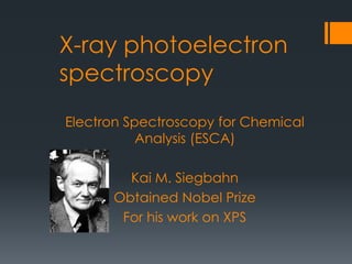
  • 1. X-ray photoelectron spectroscopy Electron Spectroscopy for Chemical Analysis (ESCA) Kai M. Siegbahn Obtained Nobel Prize For his work on XPS
  • 2. Introduction: X-ray photoelectron spectroscopy (XPS) is a surface- sensitive spectroscopic technique widely used to investigate the chemical composition of surfaces. XPS technique is based on Einstein’s idea about the photoelectric effect, developed around 1905 The concept of photons was used to describe the ejection of electrons from a surface when photons were impinged upon it
  • 3. L2,L3 L1 K Incident X-ray Ejected Photoelectron 1s 2s 2p The Photoelectric Process XPS spectral lines are identified by the shell from which electrons were emitted (1s, 2s, 2p etc.) The kinetic energy of the ejected photoelectron KE=hv-BE-F
  • 4. L2,L3 L1 K Emitted Auger Electron 1s 2s 2p Auger Relation of Core Hole L electron falls to fill core level vacancy (step1). KLL Auger electron emitted to conserve energy released in step1.
  • 5. X-Rays Irradiate the sample surface, hitting the core electrons (e-) of the atoms. The X-Rays penetrate the sample to a depth on the order of a micrometer. Useful e- signal is obtained only from a depth of around 10 to 100 Å on the surface. The X-Ray source produces photons with certain energies: MgK photon with an energy of 1253.6 eV AlK photon with an energy of 1486.6 eV Normally, the sample will be radiated with photons of a single energy (MgK or AlK). This is known as a monoenergetic X-Ray beam.
  • 6. Why the Core Electrons? An electron near the Fermi level is far from the nucleus, moving in different directions all over the place, and will not carry information about any single atom. Fermi level is the highest energy level occupied by an electron in a neutral solid at absolute 0 temperature. The core e-s are local close to the nucleus and have binding energies characteristic of their particular element. Core e- Valence e- Atom
  • 7. How Does XPS Technology Work? A monoenergetic x-ray beam emits photoelectrons from the from the surface of the sample. The x-ray photons The penetration about a micrometer of the sample The XPS spectrum contains information only about the top 10 - 100 Ǻ of the sample. Ultrahigh vacuum environment to eliminate excessive surface contamination. Cylindrical Mirror Analyzer (CMA) measures the KE of emitted e-s. The spectrum plotted by the computer from the analyzer signal. The binding energies can be determined from the peak positions and the elements present in the sample identified.
  • 9. COMPONENTS OF XPS: A source of X-rays An ultra high vacuum (UHV) An electron energy analyzer  magnetic field shielding An electron detector system A set of stage manipulators
  • 10. XPS Instrument X-Ray Source Ion Source CMA Sample introduction Chamber
  • 11. WHY WE USE UHV?  Remove adsorbed gases from the sample.  Eliminate adsorption of contaminants on the sample.  Prevent arcing and high voltage breakdown.  Increase the mean free path for electrons, ions and photons.
  • 13. Cylindrical Mirror Analyzer (CMA) Slit Detector Electron Pathway through the CMA 0 V +V 0 V 0 V 0 V +V +V +V X-Rays Source Sample Holder
  • 14. KE versus BE E E E KE can be plotted depending on BE Each peak represents the amount of e-s at a certain energy that is characteristic of some element. 1000 eV 0 eV BE increase from right to left KE increase from left to right Binding energy #ofelectrons (eV)
  • 15. Interpreting XPS Spectrum: Background The X-Ray will hit the e-s in the bulk (inner e- layers) of the sample e- will collide with other e- from top layers, decreasing its energy to contribute to the noise, at lower kinetic energy than the peak . The background noise increases with BE because the SUM of all noise is taken from the beginning of the analysis. Binding energy #ofelectrons N1 N2 N3 N4 Ntot= N1 + N2 + N3 + N4 N = noise
  • 16. XPS peak identification  Electronic Effect:  Auger lines  Chemical shifts  X-ray satellites  X-ray “Ghost”  Energy loss lines
  • 19. XPS Study of Changes in the Chemical Composition of Langasite Crystal Thin Surface Layers during Vacuum Annealing The aim of this work was to study the chemical composition of LGS La3Ga5SiO14 langasite crystal wafer surface after thermal vacuum annealing at 650°C and at elevated temperatures (1000°C). To analyze the surface and subsurface layer chemical composition we used X-ray photoelectron spectroscopy (XPS). The 1050°C, 30 min, and 1000 °C, 5 h annealing experiments showed that in both cases the crystals lost color, and the chemical composition of the wafer surface changed: the gallium concentration decreased abruptly
  • 20. Figure 1 shows the photoelectron spectra of the crystal surfaces before and after annealing from which the gallium line intensity can be seen to decrease by an order of magnitude.
  • 21. Advantages and Disadvantages  Advantages  Non-destructive technique.  Surface Sensitive (10-100 Å).  Quantitative measurements are obtained.  Provides information about chemical bonding.  Elemental mapping.  Limitations  Very expensive technique.  High vacuum is required.  Slow processing (1/2 to 8 hours/sample).  Large area analysis is required.  H and He can not be identified.  Data collection is slow 5 to 10 min.
  • 22.  XPS is used to measure:  Elemental composition of the surface (top 1–12 nm usually).  Chemical or electronic state of each element in the surface.  Uniformity of composition across the top surface (line profiling).  Applications in the industry:  Failure analysis  Polymer surface  Corrosion  Adhesion  Semiconductors  Thin film coatings Uses
  • 23. References:  Siegbahn, K.; Edvarson, K. I. Al (1956). "β-Ray spectroscopy in the precision range of 1 : 1e6". Nuclear Physics  Turner, D. W.; Jobory, M. I. Al (1962). "Determination of Ionization Potentials by Photoelectron Energy Measurement".  journals.tubitak.gov.tr  nanohub.org  srdata.nist.gov  www.eaglabs.com www.files.chem.vt.edu  Bio interface.org  www.spectroscopynow.com  www.surfaceanalysis.org  www.csma.ltd.uk

Editor's Notes

  1. What information can you obtain from XPS? •Identification of elements near the surface and surface composition •Local chemical environments •Oxidation states of transition metals •Valence band electronic structure •Morphology of thin films XPS detects all elements with (Z) >3. It cannot detect H (Z = 1) or He (Z = 2) because the diameter of these orbitals is so small, reducing the catch probability to almost zero XPS is routinely used to analyze inorganic compounds,metals,semiconductors,polymers, ceramics,etc. Organic chemicals are not routinely analyzed by XPS because they are readily degraded by either the energy of the X-rays or the heat from non-monochromatic X-ray sources
  2. X-ray photoelectron spectroscopy (XPS) is based on the photoelectric effect. Each atom in the surface has core electron with the characteristic binding energy that is conceptually, not strictly, equal to the ionization energy of that electron. When an Xray beam directs to the sample surface, the energy of the X-ray photon is adsorbed completely by the core electron of an atom. If the photon energy, hn, is large enough, the core electron will then escape from the atom and emit out of the surface. BE= Electron Binding Energy KE= Electron Kinetic Φ= Spectrometer Work Function hn is the X-ray photon energy
  3. How Does XPS Technology Work? A monoenergetic x-ray beam emits photoelectrons from the from the surface of the sample. The X-Rays either of two energies: Al Ka (1486.6eV) Mg Ka (1253.6 eV) The x-ray photons The penetration about a micrometer of the sample The XPS spectrum contains information only about the top 10 - 100 Ǻ of the sample. Ultrahigh vacuum environment to eliminate excessive surface contamination. Cylindrical Mirror Analyzer (CMA) measures the KE of emitted e-s. The spectrum plotted by the computer from the analyzer signal. The binding energies can be determined from the peak positions and the elements present in the sample identified.
  4. The sample will be introduced through a chamber that is in contact with the outside environment It will be closed and pumped to low vacuum. After the first chamber is at low vacuum the sample will be introduced into the second chamber in which a UHV environment exists
  5. The electrons ejected will pass through a device called a CMA. The CMA has two concentric metal cylinders at different voltages. One of the metal cylinders will have a positive voltage and the other will have a 0 voltage. This will create an electric field between the two cylinders. The voltages on the CMA for XPS and Auger e-s are different. When the e-s pass through the metal cylinders, they will collide with one of the cylinders or they will just pass through. If the e-’s velocity is too high it will collide with the outer cylinder If is going too slow then will collide with the inner cylinder. Only the e- with the right velocity will go through the cylinders to reach the detector. With a change in cylinder voltage the acceptable kinetic energy will change and then you can count how many e-s have that KE to reach the detector.
  6. KE Kinetic Energy (measure in the XPS spectrometer) hv photon energy from the X-Ray source (controlled) Ø spectrometer work function. It is a few eV, it gets more complicated because the materials in the instrument will affect it. Found by calibration. BE is the unknown variable The equation will calculate the energy needed to get an e- out from the surface of the solid. Knowing KE, hv and Ø the BE can be calculated.
  7. Chemical shift: change in binding energy of a core electron of an element due to a change in the chemical bonding of that element. If a charge q is added to (or removed from) the valence shell due to chemical bond formation, the electrostatic potential felt by the electron inside the atom is changed. • When atom loses valence charge (Si0 --> Si4+ ) BE increases. • When atom gains valence charge (O --> O--) BE decreases. Photoelectron core level peaks in elemental samples occur at the same binding, for example, a gold surface. In compounds, where ionic or covalent bonding occurs, the peak position may shift. For example, Si in pure Si wafer, the binding energy of Si 2p is 99 eV, In contrast, Si in SiO2, the binding energy of Si 2p is 102.3 eV Chemical shift: change in binding energy of a core electron of an element due to a change in the chemical bonding of that element. Core binding energies are determined by electrostatic interaction between it and the nucleus, and change with: • the electrostatic shielding of the nuclear charge from all other electrons in the atom (including valence electrons) • removal or addition of electronic charge as a result of changes in bonding will alter the shielding Atoms of a higher positive oxidation state exhibit a higher binding energy due to the extra coulombic interaction between the photo-emitted electron and the ion core. This ability to discriminate between different oxidation states and chemical environments is one of the major strengths of the XPS technique Binding energy of a core electron depends on number and distribution of valence electrons. •Rule of thumb: higher valence electron density = lower binding energy •Final state effects can revert this trend. •Oxidation leads to higher BE of the cation (e.g. Mn+ in metal oxide) compared to reduced element (less valence electrons). •Typically: +1eV per oxidation state An increase in oxidation state causes the binding energy to increase due to a decrease in the screening of the bound electron from the ion core. The ability of XPS to determine oxidation states is used extensively in catalysis research n principle quantum number l orbital angular quantum number s spin angular quantum number j total angular quantum number (j=l+s)
  8. Emission from non-monochromatic x-ray sources produces satellite peaks in XPS spectrum at lower BE.