RNA (gene expression) analysis of Prostate cancers and non-cancerous tissues t
Vaneet K Sharma American Chemical society meeting 2014
1. 11.801
13.586
14.760
AU
0.000
0.002
0.004
0.006
0.008
0.010
0.012
0.014
0.016
0.018
0.020
0.022
14.495
16.131
AU
-0.001
0.000
0.001
0.002
0.003
0.004
0.005
0.006
0.007
0.008
0.009
0.010
Minutes
10.00 10.50 11.00 11.50 12.00 12.50 13.00 13.50 14.00 14.50 15.00 15.50 16.00 16.50 17.00 17.50 18.00 18.50 19.00 19.50 20.00 20.50 21.00 21.50 22.00
Separation of glycoproteins clipped products attributed to residual cell protease activity using
a novel reducing size exclusion chromatography (R-SEC) method
Vaneet K Sharma, Zihao Wang, Ying Zhang and Kingman Ng
Analytical Development, Technical Development, Novartis Vaccines, Morrisville, NC 27560
Email: vaneet.sharma@novartis.com
Acknowledgment
Conclusion
References
Result & Discussion
Authors would like to thank Dipak Thakur and Kwadwo Caesar for useful discussions and Haiyan Liu for collecting SDS PAGE data. This work is funded in part by NIH and Bill and Melinda Gates Foundation (Grants &
Contracts AI066287 and HHSN266200500007C)
1. Analysis of Reduced Monoclonal Antibodies Using Size Exclusion Chromatography Coupled with Mass Spectrometry, J. Am. Soc. Mass Spectrom. 20, 2258-2264 (2009)
2. Monoclonal Antibody Fragment Separation and Characterization Using Size Exclusion Chromatography Coupled with Mass Spectrometry Application Note ZM-1008, Sepax Technologies
Cell culture productivity has increased dramatically over the last few decades. One of the most commonly used cell line for high level heterologous protein expression
is Chinese hamster ovary (CHO), which produces proteins with glycoforms that are both compatible and bioactive in humans. The higher yields are accompanied by the
protein quality issues and one of the most critical issue pertains to the protein degradation or clipping during the production of glycoproteins. Protein clipping
commonly attributed to the activity of host cell proteases cannot be avoided and needs to be monitored in order to ensure the quality of the product. Significant
proteolytic activity (>60% target product clipped) in a CHO based production process used to produce a heavily glycosylated HIV gp120 subunit vaccine candidate was
observed. To monitor this proteolytic activity induced protein clipped products, a novel reducing size exclusion chromatography (R-SEC) method was developed and
successfully used during the early stage process development for HIV gp120 subunit vaccine candidate.
1. The currently used SDS PAGE technique for determining the protein clipped products suffers from several limitations i.e. the use of neuro-toxic reagents, labor-
intensive methodology, and the lack of direct, accurate quantification. In comparison the developed R-SEC detects the clipped fragments quantitatively with
significant higher resolutions.
2. In addition to being quantitative in nature the R-SEC is a less labor intensive method and can be online coupled to multiangle light scattering (MALS) detector or
mass spectrometry for further structural characterization.
NR R
250
150
100
75
50
37
25
20
15
10
MW C19 C19
Instrument: ACQUITY® UPLC H-Class Bio System
Column: BEH200 SEC (1.7 μm, 4.6X300 mm)
Isocratic run: 10 mM sodium citrate, 300 mM sodium chloride, pH 7.0
Isocratic Run: 34 Minutes
Flow Rate: 0.15mL/min
Column Temperature: 25ºC
Channels monitored: 280nm
(a) No sample treatment†
(b) Samples treated with DTT*†
5.880
6.272
6.871
7.338
8.146
8.864
02
00
02
04
06
08
10
9.196
10.451
11.103
11.770
12.342
00
02
04
06
7.062
7.518
8.042
8.527
9.453
32
30
28
26
24
22
20
18
Minutes
5.20 5.40 5.60 5.80 6.00 6.20 6.40 6.60 6.80 7.00 7.20 7.40 7.60 7.80 8.00 8.20 8.40 8.60 8.80 9.00 9.20 9.40 9.60 9.80 10.00 10.20 10.40 10.60 10.80 11.00 11.20 11.40 11.60 11.80 12.00 12.20 12.40 12.60 12.80
5.880
6.272
6.871
7.338
8.146
8.864
04
02
00
02
04
9.195
10.451
11.103
11.770
12.342
02
01
00
01
02
03
7.062
7.518
8.042
8.527
9.453
36
34
32
30
28
26
24
Minutes
5.20 5.40 5.60 5.80 6.00 6.20 6.40 6.60 6.80 7.00 7.20 7.40 7.60 7.80 8.00 8.20 8.40 8.60 8.80 9.00 9.20 9.40 9.60 9.80 10.00 10.20 10.40 10.60 10.80 11.00 11.20 11.40 11.60 11.80 12.00 12.20 12.40 12.60 12.80
5.880
6.272
6.871
7.338
8.146
8.864
20
25
30
35
40
45
50
9.195
10.451
11.103
11.770
12.342
15
20
25
30
7.062
7.518
8.042
8.527
9.453
10.446
00
10
20
30
40
Minutes
5.20 5.40 5.60 5.80 6.00 6.20 6.40 6.60 6.80 7.00 7.20 7.40 7.60 7.80 8.00 8.20 8.40 8.60 8.80 9.00 9.20 9.40 9.60 9.80 10.00 10.20 10.40 10.60 10.80 11.00 11.20 11.40 11.60 11.80 12.00 12.20 12.40 12.60 12.80 13.00
Column: TSKgel G3000 SWXL 30 cm X 7.8 mm
Flow rate: 1.0 ml/min
Column: Zenix 3um, 300A, 7.8 X 300mm
Flow rate: 0.75 ml/min
Column: BEH200 SEC (1.7 μm, 4.6 X 300 mm)
Flow rate: 0.25 ml/ min
Wavelength: 280 nm – MBF
* Stock solutions of 8M urea and 1M dithiothreitol (DTT) was prepared in 250 mM ammonium bicarbonate
(ABC) solution. † Approximately, 9 µg of the sample was loaded onto the column
11.723
1086monomer-12.476
clipped-1-13.359
clipped-2-14.137
clipped-3-15.652
clipped-4-16.995
17.870
AU
0.000
0.002
0.004
0.006
0.008
0.010
0.012
0.014
0.016
0.018
0.020
0.022
0.024
0.026
Minutes
10.50 11.00 11.50 12.00 12.50 13.00 13.50 14.00 14.50 15.00 15.50 16.00 16.50 17.00 17.50 18.00 18.50 19.00
R² = 0.9987
R² = 0.9994
R² = 0.9979
R² = 0.9977
R² = 0.986
0
50000
100000
150000
200000
250000
300000
350000
400000
450000
500000
0.0 2.0 4.0 6.0 8.0 10.0 12.0
monomer
clipped 1
clipped 2
clipped 3
clipped 4
Amount loaded (µg)
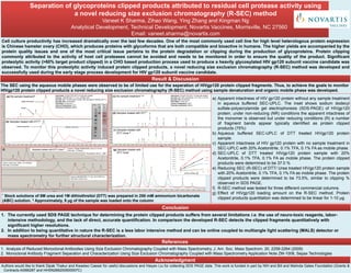
a) Apparent intactness of HIV gp120 protein without any sample treatment
in aqueous buffered SEC-UPLC. The inset shows sodium dodecyl
sulfate-polyacrylamide gel electrophoresis (SDS-PAGE) of HIVgp120
protein, under non-reducing (NR) conditions the apparent intactness of
the monomer is observed but under reducing conditions (R) a number
of fragment bands appear typically identified as protein clipped
products (75%)
b) Aqueous buffered SEC-UPLC of DTT treated HIVgp120 protein
sample.
c) Apparent intactness of HIV gp120 protein with no sample treatment in
SEC-UPLC with 20% Acetonitrile, 0.1% TFA, 0.1% FA as mobile phase.
d) SEC-UPLC of DTT treated HIVgp120 protein sample with 20%
Acetonitrile, 0.1% TFA, 0.1% FA as mobile phase. The protein clipped
products were determined to be 37.0 %
e) Reducing SEC (R-SEC) of DTT/ Urea treated HIVgp120 protein sample
with 20% Acetonitrile, 0.1% TFA, 0.1% FA as mobile phase. The protein
clipped products were determined to be 73.5%, similar to clipping %
observed in SDS PAGE.
f) R-SEC method was tested for three different commercial columns.
g) Effect of HIVgp120 loading amount on the R-SEC method. Protein
clipped products quantitation was determined to be linear for 1-10 µg.
(f) (g) Samples treated with
DTT/ Urea *
Samples treated with DTT/ Urea *†
Clipped productsIntact monomer
Intact monomer
Dimer
Intact monomer
Dimer
The SEC using the aqueous mobile phases were observed to be of limited use for the separation of HIVgp120 protein clipped fragments. Thus, to achieve the goals to monitor
HIVgp120 protein clipped products a novel reducing size exclusion chromatography (R-SEC) method using sample denaturation and organic mobile phase was developed.
11.268
11.871
12.199
12.865
AU
-0.005
0.000
0.005
0.010
0.015
0.020
0.025
0.030
0.035
11.175
11.715
12.043
1086monomer-12.592
clipped-1-13.458
clipped-2-14.274
clipped-3-15.774
clipped-4-17.030
17.844
AU
0.00
0.02
0.04
0.06
0.08
0.10
0.12
11.267
11.696
11.909
1086monomer-12.467
clipped-1-13.367
clipped-2-14.153
clipped-3-15.659
clipped-4-17.016
17.858
AU
0.000
0.010
0.020
0.030
0.040
0.050
Minutes
8.50 9.00 9.50 10.00 10.50 11.00 11.50 12.00 12.50 13.00 13.50 14.00 14.50 15.00 15.50 16.00 16.50 17.00 17.50 18.00 18.50 19.00 19.50 20.00
Instrument: ACQUITY® UPLC H-Class Bio System
Isocratic run: 20% Acetonitrile, 0.1% TFA, 0.1% FA
Isocratic Run: 30 Minutes
Flow Rate: 0.15mL/min
Column Temperature: 25ºC
Channels monitored: 280nm
(c) No sample treatment1,2,†
(d) Samples treated with DTT*1
(e) Samples treated with
DTT/ Urea *†