
Recommended
More Related Content
Similar to 2,3 acute inflammation.ppt
Similar to 2,3 acute inflammation.ppt (20)
Recently uploaded
Recently uploaded (20)
2,3 acute inflammation.ppt
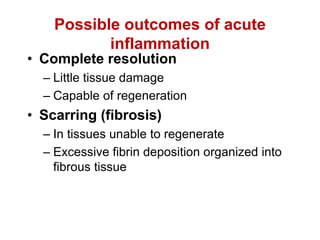
- 1. Possible outcomes of acute inflammation • Complete resolution – Little tissue damage – Capable of regeneration • Scarring (fibrosis) – In tissues unable to regenerate – Excessive fibrin deposition organized into fibrous tissue
- 2. Outcomes (cont’d) • Abscess formation occurs with some bacterial or fungal infections • Progression to chronic inflammation (next)
- 3. Morphology of acute inflammation • Pseudomembranous infla.-due to toxins of diphtheria or irritant gases. • Ulcer- in stomach, duodenum, inte. • E.g. typhoid, inte. Tb, bacillary & amoebic dysentry • Suppuration(abscess) e.g.- boil, carbuncle • Cellulitis- diffuse inflammation of soft tissue. • Bacterial infection of blood- bacteremia, septicemia, pyemia
- 4. Different morphological patterns of acute inflammation can be found depending on the cause and extend of injury and site of inflammation Serous inflammation Fibrinous inflammation Purulent inflammation ulcers
- 5. Chemical mediators Cell derived • Vasoactive amines • Arachidonic acid metabolites ( Eicosanoids) • Via cyclo-oxygenase pathway- PG, thromboxane A2, prostacyclins, resolvins • Via lipo-oxygenase pathway- 5-HETE, leucotrines,lipoxins • Lysosomal components • PAF • Cytokines- IL1,TNF- α, TNF-β, IFN-ץ, chemokines • Free radicals-Oxygen metabolites, NO Plasma derived • Kinin system • Clotting system • Fibrinolytic system • Complement system
- 6. general principles of production and actions. • Mediators are generated either from cells or from plasma proteins. 1.Cell-derived mediators are normally sequestered in intracellular granules and can be rapidly secreted by granule exocytosis (e.g., histamine in mast cell granules) or are synthesized de novo (e.g., prostaglandins, cytokines) in response to a stimulus. 2.The major cell types are platelets, neutrophils, monocytes/macrophages, and mast cells, mesenchymal cells (endothelium, smooth muscle, fibroblasts) and most epithelia can also be induced to elaborate some of the mediators.
- 7. 3. Plasma-derived mediators (e.g., complement proteins, kinins) are produced mainly in the liver and present in the circulation as inactive precursors that must be activated, usually by a series of proteolytic cleavages, to acquire their biologic properties. • One mediator can stimulate the release of other mediators.e.g. the cytokine TNF acts on endothelial cells to stimulate the production of another cytokine, IL-1, and many chemokines. • The secondary mediators may have the same actions as the initial mediators but may also have different and even opposing activities
- 8. • Active mediators are produced in response to various stimuli. like microbial products, substances released from necrotic cells, and the proteins of the complement, kinin, and coagulation systems, which are themselves activated by microbes and damaged tissues.
- 9. • Mediators vary in their range of cellular targets. They can act on one or a few target cell types, can have diverse targets, or may even have differing effects on different types of cells.
- 10. • Once activated and released from the cell, most of these mediators are short-lived. a.They quickly decay (e.g., arachidonic acid metabolites) or b. are inactivated by enzymes (e.g., kininase inactivates bradykinin), or c. they are otherwise scavenged (e.g., antioxidants scavenge toxic oxygen metabolites) or inhibited (e.g., complement regulatory proteins break up and degrade activated complement components).
- 12. Cell derived • Vasoactive amines Histamine Serotonin • Lysosomal components • Platelet activating factor • Cytokines • Chemokines • Neuropeptides • Neuropeptides • Free radicals • AA metabolites • NO
- 13. Histamine and Serotonin induce vasodilation and increased vascular permeability Mast cell : • richest source of histamine • located in connective tissue • adjacent to blood vessels • Degranulation through receptors for IgE-, IgG, histamine, bacterial products and anaphylatoxin C5a, physical injury, cold, heat • release of PAF (platelet activating factor) leads to serotonin and histamine release from activated platelets • Mast cells are very important effector cells in hypersensitivity reactions (anaphylactic reactions)
- 15. Histamine • Histamine is present in mast cell granules and is released by mast cell degranulation in response to a variety of stimuli, (1) physical injury such as trauma, cold, or heat; (2) binding of antibodies to mast cells, which underlies allergic reactions (3) fragments of complement called anaphylatoxins (C3a and C5a); (4) histamine-releasing proteins derived from leukocytes; (5) neuropeptides (e.g., substance P); and (6) cytokines (IL-1, IL-8).
- 16. Histamine • Histamine causes dilation of arterioles and increases the permeability of venules. • the principal mediator of the immediate transient phase of increased vascular permeability, producing interendothelial gaps in venules.
- 17. serotonin • release of PAF (platelet activating factor) leads to serotonin and histamine release from activated platelets • Serotonin (5-hydroxytryptamine) is a preformed vasoactive mediator • It is present in platelets and certain neuroendocrine cells, e.g. in the gastrointestinal tract, • Release of serotonin (and histamine) from platelets is stimulated when platelets aggregate after contact with collagen, thrombin, adenosine diphosphate, and antigen antibody complexes. • Thus, the platelet release reaction, which is a key component of coagulation, also results in increased vascular permeability. This is one of several links between clotting and inflammation.
- 18. Lysosomal components • Neutrophils have two main types of granules. • The smaller specific ( secondary) granules contain lysozyme, collagenase, gelatinase, lactoferrin, plasminogen activator, histaminase, and alkaline phosphatase. • The larger azurophil ( primary) granules contain myeloperoxidase, bactericidal factors (lysozyme, defensins), acid hydrolases, and a variety of neutral proteases (elastase, cathepsin G, nonspecific collagenases, proteinase 3)
- 19. Lysosomal components Granules of monocytes& tissue macrophage- • more active in chronic inflammation , • release of acid proteases, collagenase, elastase, phospholipase, and plasminogen activator etc.
- 20. • Acid proteases degrade bacteria and debris within the phagolysosomes, in which an acid pH is readily reached. • Neutral proteases are capable of degrading various extracellular components, such as collagen, basement membrane, fibrin, elastin, and cartilage, resulting in the tissue destruction • Neutral proteases can also cleave C3 and C5 complement proteins directly, releasing anaphylatoxins, and release a kinin-like peptide from kininogen. • Neutrophil elastase degrade virulence factors of bacteria and thus combat bacterial infections.
- 21. • Because of the destructive effects of lysosomal enzymes, the initial leukocytic infiltration, if unchecked, can potentiate further inflammation and tissue damage. • These harmful proteases are held in check by a system of antiproteases in the serum and tissue fluids. • E.g. α1-antitrypsin, which is the major inhibitor of neutrophil elastase • α2-Macroglobulin is another antiprotease found in serum and various secretions.
- 22. Platelet activating factor • released from IgE sensitised basophils or mast cells , neutrophils,other leucocytes, endothelium, platelets • Actions- platelet aggregation, release reaction Vasodilatation in low conc., vasoconstriction otherwise Increased vascular permeability, adhesion of leucocytes to endothelium. Chemotaxis Bronchoconstriction Increase O2 production (oxidative burst) & degranulation in macrophage. boosts the synthesis of other mediators, particularly eicosanoids, by leukocytes and other cells
- 23. Cytokines • Cytokines are proteins produced by many cell types (activated lymphocytes and macrophages, endothelial, epithelial, and connective tissue cells) that modulate the functions of other cell types • TABLE
- 25. TNF & IL1 • In endothelium they induce a spectrum of changes referred to as endothelial activation a) they induce the expression of endothelial adhesion molecules; b) synthesis of chemical mediators, including other cytokines, chemokines, growth factors, eicosanoids, and NO; c) production of enzymes associated with matrix remodeling; and d) increases in the surface thrombogenicity of the endothelium e) TNF also augments responses of neutrophils to other stimuli such as bacterial endotoxin.
- 26. • TNF also regulates energy balance by promoting lipid and protein mobilization and by suppressing appetite. • sustained production of TNF leads to cachexia, a pathologic state characterized by weight loss and anorexia that accompanies some chronic infections and neoplastic diseases
- 27. Chemokines • Chemokines are a family of small (8 to 10 kD) proteins that act primarily as chemoattractants for specific types of leukocytes • classified into four major groups 1. C-X-C chemokines (α chemokines)- • e.g. IL-8 -secreted by activated macrophages, endothelial cells, and other cell types by microbial products and other cytokines, mainly IL-1 and TNF. • causes activation and chemotaxis of neutrophils, with limited activity on monocytes and eosinophils.
- 28. 2. The C-C chemokines, (β chemokines) -e.g. a.monocyte chemoattractant protein (MCP-1), b. eotaxin, c.macrophage inflammatory protein-1α (MIP-1α), d.RANTES (regulated and normal T-cell expressed and secreted), •generally attract monocytes, eosinophils, basophils, and lymphocytes but not neutrophils. •eotaxin selectively recruits eosinophils.
- 29. 3. C chemokines (γ chemokines)- • e.g., lymphotactin- are relatively specific for lymphocytes 4. CX3C chemokines – • E.g. fractalkine-exists in two forms: • the cell surface-bound protein can be induced on endothelial cells by inflammatory cytokines and promotes strong adhesion of monocytes and T cells, • a soluble form, derived by proteolysis of the membrane- bound protein, has potent chemoattractant activity for the same cells.
- 30. Chemokines have two main functions: 1. they stimulate leukocyte recruitment in inflammation and 2.control the normal migration of cells through various tissues
- 31. Other Cytokines in Acute Inflammation. • IL-6, made by macrophages and other cells, is involved in local and systemic reactions • IL-17, produced mainly by T lymphocytes, promotes neutrophil recruitment
- 32. Neuropeptides • Neuropeptides are secreted by sensory nerves and various leukocytes, and play a role in the initiation and propagation of an inflammatory response • The small peptides, such as substance P and neurokinin A, produced in the central and peripheral nervous systems • Substance P has many biologic functions, like the transmission of pain signals, regulation of blood pressure, stimulation of secretion by endocrine cells, and increasing vascular permeability
- 33. Free radicals • Oxygen metabolites- superoxide oxygen, H2O2, OH & toxic NO products • Actions- • endo. cell damage & thereby increased vascular permeability • Activation of protease & inactivation of anti- protease causing tissue matrix damage • Damage to other cells • These actions are counteracted by antioxidants present in tissue & serum.
- 34. • the physiologic function of these ROS in leukocytes is to destroy phagocytosed microbes, but release of these potent mediators can be damaging to the host . • They are implicated in the following responses in inflammation: • Endothelial cell damage, with resultant increased vascular permeability. Adherent neutrophils, when activated, not only produce their own toxic species but also stimulate production of ROS in the endothelial cells. • Injury to other cell types (parenchymal cells, red blood cells). • Inactivation of antiproteases, such as α1-antitrypsin. This leads to unopposed protease activity, with increased destruction of extracellular matrix. In the lung, such inhibition of anti-proteases contributes to destruction of elastic tissues, as in emphysema
- 35. • Serum, tissue fluids, and host cells possess antioxidant mechanisms that protect against these potentially harmful oxygen-derived radicals. (1) the enzyme superoxide dismutase, which is found in or can be activated in a variety of cell types; (2) the enzyme catalase, which detoxifies H2O2; (3) glutathione peroxidase, another powerful H2O2 detoxifier; (4) the copper-containing serum protein ceruloplasmin; (5) the iron-free fraction of serum transferrin. • Thus, the influence of oxygen-derived free radicals in any given inflammatory reaction depends on the balance between production and inactivation of these metabolites by cells and tissues.
- 36. Metabolites of Arachidonic Acid (eicosanoids) •Membrane lipids of activated cells can be transformed into biological active lipid mediators •All mammalian cells except erythrocytes can produce eicosanoids •They are autocoids = short-range hormones (very short range and half-life) • Arachidonic acid is derived from conversion of linoleic acid
- 37. Arachidonic Acid (eicosanoids) • AA is a 20-carbon polyunsaturated fatty acid (5,8,11,14- eicosatetraenoic acid) derived from dietary sources or by conversion from the essential fatty acid linoleic acid. • It does not occur free in the cell but is normally esterified in membrane phospholipids. • Mechanical, chemical, and physical stimuli or other mediators (e.g., C5a) release AA from membrane phospholipids through the action of cellular phospholipases, mainly phospholipase A2. • AA-derived mediators, (eicosanoids,) are synthesized by two major classes of enzymes: cyclooxygenases (which generate prostaglandins) and lipoxygenases (which produce leukotrienes and lipoxins) • Eicosanoids bind to G protein–coupled receptors on many cell types and can mediate virtually every step of inflammation
- 40. • Prostaglandins (PGs) are produced by mast cells, macrophages, endothelial cells, and many other cell types, and are involved in the vascular and systemic reactions of inflammation • They are produced by the actions of two cyclooxygenases COX1 & COX2. • TxA2 -platelets contain the enzyme thromboxane synthetase, TxA2 is the major product in these cells. TxA2, a potent platelet- aggregating agent and vasoconstrictor, • is itself unstable and rapidly converted to its inactive form TxB2
- 41. • prostacyclin (PGI2)-Vascular endothelium possesses prostacyclin synthetase, which leads to the formation of prostacyclin (PGI2) and its stable end product PGF1α. • Prostacyclin is a vasodilator, a potent inhibitor of platelet aggregation and markedly potentiates the permeability- increasing and chemotactic effects of other mediators • PGD2 is the major prostaglandin made by mast cells; along with PGE2 , it causes vasodilation and increases the permeability of post-capillary venules, thus potentiating edema formation. • PGD2 is a chemoattractant for neutrophils. • PGF2α stimulates the contraction of uterine and bronchial smooth muscle and small arterioles
- 42. • Leukotrienes-The lipoxygenase enzymes are responsible for the production of leukotrienes, which are secreted mainly by leukocytes, • are chemoattractants for leukocytes, and also have vascular effects. • There are three different lipoxygenases, 5- lipoxygenase being the predominant one in neutrophils. • This enzyme converts AA to 5- hydroxyeicosatetraenoic acid, which is chemotactic for neutrophils, and is the precursor of the leukotrienes
- 43. • LTB4 is a potent chemotactic agent and activator of neutrophils, causing aggregation and adhesion of the cells to venular endothelium, generation of ROS, and release of lysosomal enzymes. • The leukotrienes C4, D4, and E4 (LTC4, LTD4, LTE4) cause intense vasoconstriction, bronchospasm (important in asthma), and increased vascular permeability. • Leukotrienes are much more potent than is histamine in increasing vascular permeability and causing bronchospasm.
- 44. • Lipoxins are generated from AA by the lipoxygenase pathway, • the lipoxins are inhibitors of inflammation. • Leukocytes, (particularly neutrophils), produce intermediates in lipoxin synthesis, and these are converted to lipoxins by platelets interacting with the leukocytes. • The principal actions of lipoxins are a. to inhibit leukocyte recruitment and the cellular components of inflammation. b. inhibit neutrophil chemotaxis and adhesion to endothelium
- 45. • There is an inverse relationship between the production of lipoxin and leukotrienes, • the lipoxins may be endogenous negative regulators of leukotrienes and may thus play a role in the resolution of inflammation.
- 46. Eicosanoids can mediate virtually every step of inflammation Action Metabolite Vasoconstriction Thromboxane A2, Leukotrien C4, D4, E4 Vasodilation PGI2, PGE1, PGE2, PGD2 Increased vascul. permeab. LTC4, LTD4, LTE4 Chemotaxis, Leuko. adhesion LTB4, 5-HETE Bronchospasm Leukotrien C4, D4, E4 Platelet aggregation Thromboxane A2 Pain mediation, Fever induction PGE2
- 47. Mediator • PGD2,PGE2 • PGF2α • TXA2 • PGI2 • Resolvin • LTB4 • LTC4,LTD4, LTE4,lipoxins Action • Brochodilator, vasodilator,increased permeability • Vasodilator, bronchoconstrictor • Vasoconstrictor,bronchoconstrictor,plt. Aggregation • Vasodilator,bronchodilator,antiaggregating agent • Inhibitor of pro-inflammatory cytokines • Chemotactic, cell adherence • Smooth muscle constrictor,vasoconstrictor,bronchoconstrictor, increased vas. permeability
- 48. Nitric Oxide (NO) NO was initially discovered as endothelium derived relaxing factor NO is a soluble gas NO is produce by many cells including: 1. endothelial cells 2. some neurons 3. phagocytes synthesized from L-arginine by: nitric oxide synthase (NOS) Three different NOS: endothelial eNOS neuronal nNOS inducible iNOS (phagocytes) *
- 49. • eNOS and nNOS are constitutively expressed at low levels and can be activated rapidly by an increase in cytoplasmic Ca2+. • iNOS,, is induced when macrophages and other cells are activated by cytokines (e.g., TNF, IFN- γ) or microbial products. •
- 52. NO has dual actions in inflammation: • it relaxes vascular smooth muscle and promotes vasodilation, thus contributing to the vascular reaction, • an inhibitor of the cellular component of inflammatory responses • reduces platelet aggregation and adhesion • inhibits several features of mast cell–induced inflammation, • inhibits leukocyte recruitment. • NO is thought to be an endogenous mechanism for controlling inflammatory responses. • NO and its derivatives are microbicidal, and thus NO is a mediator of host defense against infection
- 53. Plasma derived mediators of acute inflammation Plasma factors synthesized mainly in liver Plasma proteins Factor XII = coagulation system (Hageman factor) activation Kinin system Coagulation system Complement activation C3a C5a C3b C5b-C9 anaphylatoxins opsonin MAC
- 54. Complement system • functions in both innate and adaptive immunity for defense against microbial pathogens. • In the process of complement activation several cleavage products of complement proteins are elaborated that cause increased vascular permeability, chemotaxis, and opsonisation • C3 and C5 can be cleaved by several proteolytic enzymes present within the inflammatory exudate(plasmin and lysosomal enzymes released from neutrophils)
- 56. • The biologic functions of the complement system fall into three general categories • Inflammation. C3a, C5a, and C4a are cleavage products of the corresponding complement components that stimulate histamine release from mast cells and thereby increase vascular permeability and cause vasodilation. • They are called anaphylatoxins because they have effects similar to those of mast cell mediators that are involved in the reaction called anaphylaxis • C5a is also a powerful chemotactic agent for neutrophils, monocytes, eosinophils, and basophils. • C5a activates the lipoxygenase pathway of AA metabolism in neutrophils and monocytes, causing further release of inflammatory mediators. •
- 57. • Phagocytosis- C3b and its cleavage product iC3b (inactive C3b), when fixed to a microbial cell wall, act as opsonins and promote phagocytosis by neutrophils and macrophages, which bear cell surface receptors for the complement fragments. • Cell lysis-The deposition of the MAC on cells makes these cells permeable to water and ions and results in death (lysis) of the cells.
- 58. Kinin-Bradykinin System (HMWK) Bradykinin increases vascular permeability, contraction of smooth muscles, vasodilation and pain Kallikrein is a potent activator of factor XII, is chemotactic and can directly convert C5 to C5a
- 59. Coagulation system a cascade of serine proteases • Thrombin provides the main link between coagulation and inflammation by binding to protease activated receptors (PARs) on platelets, endothelium and smooth muscle and leukocytes • PAR-signaling induces: • Chemokines • Endothelial adhesion molecules (ICAM, VCAM) • P selectin mobilization from Weibel Palade bodies • COX-2 • PAF • NO
- 62. Complement System Immune complex Mannose on microbe Microbes
- 64. Interaction of Kinin-, Coagulation- and Complement system during acute inflammation Kallikrein HMWK Plasminogen Prekallikrein Factor XII (Hageman) XIIa Collagen, basement membrane, platelets and microbial surfaces Kinin cascade Clotting cascade Fibrinolysis Plasmin Complement Bradykinin Acute Inflammation Fibrin Fibrinogen C3 C3a Prothrombin Thrombin PAR* * Protease activated receptors
- 65. Outcome of acute inflammation • Complete resolution • Abscess formation (encapsulation and pus)- • Chronic inflammation- • Healing with scar formation- when tissue destruction is extensive so that there is no tissue regeneration but healing by fibrosis occurs. • Suppuration- pyogenic bacteria causing acute infla. results in severe tissue necrosis, the process progress to suppuration .
- 66. Fate of acute inflammation • Resolution • Healing • Suppuration • Chronic inflammation
- 67. Factors determining infla. responce Involving organisms- • Type of injury & infection • Virulence • Dose • Portal of entry • Product of organisms Involving host • Systemic disease • Immune status of host • Cong. Neutrophil defect • Leukopenia • Site or type of tissue involved • Local host factors
- 68. • Type of exudation • Serous • Fibrinous • Purulent or suppurative • Hemorrhagic • catarrhal
- 69. • Acute infla.etiologic agents removed • if no tissue lossresolution or with tissue loss healing by fibrosis or regeneration. • If etiologic agent persists chronic infla. or discharge of pus suppuration
- 70. Systemic effects of acute inflammation acute phase response • Fever (temperature > 37.8oC or >100 F) • Increased pulse, blood pressure • Chills • Anorexia • Leukocytosis • Neutrophilia and left shift of neutrophils points to bacterial infection • Lymphocytosis points to viral infection • Eosinophilia point to allergy or parasitic infection • Acute phase protein production in liver • fibrinogen, CRP,SAA leads to increased ESR • Lymphangitis & lymphadenitis • Shock in severe cases
- 71. Increased Erythrocyte Sedimentation Rate as a result of the presence of acute phase reactants ESR = rate at which erythrocytes settle out of unclotted blood in one hour Normally, Erythrocytes are very buoyant and settle slowly Erythrocytes are negatively charged and repel each other (no aggregation occurs) In presence of acute phase reactants (fibrinogen) erythrocytes aggregate due to loss of their negative charge resulting in increased sedimentation ESR is a widely performed test to detect occult processes and monitor inflammatory conditions
- 72. Granulocytosis with “left shift” of neutrophil population are a good indicator for a severe bacterial infection Leukocyte release results from a direct effect of IL-1 and IL-6 on bone marrow neutrophil stores. Exaggeration of this can result in a “Leukemoid reaction” release of very immature precursors and cell counts >25-30 x 106/ml
- 73. Examples of acute inflammatory diseases of different origin • Allergic reaction • Bacterial pneumonia • Peptic ulcer • Sepsis
- 74. Allergic Reaction with swelling of the larynx Or mucosa Asthma symptoms when affecting the lung
- 75. Pneumonia = infection of the lung • Most community acquired Pneumonias are bacterial of origin • Often the infection follows a viral upper respiratory tract infection • Acute bacterial pneumonias present as two anatomical patterns: – Bronchopneumonia – Lobar pneumonia
- 76. Abscess formation • is the result of a suppurative (purulent) necrosis of the parechyma resulting in the formation of one or more cavities • it has a central necrosis, rimmed by neutrophils and surrounded by fibroblasts Occurs in the lung due to: • Aspiration of infective material • Aspiration of gastric content • Complication of necrotizing bacterial pneumonia (e.g Staphylococcus) • Septic embolism
- 77. Peptic ulcer An ulcer is a local defect of mucosal lining produced by shedding of necrotic tissue Peptic ulcers are produced by an imbalance between gastro- duodenal defense mechanisms and the damaging force 70% of all ulcers are due to H. pyolri infection which initiates a strong inflammatory response
- 78. Septicemia with disseminated intravascular coagulation due to Meningococcal Infection Invasion of the bloodstream by Neisseria meningitides leads to widespread vascular injury with endothelial necrosis, thrombosis and peri-vascular hemorrhage. Hemorrhage as it is seen in the skin can occur in all organs