High Profile Call Girls Jaipur Vani 8445551418 Independent Escort Service Jaipur
Presentation1 anatomy, eyeball.pptx
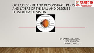
1. OP 1.1,DESCRIBE AND DEMONSTRATE PARTS
AND LAYERS OF EYE BALL AND DESCRIBE
PHYSIOLOGY OF VISION
DR SARITA AGGARWAL
PROF AND HOD
OPHTHALMOLOGY
2. GROSS ANATOMY OF EYEBALL
• Eyeball is a cystic structure kept distended by pressure inside it
Each eyeball is suspended by extra ocular muscle and their facial sheaths.
It is not a true sphere but consist of two modified spheres .
• .
3. Dimensions of eyeball
AP /axial diameter---24 mm in adult / 17.5 mm at birth
Vertical diameter---23 mm
Horizontal diameter----23.5 mm
Volume -6.5 ml. Weight-7 g
Circumference -75 mm
4. • Eyeball is not a true sphere ,but consist of segments of two modified spheres ,cornea is a
part of ant smaller sphere with R 7.8 mm, sclera is a part of posterior sphere with R of 12
mm.
5. • Globe has
• 1. Three layers.
• a. outer coat –cornea, sclera and limbus
• ant 1/6th is transparent called cornea ,post 5/6 th is opaque called sclera
• centre point of ant convexity is ant pole, centre point on post scleral curvature is
post pole.
• b. middle Vascular coat -uveal tissue consist of iris, ciliary body and choroid
• provides nutrition to eyeball
• c. inner neural layer—retina
• concerned with visual fn
6. • 2.Two chambers
• a. anterior chamber—
• bounded ant by post surface of cornea and posteriorly by iris and lens.
• depth-2.5 to 3 mm ,vol-0.25 mm
• contains clear fluid called aqueous.
• b. posterior chamber-
• triangular space bounded ant by post surface of iris ,anterio laterally by a part of
ciliary
• and post by ciliary body, lens and zonules.
• vol-0.06 ml
• Both chambers contains aqueous and communicate through pupil.
•
7. • Causes of deep anterior chamber
• high myopia
• aphakia
• post dislocation of lens
• buphthalmos
• Causes of shallow anterior chamber
• high hypermetropes
• closed angle glaucoma
• extremes of ages
•
8. • 3. Two Segments
• a. anterior segment-consist of cornea, ant chamber, iris, post chamber,lens and
part of ciliary body.
• b posterior segment-includes vitreous,retina,choroid,optic nerve.
•
9. • 4. Lens-
• situated behind the iris and supported by fine fibres called zonules ,arise from ciliary
process..
• transparent biconvex body
• dioptric power- -+15 D
• 5.vitreous cavity
• largest and located behind the lens and zonules
• contains transparent gel like str called vitreous humor
10. • Cornea
• forms ant 1/6 th of eyeball,transparent layer
• main refracting surface of eye
• Dioptric power- +43 to +45 D
• consist of 6 layers
• Sclera
• forms post 5/6 th of eyeball
• opaque
•
15. PHYSIOLOGY OF VISION
• The process of vision can be divided into three stages
• 1. initiation of visual sensations
• 2. transmission of visual sensations
• 3. visual perception
16. • 1. initiation of visual symptoms
• There are 125 million rods and 6 to 7 million cones in retina serving the function of
photoreception.
• When light falls on retina ,two changes occur
a. photochemical –occur in pigment of rods and cones
Rhodopsin is a chromoprotein,present in outer segment of rods, which absorbs light and
triggers electrical impulses in retina.
it is responsible for night vision [scotopic vision]
• b. electrical
• The photochemical reaction coverts light energy into electrical
potential ,transmitted through visual pathway to brain.
the electrical changes in retina can be demonstrated by electroretinography[ERG]
17. • 2. Transmission of visual impulse
• The potential generated in the photoreceptors transmits signals to retinal ganglionic cells
through bipolar cells
• Some cells of retina are stimulated when light is switched on ,while others when light turned off.
• 3.Visual perception
• it is the ability to interpret information and surroundings from the effect of visible light
reaching the eye.
• The stimulation of retina with light yields four types of sensations—
• a.Light sense. b. Form sense
• c. sense of contrast d. color sense
•
•
18. Dark adaptation-it is the ability of the eye to adapt to low illumination
Rods are responsible for this ,k/as scotopic vision.
The time required to see in dark light is called dark adaptation time.[20 to 30 min]
19. • Light adaptation- It is the ability to adapt in bright illumination.
Cones are responsible for this ,called as photopic vision.
adaptation to bright light occurs more rapidly, [3 to 5 min]
2. Form sense
Ability to discriminate between the shape of the object.
Cones are responsible for this, therefore most acute at fovea.
Visual acuity is a measure of form sense.
3.Contrast sensitivity-
It is the ability to distinguish between luminance of different levels.
It is max at the age of 20 yrs, then declines.
Reduced in cataract, glaucoma, DR and optic nerve ds
20. • 3. Color sense
It is the ability by which eye distinguishes different colors and color tones.
there are three primary colors-red , green and blue.
The cones are responsible for color sense.
THEORIES OF COLOR VISION
1. The young Helmholtz trichromatic theory
there are three types of cone photopigments and are sensitive to one of the 3 prim
ary colors
a. blue cone system-contains s pigment, responds to short wavelength [440nm senses blue
color]
b. green cone system-contains M pigment, responds to middle wave length[535nm senses
green ]
c. red cone system-contains L pigment , responds to large wave length [>535nm]
21. • 2. Hering opponent color theory
It suggested that there are three sets of receptor system,
a. red-green
b. Blue-yellow
c. Black-white
The stimulation of one results in inhibition of opposite receptors in the pair.
3. Stage theory
most widely accepted theory
it incorporates both the theory into 2 stages
1st stage-receptor stage-consist of 3 photopigments
2nd stage-neural processing stage-color occupancy occurs at post reception level.