
Recommended
More Related Content
What's hot
What's hot (20)
Similar to NeuroLocalisation.ppt
Similar to NeuroLocalisation.ppt (20)
Recently uploaded
Recently uploaded (20)
NeuroLocalisation.ppt
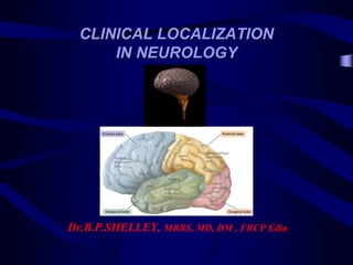
- 1. CLINICAL LOCALIZATION IN NEUROLOGY Dr.B.P.SHELLEY, MBBS, MD, DM , FRCP Edin
- 2. Correlative clinical neurology, imaging findings and surgical results run hand in glove. There are, however, at times we do face discordance/unexplainable clinical parameters or radiological features. OCCAM'S RAZOR Suppose there exist two explanations for an occurrence. In this case, the simpler one is usually better. Another way of saying it is that the more assumptions you have to make, the more unlikely an explanation is. Occam's razor applies especially in the philosophy of science, but also more generally. HICKAM’S DICTUM HICKAM'S DICTUM IS a counterargument to the use of Occam's razor in the medical profession. The principle is commonly stated: "Patients can have as many diseases as they damn well please". The principle is attributed to John Hickam, MD. CLINICAL RATIONALISM-WISDOM Clinical medicine sometimes is not like mathematics
- 5. Ann Indian Acad Neurol 2018;21:9-18. Arch Med Health Sci 2017;5:1-8. Dr. Ralph Jozefowicz
- 6. PRINCIPLES OF NEUROLOGICAL DIAGNOSIS • Symptom profile and symptom analysis - HISTORY • Grouping of signs ( by EXAMINATION) and symptoms – SYNDROMIC FORMULATION & PATTERN RECOGNITION • Mode of ONSET and COURSE • Anatomic Localization – WHERE IS THE LESION? • Pathology – WHAT IS THE LESION? • Aetiology – WHY IS THE LESION? • Investigations • Final Diagnosis
- 7. GENERAL PRINCIPLES: NEUROLOGICAL LOCALISATION Observe each symptom carefully, and consider its significance. Then put the several symptoms together and consider the meaning of their combination especially whether there is any one part of the nervous system at which disease might cause them all Lastly, consider the way they came on as indicating the nature of the lesion, comparing this with the evidence of their seat and remembering also that their character may in itself tell you something of their probable nature. When described in the abstract, this may seem a lengthy process. It may even seem a formidable process . As a rule it is neither” Sir William Gowers
- 8. PROBLEM SOLVING IN NEUROLOGY Symptom recognition, symptom cluster & symptom analysis Pattern Recognition Logical Analysis-Inductive reasoning; based on neuroanatomy, neurological disorders knowledge database (clinical pattern, temporal profile, etiology, and neuropathology Longitudinal (vertical extent) vs. Transverse localization (horizontal extent) Unilateral; Bilateral; Symmetrical; Asymmetrical; Multifocal; Diffuse To define the level of the lesion: CNS vs. PNS – Supratentorial : cerebral hemispheres – Infratentorial : brainstem, cerebellum – Spinal : spinal cord, nerve roots – Multiple levels (multifocal)
- 9. PROBLEM SOLVING-PATHOLOGY WHAT IS THE LESION? • Define the most likely pathology: – Infective – Vascular : Acute – Inflammatory : Subacute – Neoplastic : Focal and chronic – Heredofamial Degenerative : Diffuse and chronic – Traumatic – Toxic-metabolic-Physical agents (radiation etc) – Demyelinating – Dysmyelinating – Post infective/Para-infective – Endocrine
- 10. LOGICAL REASONING : THE BASICS • Define Level of the lesion: – Supratentorial : cerebral hemispheres (lobology) – Infratentorial : brainstem, cerebellum – Spinal : spinal cord, nerve roots – Multiple levels (multifocal- encephalomyeloradiculopathy (TB, CHIKC, CMV,HSV,EBV,ADEM; metastasis, neurofirbromatosis)
- 11. STEPS IN THE DIAGNOSIS OF NEUROLOGIC DISEASE • History • Neurologic examination • Anatomic diagnosis [where-localisation, regional, point, vertical, horizontal] • Pathologic diagnosis [what] • Etiologic diagnosis [why]
- 12. NERVOUS SYSTEM Nervous System CNS PNS Brain Spinal cord Peripheral nerves Nerve ganglia Cr N (12) Sp N (31) Splanchnic N
- 14. Supratentorial Level Motor deficits with neighborhood lobology signs – Weakness and/ or loss of sensation on the same side of the face and body (Uncrossed weakness & COPS) – Contralateral to lesion – Seizures, MMSE abnormal; Behavioural alterations, Psychiatric disturbances; Lobe functions-abnormal; Altered level/content of consciousness
- 15. Supratentorial Level Lobology Signs – Visual field defect – Olfactory symptoms – Language disturbance (aphasia) – Memory deficit – Attention deficit ( amnesia) – Affect and emotional disturbance – Impaired performance of learned motor skills – Seizures
- 16. Infratentorial Level – Weakness and /or cranial nerve deficits on the side of lesion (ipsilateral to lesion) – Long tracts signs on the opposite side of the body (contralateral to lesion) – Crossed hemiplegia (at brainstem-pons-ipsilateral LMN VII and contralateral hemiplegia & COPS) – Above pons- Uncrossed hemiplegia (UMN VII and hemiplegia on the same side, contralateral to lesion)
- 17. Infratentorial Level • Segmental: Ipsilateral to lesion – Weakness of upper and lower face muscles – Double vision (Diplopia) – Difficulty with articulation (Dysarthria) – Difficulty swallowing (Dysphagia) – Incoordination (Ataxia)
- 18. Spinal level – Weakness or sensory loss in both sides of the body but not the face (cranial nerves spared-below FM) – Upper motor neuron weakness in: • Quadriparesis or paraparesis • Monoparesis(leg) or hemiparesis – Sensory loss with sensory level (myelopathic level) – Sphincteric involvement – Brown Sequard, Transverse myelopathy – Red Flag: Cortical paraplegia; Pseudospinal stroke
- 19. PATTERN RECOGNITION • Mode of presentation: acute, subacute, chronic • Temporal profile: intermittent, paroxysmal, partially/completely reversible, residual neurological disability, remitting-relapsing, recurrent, progressive, progressive with intermittent relapses • UMN vs. LMN • Aetiology • Upper motor neuron weakness – In the upper limbs-Extensors weaker than flexors – In the lower limbs-Flexors weaker than extensors – Muscle stretch reflexes are brisk- Hyperreflexia
- 20. Divisions of the Neuraxis • Cortical Brain • Subcortical Brain • Brainstem • Cerebellum • Spinal Cord • Root • Peripheral Nerve • Neuromuscular Junction • Muscle
- 21. PATTERN RECONITION-PARKINSONISM • Bradykinesia • Tremor at rest • “Cogwheel rigidity” • Postural instability • Shuffling gait • Monotonous speech, hypophonia • Stooped posture
- 22. PATTERN RECONITION-PERIPHERAL NEUROPATHY • Motor Weakness predominantly distal – Hypotonia • Glove and stocking sensory loss • Diminished reflexes-Hyporeflexia • Pan-sensory modalities affected (large vs. small fiber neuropathy) • Exceptions: Proximal neuropathies: Guillain- Barre syndrome, CIDP, Porphyric neuropathy, GB mimics, Diabetic proximal neuropathy (PDN)
- 23. PATTERN RECONITION-MYOPATHY • Proximal greater than distal muscle weakness • Neck muscles often affected • Sensations are intact; Pure motor presentation • Muscle stretch reflexes are normal (Ankle DTRs last to be lost) • Presence of pseudohypertrophy • Duchenne muscular dystrophy, polymyositis • HIMET acronym • Exceptions: Distal myopathies: Welander; Miyoshi, Nonaka, Udd, Markesberg-Griggs, Laing distal myopathy
- 24. PATTERN RECONITION-RADICULOPATHY • Proximal (upper lumbar roots) as well as distal weakness (lower lumbo-sacral roots) innervated by a common nerve root • Vertebral pain (extradural compression) • Radicular pain (sensory root) • Dermatomal sensory loss • Segmental reflex alteration: – C5,6: Biceps and brachioradialis – C7: Triceps jerk – L3,4: Knee jerk – S1: Ankle jerk
- 25. PATTERN RECONITION-PARTIAL MYELOPATHY (Brown Sequard syndrome) • At the level of the lesion: Segmental Lower motor neuron weakness • Upper motor neuron weakness, hyperreflexia and Babinski sign ipsilateral to the lesion • Dissociated sensory loss: – Vibration and proprioception- ipsilateral – Pain and temperature-Contralateral – Sensory level
- 27. MENTAL STATUS EXAMINATION • Level of consciousness • Speech • Vocabulary • Orientation • Knowledge of current events • Judgment • Emotional response • Memory • Object recognition • Calculation
- 28. BRIEF MENTAL FUNCTION – CORTICAL LOCALISATION MMSE • Orientation • Registration • Attention & Calculation • Recall [Memory] • Language
- 29. MMSE Domain Test Orientation Year,season,date,day,month / state,country,state/city,hospital,ward Registration [STM] 3 common objects [Glass,pen,watch] Attention Calculation Spell ‘WORLD’ backwards 100-7/40-7 serial 5 steps 5 minute Recall 3 objects repeated above Language Naming:2 objects;Repetition;Ideational apraxia-3 stage command;Read & Obey; Write a sentence; Copy intersecting pentagons Localization 5 minute recall – Memory; Left medial temporal) Naming/Repetition/Read/Writing – Left language areas. Calculation / Ideational apraxia – Left parietal Constructional praxis – Parietal (Rt>Lt parietal)
- 32. FRONTAL LOBE • Anosmia – orbitofrontal • Motor deficit – contralateral hemiplegia [hands,tongue,lips,foot] • Language (Lt frontal) – Broca’s motor aphasia • Kennedy Foster syndrome • Apathy,Depressed (Left) • Frontal bladder deficits • Primitive release reflexes – Prefrontal lobe • Paratonic rigidity (Geganhalten) • Cognitive deficits – Dorsolateral frontal • Abulia / Akinetic mute state • Behavioural disinhibition • Pseudopsychopathic behaviour Rt Orbitofrontal • No sensory deficits, No visual field deficits
- 33. FRONTAL LOBOLOGY – PHINEAS GAGE 1868 Phineas Gage, the science of brain localization; phrenology-functional specialization-anatomical correlation-’modularity of the mind’
- 34. Neurological stamp Phineas Gage and the science of brain localization Commemorating the 150th anniversary of the event; the first day cover shows Dr John Martin Harlow J Neurol Neurosurg Psychiatry 2001;71:761
- 35. TEMPORAL LOBE • Medial temporal – Hippocampus, Taste & Smell (Uncus;Insula) • Lateral temporal – Wernicke’s aphasia • Auditory cortex (Heschl transverse gyri) – auditory hallucinations • Wernicke aphasia – Sensory aphasia Left STG • Visual field deficits – Upper homonymous quadrantanopia • Memory deficits – Medial temporal [Rt- visual memory / Lt- verbal memory] • Temporo occipital – Visual hallucinations • Korsakoff amnesic defect – memory deficits in chronic alcoholics • Kluver-Bucy syndrome [Bilateral temporal]
- 36. PARIETAL LOBE • Wernicke’s aphasia – Left angular and supramarginal gyrus • Homonymous hemianopia ( inferior quadrantanopia) • Cortical sensory loss – 2 point discrimination; stereognosis; tactile localisation; graphesthesia • Gerstmann syndrome Lt parietal – Lt/Rt confusion, acalculia, agraphia, finger agnosia • Hemi Neglect rt parietal • Ideational / Ideomotor apraxia Lt parietal • Dressing apraxia Rt parietal • Topographical disorientation Rt parietal • Anosognosia Rt parietal • Constructional apraxia Rt>Lt parietal
- 37. OCCIPITAL LOBE • Visual field defects – Homonymous hemianopia • Visual hallucinations • Cortical blindness [Bilat] • Visual agnosia – Object agnosia [Lt]; Simultanagnosia [Bilat], Colour anomia [Lt] ; Alexia [Lt]
- 42. CEREBRAL CORTICAL REPRESENTATION – BODY SCHEMA
- 43. MOTOR / SENSORY CORTICAL REPRESENTATION
- 45. SUBCORTICAL - BASAL GANGLIA Internal capsule
- 46. Motor pathway – UMN and LMN Internal capsule localization – 3 Hs •Hemiplegia – dense (UL= LL) •Hemianesthesia •Hemianopia
- 47. PATTERN OF MOTOR WEAKNESS • Cortical Paraplegia • UL>LL = Cortical Watershed zone stroke (Man in a Barrel) • LL>UL = Subcortical Watershed zone stroke (Pseudospinal stroke) • Mute + bilateral UMN VII + Pseudobulbar Palsy = Opercular Syndrome • Faciobrachial weakness = Anterior Limb IC (Heubner’s artery) • Crossed hemiplegia; UL>LL weakness = Cruciate hemiplegia (Medullary pyramidal decussation to upper cervical cord C23 level) • Stroke picture, somnolent mutism, agitated delirium with hallucinations = Top of Basilar Syndrome (“hidden eye signs”) • Stroke, Horner’s syndrome, Anterior neck pain (carotidynia) = Carotid dissection • Ipsilateral cranial nerve involvement + contralateral pyramidal signs = Brain stem lesions • COPS: Cortical lesion= head deviated Opposite to plegia / Pontine lesion = head/eye deviated to Same side of plegia
- 49. Brain stem Localization – Crossed syndromes Midbrain syndrome Pontine syndrome Medullary syndrome
- 51. BRAINSTEM LOCALIZATION Weber syndrome (Base of Midbrain) Ipsilateral III + Contralateral hemiplegia Claude syndrome Ipsilateral III + Contralateral cerebellar ataxia/red nucleus tremor Benedikt syndrome Ipsilateral III + Contralateral long tract signs (CST,Cerebellar,Tremor) Millard Gubler syndrome (Base of Pons) Ipsilateral VI and VII + Contralateral hemiplegia Wallenberg syndrome (Lateral Medulla) Ipsilateral V,IX,X,ataxia,face pain loss + contralateral spinothalamic loss Medial medullary syndrome Ipsilateral XII + contralateral hemiplegia
- 52. MOTOR WEAKNESS PATTERN • CORTICAL Monoparesis, body regions that have maximal cortical representation is most affected + Cortical signs • SUBCORTICAL Monoparesis • INTERNAL CAPSULE Hemiplegia, Hemianesthesia, Homonymous hemianopia • BRAINSTEM Ipsilateral cranial nerve signs + Contralateral hemiplegia
- 53. MOTOR TRACT SOMATOTOPIC LAMINATIONS
- 54. CST LAMINATIONS
- 55. UMN vs LMN
- 56. 1st Step - UMNL vs. LMNL Cerebral cortex to spinal cord (excluding AHC) UMN distribution of motor weakness (Pyramidal weakness) Spasticity DTRs – hyper reflexic Absent superficial reflexes Normal muscle bulk Paralysis of movement Plantar response – Babinski sign positive No fasciculation AHC level to muscle LMN distribution of weakness Hypotonia DTRs – areflexic / hyporeflexic Plantar response – No Babinski sign Paralysis of muscle Muscle bulk – wasting / atrophy (3 weeks) [Parietal wasting] Focal / Segmental myotomal muscle weakness Fasciculation present
- 57. MOTOR CORTEX REPRESENTATION Pyramidal distribution of motor weakness
- 59. CEREBRAL LOCALIZATION Site How to localise Cerebral cortex [Cortical] [Grey matter] Lobe functions / Lobe function tests [Aphasia,apraxia,agnosia,behavioural & personality changes,h/o seizures,MMSE alterations] Motor weakness - monoparesis Subcortical [White matter] Hemiplegia,Hemianesthesia,Hemiano piaDenser weakness pattern,movement disorders [Basal ganglia],No seizures,No cognitive deficits usually Diencephalon Thalamic syndrome,Asc RAS – altered level of consciousness Brain stem The company of cranial nerve deficits Ipsilateral cranial nerve + contralateral long tract signs [Crossed signs]
- 60. MOTOR WEAKNESS LOCALIZATION Monoparesis + Cortical signs Monoparesis;No cortical signs 3 H’s Ipsilat III + Contralateral H Ipsilat VI/VII + Contralateral H Ipsilat V,IX,X,XII + Contra Long tract signs Only weakness, No Cr N signs
- 61. LMN LESIONS • Anterior Horn Cells (AHC) • Roots – Radiculopathy • Plexus – Plexopathy • Peripheral Nerves – Neuropathy • NMJ • Muscle - Myopathy
- 62. CLUES FOR LMN LOCALIZATION Type/Site of Lesion Diagnostic Features Anterior Horn Cell Focal/Segmental LMN type of weakness; Pure Motor signs; Fasciculations Roots Root pains; Localization according to root affected [Motor & Sensory signs] Plexus No root pains; Plexus anatomy & localization [Mainly motor signs] Peripheral Nerves Motor + Sensory signs Neuromuscular Junction Pure motor signs; Fatigability; Diurnal variation Muscle Pure motor signs
- 63. RADICULOPATHY • Motor signs – DTRs – hypo/areflexic Biceps – C5 Triceps – C7 Supinator – C6 Knee – L4 Ankle – S1 • Sensory signs – Dermatomal loss
- 65. SENSORY LOCALIZATION CN V Face T4: Nipple C2-3: Neck T10: Umbilicus C4: Shoulder L1: Inguinal C6: Thumb L3: Knee C7: M. finger L5: Ant. Leg and foot C8: L. finger S1: Heel
- 66. NEUROPATHY MYOPATHY Distal Symmetrical Motor; Glove & Stocking sensory signs Pure Motor; No Sensory Symmetrical Proximal weakness; Selective muscle involved with hypertrophy/pseudohypertrophy DTRs – Hyporeflexic; Distal Ankle jerks – lost first/early DTRs – Normal; Proportional to muscle bulk/weakness;Ankle jerks – preserved till late/last to be lost Proximal Neuropathy AIDP/GB Syndrome Diabetic Amotrophy Porphyric Neuropathy Distal Myopathy - Genetic Extensor Digitorum Brevis (EDB) - wasted EDB - preserved JPS/Vibration Sense - affected JPS/Vibration Sense – Normal
- 72. EOM SO4 / LR6 /OM3
- 75. LEFT GAZE PALSY LEFT INO
- 79. Upper half of face – bilateral cortical representation
- 81. UMN VII PALSY
- 82. LMN VII PALSY
- 83. EMOTIONAL FACIAL PARESIS • Emotional facial paresis (EFP) is characterized by impaired activation of face muscles with emotion [smiling; mimetic smile)] but normal voluntary activation. • Volitional facial paresis affects facial movements with voluntary effort, sparing activation on emotion. • Volitional facial paresis (VFP) refers to weakness of facial muscles on voluntary effort while emotional movements are preserved. • Separate pathways for volitional and emotional facial muscle control • Sites EFP: frontal lobe white matter, SMA, striatocapsular territory, anterolateral thalamus, insula, posterior thalamus, subthalamus, hypothalamus, operculum, mesial temporal lobe and insula, dorsal midbrain • Sites for VFP: involvement of motor cortex/descending pyramidal tract/corticobulbar pathways; sparing BG, frontal & temporal • Aetiology: stroke, lacunar infarcts, tumours, demyelinating lesions, hematoma, ICA dissection
- 84. Emotional Facial Palsy (EFP) (A) Symmetric voluntary activation (showing the teeth) (B) right EFP while smiling (C) MRI showing left striatocapsular infarction A B C
- 85. Hge infarct-lat thalamus,BG, insula; Rt ICA thrombus Left frontal WM edema; sparing thalamus & BG Hematoma- left posterior SITES/AETIOLOGY-EFP
- 86. (A) “Showing the teeth” demonstrates symmetric innervation on volition. (B) Smiling discloses left emotional facial paresis. Right Horner’s syndrome is demonstrated by the small palpebral fissure. (C) Contrastenhanced T1-weighted MRI. Section at the upper pontine level documenting involvement of the cerebellum and the right dorsal pontine tegmentum. For C and D, right is on the right-hand side. (D) Drawing of the upper pontine level. The hatched area corresponds to the area involved as shown in C. This area is quite distinct from the corticobulbar tract mediating voluntary facial innervation EFP OF PONTINE ORIGIN
- 87. VOLUNTARY FACIAL PARESIS (A) VFP (showing the teeth), (B) symmetric activation with emotion (laughing). (C) MRI after intravenous gadolinium (0.1 mmol lkg body weight) showing enhancement of the right cortex. A B C
- 88. MYELOPATHY Sensory level Motor level Reflex level Vibration level
- 89. Cervical – Add 1 T1-T6 – Add 2 T789 – Add 3 T10 – L1L2 T11 – L3L4 T12 – L5 L1- Sacral segments
- 90. Conus Medullaris Cauda Equina Spontaneous pain Bilateral;Symmetric S234 (Perineum/Thighs) Radicular pain;Unilateral;Asymme tric at S234 Sensory deficit Saddle area,Bilateral,Symmetrical, Dissociative Saddle,Unilateral,Asym metric,No dissociation,All sensations affected Motor loss Symmetric,Less marked Asymmetric,Marked Reflex loss Ankle lost Knee/Ankle lost Sphincters Early and marked Late and less marked Trophic changes Common Less marked Sexual functions Erection impaired Less impaired Onset Sudden and Bilateral Gradual and Unilateral
- 91. REFLEX LEVEL LOCALIZATION - MYELOPATHY
- 92. Motor Eye Field Area CO PS CO Cortical – Eyes deviated to opposite to side of weakness PS Brainstem – Eyes deviated to same side of weakness
- 99. The Holmesian logician: Sherlock Holmes’ “Science of Deduction and Analysis” Cognitive heuristics, Thinking and Deducing Logical Proofs