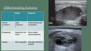
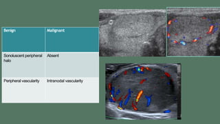
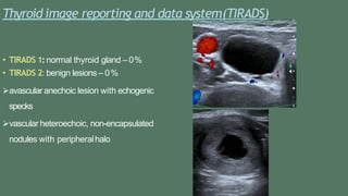
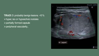
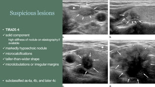
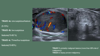
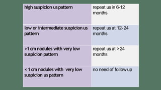
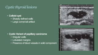
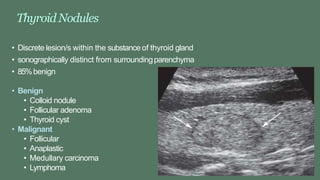
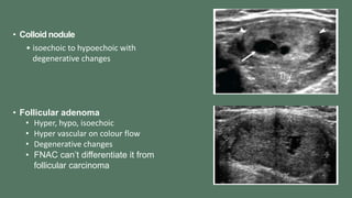
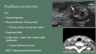
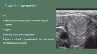
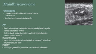
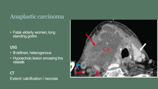
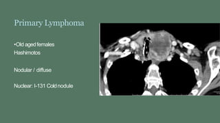
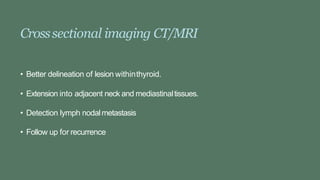
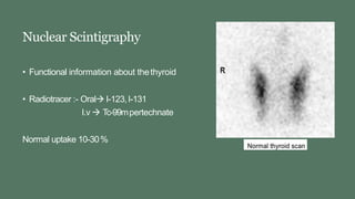
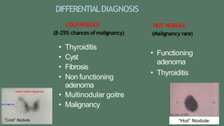
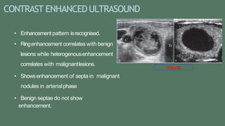
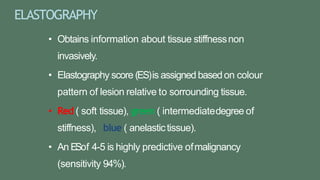
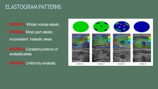
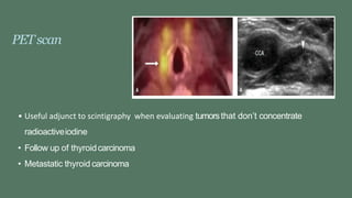
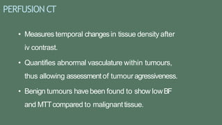
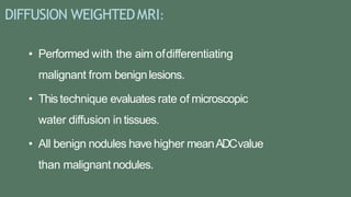
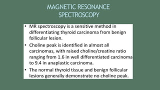
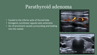
IMAGINGOFTHYROIDLESIONS
DR.PrashanthiK
JuniorResident
IMAGING
• Plain Radiography
• USG
• CT/MRI
• Nuclear scintigraphy
Plain radiograph
• Paratracheal soft tissuemass
• Tracheal shift/narrowing
• Calcification
• Retrosternal extension
• Bony destruction
• Pulmonary metastasis
Roleof USGin thyroid diseases
• Solid vscystic lesions.
• Benign vsmalignant lesions
• Nodule detection when physical examination isunequivocal.
• Thyroid nodules from other cervicalmasses
Differentiating features
Benign Malignant
Internal
consistenc
y
Cystic
componen
t
Predominantly solid
composition
Echogenicity Hypoechoic /iso
/hyper
More marked
Hypoechogenicity
Margins Well marginated Spiculated, illdefined,
irregular
Benign Malignant
Sonoluscent peripheral
halo
Absent
Peripheral vascularity Intranodal vascularity
Benign Malignant
Wider than taller Taller than wider
Peripheralcalcification
Scattered echogenic
Micro calcification
Histopathology -colloid goiter
Thyroid image reporting and data system(TIRADS)
• TIRADS 1:normal thyroid gland – 0%
• TIRADS 2: benign lesions – 0%
avascular anechoic lesion with echogenic
specks
vascular heteroechoic, non-encapsulated
nodules with peripheralhalo
TIRADS 3: probably benign lesions <5%
hyper, iso or hypoechoicnodules
partially formed capsule
peripheral vascularity..
Suspicious lesions
• TIRADS 4:
solid component
high stiffness of nodule on elastographyif
available
markedly hypoechoic nodule
microcalcifications
taller-than-wider shape
microlobulations or irregularmargins
• subclassified as4a, 4b, and later 4c
TIRADS 4a: one suspiciousfeature
(5-10%)
TIRADS 4b: two suspicious
features(10-80 %)
TIRADS 4c:Three/foursuspicious
features(10-80%)
TIRADS 5: probably malignant lesions (more than 80%riskof
malignancy)
TIRADS 6: biopsy proven malignancy
high suspicion uspattern repeat usin 6-12
months
low or intermediate suspicionus
pattern
repeat usat 12-24
months
>1cm nodules with verylow
suspicion pattern
repeat usat >24
months
<1cm nodules with verylow
suspicion uspattern
no need of followup
Cystic thyroid lesions
• Colloid cyst
• Sharply defined walls
• Large comet tail artifact
• Cystic Variant of papillary carcinoma
• Irregular walls
• Lobulated margins
• Presence of blood vessels in solid component
ThyroidNodules
• Discrete lesion/s within the substance of thyroid gland
• sonographically distinct from surroundingparenchyma
• 85%benign
• Benign
• Colloid nodule
• Follicular adenoma
• Thyroid cyst
• Malignant
• Follicular
• Anaplastic
• Medullary carcinoma
• Lymphoma
• Colloid nodule
• isoechoic to hypoechoic with
degenerative changes
• Follicular adenoma
• Hyper, hypo, isoechoic
• Hyper vascular on colour flow
• Degenerative changes
• FNAC can’t differentiate it from
follicular carcinoma
Papillary carcinoma
USG
• Hypoechogenicity
• Microcalcification -Finepunctate
• Primary lesion and lymph nodes
• Hypervascularity
• Lymph node – cystic, thick nodular walls,
calcification
• Lateral aberrant thyroid
• CECT: Heterogeneousenhancement
Follicular carcinoma
USG
• Hypoechoic ill defined lesion withThick irregular
capsule
• Types:
Minimally invasiveEncapsulated
Invasive  Not well encapsulated with vascularinvasion
Central chaotic vascularity
Medullary carcinoma
Ultrasound
• Hypoechoic solid nodules with coarse internal
calcifications.
• Involved lymph nodestypicallycalcify.
CT
• Both primary and metastatic lesions usually haveirregular
densecalcific foci within.
• In the chest, bullae formation and pulmonaryfibrosis –
desmoplastic reaction
Nuclearimaging
• do not concentrate radioactiveiodine – doesn’t arise from
thyroid follicular cells
FDG-PET
• ~75%(range60-95%)sensitive for metastatic disease6
Anaplastic carcinoma
• Fatal- elderly women,long
standing goitre
USG
• Ill defined, heterogenous
• Hypoechoic lesion encasing the
vessels
CT
Extent/ calcification / necrosis
Primary Lymphoma
•Old agedfemales
Hashimotos
Nodular / diffuse
Nuclear: I-131 Coldnodule
Crosssectional imaging CT/MRI
• Better delineation of lesion withinthyroid.
• Extension into adjacent neck and mediastinaltissues.
• Detection lymph nodalmetastasis
• Follow up for recurrence
Nuclear Scintigraphy
• Functional information about thethyroid
• Radiotracer :- Oral I-123,I-131
I.v  Tc-99mpertechnate
Normal uptake 10-30%
Nuclear scintigraphy indications
• Assessment of anatomy
• Assessment of function
• Postoperative assessment
• Detection of nodule– hot or cold orwarm
• Detection offunctional metastatic tissue in known
caseof thyroid ca.
• Detection of retrosternalgoitre.
DIFFERENTIALDIAGNOSIS
COLD NODULE
(8-25% chances ofmalignancy)
• Thyroiditis
• Cyst
• Fibrosis
• Non functioning
adenoma
• Multinodular goitre
• Malignancy
HOT NODULE
(Malignancy rare)
• Functioning
adenoma
• Thyroiditis
RECENTDEVELOPMENTS
CONTRAST ENHANCEDULTRASOUND
• Enhancement pattern isrecognised.
• Ringenhancement correlates with benign
lesions while heterogenousenhancement
correlates with malignantlesions.
• Showsenhancement of septa in malignant
nodules in arterialphase
• Benign septae do not show
enhancement.
COLLOID
ELASTOGRAPHY
• Obtains information about tissue stiffnessnon
invasively.
• Elastography score (ES)is assigned basedon colour
pattern of lesion relative to sorrounding tissue.
• Red ( soft tissue), green ( intermediatedegree of
stiffness), blue ( anelastictissue).
• An ESof 4-5 is highly predictive ofmalignancy
(sensitivity 94%).
ELASTOGRAMPATTERNS
• P
A
T
T
E
R
N1: Whole nodule elastic
• P
A
T
T
E
R
N2: Most part elastic,
inconsistent inelastic areas
• P
A
T
T
E
R
N3: Constantportions of
anelasticareas
• P
A
T
T
E
R
N4: Uniformlyanelastic
PETscan
• Useful adjunct to scintigraphy when evaluating tumorsthat don’t concentrate
radioactiveiodine
• Follow up of thyroidcarcinoma
• Metastatic thyroid carcinoma
PERFUSION CT
• Measures temporal changesin tissue density after
iv contrast.
• Quantifies abnormal vasculature within tumours,
thus allowing assessment of tumouragressiveness.
• Benign tumours havebeen found to show lowBF
and MTTcompared to malignanttissue.
DIFFUSION WEIGHTEDMRI:
• Performed with the aim ofdifferentiating
malignant from benignlesions.
• This technique evaluates rate of microscopic
water diffusion in tissues.
• All benign nodules havehigher meanADCvalue
than malignant nodules.
MAGNETIC RESONANCE
SPECTROSCOPY
Spotters
Lingual Thyroid
Toxic Multinodular Goiter
Parathyroid adenoma
• Caudal to the inferior pole of thyroid lobe
• Echogenic curvilinear capsule seen anteriorly
• Arc of prominent vessels surrounding and leading
into the nodule
Thank you

Imaging of Thyroid Lesions.pptx

  • 1.
  • 2.
    IMAGING • Plain Radiography •USG • CT/MRI • Nuclear scintigraphy
  • 3.
    Plain radiograph • Paratrachealsoft tissuemass • Tracheal shift/narrowing • Calcification • Retrosternal extension • Bony destruction • Pulmonary metastasis
  • 4.
    Roleof USGin thyroiddiseases • Solid vscystic lesions. • Benign vsmalignant lesions • Nodule detection when physical examination isunequivocal. • Thyroid nodules from other cervicalmasses
  • 5.
    Differentiating features Benign Malignant Internal consistenc y Cystic componen t Predominantlysolid composition Echogenicity Hypoechoic /iso /hyper More marked Hypoechogenicity Margins Well marginated Spiculated, illdefined, irregular
  • 6.
  • 7.
    Benign Malignant Wider thantaller Taller than wider Peripheralcalcification Scattered echogenic Micro calcification Histopathology -colloid goiter
  • 8.
    Thyroid image reportingand data system(TIRADS) • TIRADS 1:normal thyroid gland – 0% • TIRADS 2: benign lesions – 0% avascular anechoic lesion with echogenic specks vascular heteroechoic, non-encapsulated nodules with peripheralhalo
  • 9.
    TIRADS 3: probablybenign lesions <5% hyper, iso or hypoechoicnodules partially formed capsule peripheral vascularity..
  • 10.
    Suspicious lesions • TIRADS4: solid component high stiffness of nodule on elastographyif available markedly hypoechoic nodule microcalcifications taller-than-wider shape microlobulations or irregularmargins • subclassified as4a, 4b, and later 4c
  • 11.
    TIRADS 4a: onesuspiciousfeature (5-10%) TIRADS 4b: two suspicious features(10-80 %) TIRADS 4c:Three/foursuspicious features(10-80%) TIRADS 5: probably malignant lesions (more than 80%riskof malignancy) TIRADS 6: biopsy proven malignancy
  • 12.
    high suspicion uspatternrepeat usin 6-12 months low or intermediate suspicionus pattern repeat usat 12-24 months >1cm nodules with verylow suspicion pattern repeat usat >24 months <1cm nodules with verylow suspicion uspattern no need of followup
  • 13.
    Cystic thyroid lesions •Colloid cyst • Sharply defined walls • Large comet tail artifact • Cystic Variant of papillary carcinoma • Irregular walls • Lobulated margins • Presence of blood vessels in solid component
  • 14.
    ThyroidNodules • Discrete lesion/swithin the substance of thyroid gland • sonographically distinct from surroundingparenchyma • 85%benign • Benign • Colloid nodule • Follicular adenoma • Thyroid cyst • Malignant • Follicular • Anaplastic • Medullary carcinoma • Lymphoma
  • 15.
    • Colloid nodule •isoechoic to hypoechoic with degenerative changes • Follicular adenoma • Hyper, hypo, isoechoic • Hyper vascular on colour flow • Degenerative changes • FNAC can’t differentiate it from follicular carcinoma
  • 16.
    Papillary carcinoma USG • Hypoechogenicity •Microcalcification -Finepunctate • Primary lesion and lymph nodes • Hypervascularity • Lymph node – cystic, thick nodular walls, calcification • Lateral aberrant thyroid • CECT: Heterogeneousenhancement
  • 17.
    Follicular carcinoma USG • Hypoechoicill defined lesion withThick irregular capsule • Types: Minimally invasiveEncapsulated Invasive  Not well encapsulated with vascularinvasion Central chaotic vascularity
  • 18.
    Medullary carcinoma Ultrasound • Hypoechoicsolid nodules with coarse internal calcifications. • Involved lymph nodestypicallycalcify. CT • Both primary and metastatic lesions usually haveirregular densecalcific foci within. • In the chest, bullae formation and pulmonaryfibrosis – desmoplastic reaction Nuclearimaging • do not concentrate radioactiveiodine – doesn’t arise from thyroid follicular cells FDG-PET • ~75%(range60-95%)sensitive for metastatic disease6
  • 19.
    Anaplastic carcinoma • Fatal-elderly women,long standing goitre USG • Ill defined, heterogenous • Hypoechoic lesion encasing the vessels CT Extent/ calcification / necrosis
  • 20.
    Primary Lymphoma •Old agedfemales Hashimotos Nodular/ diffuse Nuclear: I-131 Coldnodule
  • 21.
    Crosssectional imaging CT/MRI •Better delineation of lesion withinthyroid. • Extension into adjacent neck and mediastinaltissues. • Detection lymph nodalmetastasis • Follow up for recurrence
  • 22.
    Nuclear Scintigraphy • Functionalinformation about thethyroid • Radiotracer :- Oral I-123,I-131 I.v  Tc-99mpertechnate Normal uptake 10-30%
  • 23.
    Nuclear scintigraphy indications •Assessment of anatomy • Assessment of function • Postoperative assessment • Detection of nodule– hot or cold orwarm • Detection offunctional metastatic tissue in known caseof thyroid ca. • Detection of retrosternalgoitre.
  • 24.
    DIFFERENTIALDIAGNOSIS COLD NODULE (8-25% chancesofmalignancy) • Thyroiditis • Cyst • Fibrosis • Non functioning adenoma • Multinodular goitre • Malignancy HOT NODULE (Malignancy rare) • Functioning adenoma • Thyroiditis
  • 25.
  • 26.
    CONTRAST ENHANCEDULTRASOUND • Enhancementpattern isrecognised. • Ringenhancement correlates with benign lesions while heterogenousenhancement correlates with malignantlesions. • Showsenhancement of septa in malignant nodules in arterialphase • Benign septae do not show enhancement. COLLOID
  • 27.
    ELASTOGRAPHY • Obtains informationabout tissue stiffnessnon invasively. • Elastography score (ES)is assigned basedon colour pattern of lesion relative to sorrounding tissue. • Red ( soft tissue), green ( intermediatedegree of stiffness), blue ( anelastictissue). • An ESof 4-5 is highly predictive ofmalignancy (sensitivity 94%).
  • 28.
    ELASTOGRAMPATTERNS • P A T T E R N1: Wholenodule elastic • P A T T E R N2: Most part elastic, inconsistent inelastic areas • P A T T E R N3: Constantportions of anelasticareas • P A T T E R N4: Uniformlyanelastic
  • 29.
    PETscan • Useful adjunctto scintigraphy when evaluating tumorsthat don’t concentrate radioactiveiodine • Follow up of thyroidcarcinoma • Metastatic thyroid carcinoma
  • 30.
    PERFUSION CT • Measurestemporal changesin tissue density after iv contrast. • Quantifies abnormal vasculature within tumours, thus allowing assessment of tumouragressiveness. • Benign tumours havebeen found to show lowBF and MTTcompared to malignanttissue.
  • 31.
    DIFFUSION WEIGHTEDMRI: • Performedwith the aim ofdifferentiating malignant from benignlesions. • This technique evaluates rate of microscopic water diffusion in tissues. • All benign nodules havehigher meanADCvalue than malignant nodules.
  • 32.
  • 33.
  • 35.
  • 37.
  • 39.
    Parathyroid adenoma • Caudalto the inferior pole of thyroid lobe • Echogenic curvilinear capsule seen anteriorly • Arc of prominent vessels surrounding and leading into the nodule
  • 40.