JG College Obstetrical Emergencies Seminar
•Download as DOCX, PDF•
2 likes•409 views

This document outlines an obstetrical emergency seminar on shock, vasa previa, and uterine inversion. It defines each condition and discusses causes, types, diagnosis, symptoms, and management. Shock is summarized as a condition resulting from circulatory system inability to provide tissues with oxygen and nutrients. Types of shock include hemorrhagic, neurogenic, endotoxic, and anaphylactic. Vasa previa is defined as babies' blood vessels crossing near the uterus' internal opening, putting them at risk of rupture during membrane rupture. Risk factors include velamentous cord insertion and IVF pregnancy. Uterine inversion occurs when the uterus turns inside out, and can be acute or chronic with causes including uterine atony

Recommended
More Related Content
What's hot
What's hot (20)
Similar to JG College Obstetrical Emergencies Seminar
Similar to JG College Obstetrical Emergencies Seminar (20)
More from sonal patel
More from sonal patel (20)
Recently uploaded
Recently uploaded (20)
JG College Obstetrical Emergencies Seminar
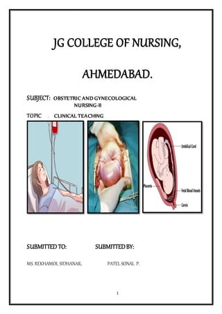
- 1. 1 JG COLLEGE OF NURSING, AHMEDABAD. SUBJECT: OBSTETRIC AND GYNECOLOGICAL NURSING-II TOPIC : CLINICAL TEACHING SUBMITTED TO: SUBMITTEDBY: MS. REKHAMOL SIDHANAR, PATEL SONAL P.
- 2. 2 ASSISTANT PROESSOR, s.Y M.SC NURSING, J.G COLLEGE OF NURSING J.G COLLEGE OF NURSING AHMEDABAD. AHMEDABAD. OUTLINE OF SEMINAR ON OBSTETRICAL EMERGENCY ( SHOCK, VASA PREVIA,UTERINE INVERSION) 1. INTRODUCTION 2. DEFINITION 3. OBSTETRICAL EMERGENCY SHOCK VASA PREVIA UTERINE INVERSION 4. SHOCK DEFINITION CAUSES TYPES HAEMORRHAGIC SHOCK NEUROGENIC SHOCK ENDOTOXIC SHOCK ANAPHYLATIC SHOCK 5. MANAGEMENT 6. VASA PREVIA DEFINITION CAUSES DIAGNOSIS SYMPTOMES TYPES MANAGEMENT 7. UTERINE INVERSION DEFINITION
- 3. 3 CAUSES CLASSIFICATION MANAGEMENT BIBLIOGRAPY Obstetrical Emergencies Obstetrical emergencies are lifethreatening medical conditions that occur in pregnancy or during or after labor and delivery. There are a number of illnesses and disorders of pregnancy that can threaten the wellbeing of both mother and child.Obstetrical emergencies may also occur during active labor, and after delivery (postpartum). Definition Shock is a condition resulting from inability of the circulatory system to provide the tissues requirements from oxygen and nutrients and to remove metabolites. Types and Causes Haemorrhagic shock excessive blood loss may be due to: o Causes of bleeding early in pregnancy. o Causes of antepartum haemorrhage. o Causes of postpartum haemorrhage. Neurogenic shock painful conditions my be due to: o Disturbed ectopic pregnancy. o Concealed accidental haemorrhage. o Forceps or breech extraction before full cervical dilatation. o Rough internal version. o Crédé’s method. o Rupture uterus. o Acute inversion of the uterus.
- 4. 4 o Rapid evacuation of the uterine contents as in precipitate labour and rupture of membranes in polyhydramnios. This is accompanied by rapid accumulation of blood in the splanchnic area due to sudden relief of pressure (splanchnic shock). Neurogenic shock: ineffective contraction of the cardiac muscle due to o Myocardial infarction. o Heart failure. Endotoxic shock: generalised vascular disturbance due to release of toxins. Anaphylactic shock: caused by sensitivity to drugs. Other causes: o Embolism: amniotic fluid, air or thrombus. o Anaesthetic complications: as Mendelson's syndrome. o The shock may be caused by more than one factor as: o Incomplete abortion: leads to haemorrhagic and endotoxic shock. o Disturbed ectopic and rupture uterus: lead to haemorrhagic and neurogenic shock. Classic Clinical Picture of Shock Low blood pressure. Rapid weak (thready) pulse. Pallor. Cold clammy sweat. Cyanosis of the fingers. Air hunger. Dimness of vision. Restlessness. Oliguria or anuria. HAEMORRHAGIC SHOCK Definition: Hypovolemic shock, also known as hemorrhagic shock, is a life-threatening condition that results when you lose more than 20 percent (one-fifth) of your body's blood or fluid supply. This severe fluid loss makes it impossible for the heart to pump a sufficient amount of blood to your body. Classification of Haemorrhage Class Blood Loss% Clinical Picture I 15% Normal pulse & blood pressure. Tilt test +ve II 20-25% Tachycardia. Tachypnoea. Pulse pressure (<30mmHg). Low systolic pressure. Delayed capillary filling III 30-35% Skin: cold, clammy and pale. Severe drop in blood pressure. Restlessness. Oliguria (<30 ml/hour). Metabolic acidosis (blood pH <7.5) IV 40-45% Profound hypotension. The carotid pulse is the only felt one.
- 5. 5 Irreversible shock Tilt test It is done in patient with considerable bleeding but the blood pressure and/ or pulse rate are normal. When this patient is in a sitting position, she develops hypotension and / or tachycardia. Phases of Haemorrhagic Shock The normal pregnant woman can withstand blood loss of 500 ml and even up to 1000 ml during delivery without obvious danger due to physiological cardiovascular and haematological adaptations during pregnancy. Phase of compensation Sympathetic stimulation: It is the initial response to blood loss leading to peripheral vasoconstriction to maintain blood supply to the vital organs. Clinical picture: Pallor, tachycardia, tachypnoea. Phase of decompensation Blood loss exceeds 1000 ml in normal patient or less if other adverse factors are operating. Clinical picture: is the classic clinical picture of shock (see before). Adequate treatment at this phase improves the condition rapidly without residual adverse effects. Phase of cellular damage and danger of death Inadequately treated haemorrhagic shock results in prolonged tissue hypoxia and damage with the following effects: Metabolic acidosis: due to anaerobic metabolism initiated after lack of oxygen. Arteriolar dilatation: caused by accumulation of metabolites leading to pooling and stagnation of blood in the capillaries and leakage of fluid into the tissues. Disseminated intravascular coagulation: caused by release of thromboplastin from the damaged tissues. Cardiac failure: due to diminished coronary blood flow. In this phase death is imminent, transfusion alone is inadequate and if recovery from acute phase occurs residual tissue damage as renal and/ or pituitary necrosis will occur. Management Urgent interference is indicated as follow: Detect the cause and arrest haemorrhage. Establish an airway and give oxygen by mask or endotracheal tube. Elevate the legs to encourage return of blood from the limbs to the central circulation. Two or more intravenous ways are established for blood, fluids and drugs infusion which should be given by IV route in shocked patient. If the veins are difficult to find a venous cut down or intrafemoral canulation is done. Restoration of blood volume by: o Whole blood: cross-matched from the same group if not available group O-ve may be given as a life -saving. o Crystalloid solutions: as ringer lactate, normal saline or glucose 5%. They have a short half life in the circulation and excess amount may cause pulmonary oedema. o Colloid solutions: as dextran 40 or 70, plasma protein fraction or fresh frozen plasma. Drug therapy: o Analgesics: 10-15 mg morphine IV if there is pain, tissue damage or irritability. o Corticosteroids: Hydrocortisone 1gm or dexamethasone 20 mg slowly IV. Its mode of action is controversial; it may decrease peripheral resistance and potentiate cardiac response so it improves tissue perfusion. o Sodium bicarbonate: 100 mEq IV if metabolic acidosis is demonstrated.
- 6. 6 o Vasopressors: to increase the blood pressure so maintain renal perfusion. Dopamine: 2.5m g/ kg/ minute IV is the drug of choice. ß -adrenergic stimulant: isoprenaline 1mg in 500 ml 5% glucose slowly IV infusion. Monitoring: o Central venous pressure (CVP): normal 10-12 cm water. o Pulse rate. o Blood pressure. o Urine output: normal 60 ml/hour. o pulmonary capillary wedge pressure: Normal 6-18 Torr. o Clinical improvement in the: pallor, cyanosis, air hunger, sweating and consciousness. Complications Acute renal failure. Pituitary necrosis (Sheehan’s syndrome). Disseminated intravascular coagulation. ENDOTOXIC (SEPTIC OR BACTERAEMIC) SHOCK Definition: Septic shock is a serious medical condition that occurs when sepsis, which is organ injury or damage in response to infection, leads to dangerously low blood pressure and abnormalities in cellular metabolism. Obstetric Causes Septic abortion. Prolonged rupture of membranes. Manipulations and instrumentations. Trauma. Retained placental tissues. Puerperal sepsis. Severe acute pyelonephritis. Causative Organisms Gram-negative bacilli: E.coli, proteus, pseudomonas and bacteroids. The endotoxin is a phospholipopolysaccharide released by lysis of its cell envelope. A similar picture is produced from exotoxin of ß-haemolytic streptococci, anaerobic streptococci and clostridia. Pathology Release of endotoxin results in increased lysosomal permeability and cytotoxicity. The sequence of events thereafter may occur in few minutes and include: Stimulation of the adrenal medulla and sympathetic nervous system → constriction of arterioles and venules → local acidosis → arteriolar dilatation but with continuing constriction of the venules → capillary pooling of blood → haemorrhagic engorgement of bowel, liver, kidneys and lungs. There is associated extensive disseminated intravascular coagulation due to sudden massive plasmin generation with which the antiplasmins cannot cope. Clinical Features Endotoxic shock passes with 2 main stages: Reversible stage It has 2 phases: Early (warm) phase: there are; o hypotension,
- 7. 7 o tachycardia, o pyrexia, o rigors, o flushed skin, o patient is alert, o leucocytosis develops within hours. Late (cold) phase: there are; o cold and clammy skin, o mottled cyanosis, o purpura, o jaundice, o progressive mental confusion, o coma. Irreversible stage Prolonged cellular hypoxia leads to: metabolic acidosis, acute renal failure, cardiac failure, pulmonary oedema, adrenal failure and ultimately death. Differential Diagnosis Amniotic fluid embolism. Pulmonary embolism. Pulmonary aspiration syndrome. Myocardial infarction. Incompatible blood transfusion. Management It includes 3 major lines of treatment: Restoration of circulatory function and oxygenation Replacement of blood loss: by whole blood, if not available start with colloids or crystalloids. The CVP measurement is essential to guard against circulatory overload. Corticosteroids: as; o Hydrocortisone 1gm IV / 6 hours or, o Dexamethasone 20 mg initially followed by 200 mg/day by IV infusion. β-adrenergic stimulants: as isoprenaline cause arteriolar dilatation, increase heart rate and stroke volume improving tissue perfusion. Blood volume must be normal prior to its administration. Oxygen: if respiratory function is impaired. Aminophylline: improves respiratory function by alleviating bronchospasm. Eradication of infection Antibiotic therapy: Swabs for culture and sensitivity are taken first. Antibiotic therapy is starting immediately till the result of culture and given by IV route. The therapy should cover the wide range of organisms: Ampicillin or Cephalosporines Gentamycin Metronidazole Clindamycin Gentamycin
- 8. 8 Surgical treatment: is indicated when there is retained infected tissues as in septic abortion. It should be removed as soon as antibiotic therapy and resuscitative measures have been started by: suction evacuation, digital evacuation, or hysterectomy in advanced infection with a gangrenous (clostridium welchii) or traumatised uterus. Correction of fluid and electrolyte deficits Disseminated intravascular coagulation Heparin therapy (see DIC) except if there is active bleeding where the condition is best treated by fresh blood transfusion. CARDIOGENIC SHOCK Can also occur in the setting of septic shockor hemorrhagic shock, especially in patientswho have baseline cardiovascular disease.Treatment requires invasive monitoring anddealing with the underlying disorder. NEUROGENIC SHOCK Definition: Neurogenic shock is a distributive type of shock resulting in low blood pressure, occasionally with a slowed heart rate, that is attributed to the disruption of the autonomic pathways within the spinal cord. It can occur after damage to the central nervous system such as spinal cord injury. Etiology: trauma and tissue damage as in cases of: Disturbed extrauterine pregnancy. Concealed accidental hemorrhage. Difficult forceps delivery or breech extraction (especially if the cervixisn’t fully dilated). Difficult internal version. Repeat rough attempts at Crede’s method. Rupture of the uterus or cervical tears extending into the lower uterinesegment. Acute inversion of the uterus. Rapid evacuation of the uterus as in precipitate labor andpolyhydramnios Retained placenta especially for more than 2 hours. TREATMENT: General measure: mentioned earlier. Fluid replacement. Vasopressor and inotropic agents. Dealing with the cause. Vasa praevia Vasa praevia, also spelled vasa previa, is a complication of pregnancy in which babies blood vessels cross or run near the internal opening of the uterus. These vessels are at risk of rupture when the supporting membranes rupture, as they are unsupported by the umbilical cord or placental tissue.
- 9. 9 Causes: Vasa previa is present when unprotected fetal vessels traverse the fetal membranes over the internal cervical os. These vessels may be from either a velamentous insertion of the umbilical cord or may be joining an accessory (succenturiate) placental lobe to the main disk of the placenta. If these fetal vessels rupture the bleeding is from the fetoplacental circulation, and fetal exsanguination will rapidly occur, leading to fetal death. It is thought that vasa previa arises from an early placenta previa. As the pregnancy progresses, the placenta tissue surrounding the vessels over the cervix undergoes atrophy, and the placenta grows preferentially toward the upper portion of the uterus. This leaves unprotected vessels running over the cervix and in the lower uterine segment. This has been demonstrated using serial ultrasound. Oyelese et al. found that 2/3 of patient with vasa previa at delivery had a low-lying placenta or placenta previa that resolved prior to the time of delivery. Types: There are three types of vasa previa. Types 1 and 2 were described by Catanzarite et al. In Type 1, there is a velamentous insertion with vessels running over the cervix. In Type 2, unprotected vessels run between lobes of a bilobed or succenturiate lobed placenta. In Type 3, a portion of the placenta overlying the cervix undergoes atrophy. In this type, there is a normal placental cord insertion and the placenta has only one lobe. However, vessels at a margin of the placenta are exposed. Risk factors: Vasa previa is seen more commonly with velamentous insertion of the umbilical cord, accessory placental lobes (succenturiate or bilobate placenta), multiple gestation, IVF pregnancy. In IVF pregnancies incidences as high as one in 300 have been reported. The reasons for this association are not clear, but disturbed
- 10. 10 orientation of the blastocyst at implantation, vanishing embryos and the increased frequency of placental morphological variations in in vitro fertilisation pregnancies have all been postulated. Diagnosis: The classic triad of the vasa praevia is: membrane rupture, painless vaginal bleeding and fetal bradycardia or fetal death. Prior to the advent of ultrasound, this diagnosis was most often made after a stillbirth or neonatal death in which the mother had ruptured her membranes, had some bleeding, and delivered an exsanguinated baby. In these cases, examination of the placenta and membranes after delivery would show evidence of a velamentous cord insertion with rupture of the vessels. However, with almost universal use of ultrasound in the developed world, many cases are now detected during pregnancy, giving the opportunity to deliver the baby before this catastrophic rupture of the membranes occurs. Vasa previa is diagnosed with ultrasound when echolucent linear or tubular structures are found overlying the cervix or in close proximity to it. Transvaginal ultrasound is the preferred modality. Color, power and pulsed wave Doppler should be used to confirm that the structures are fetal vessels. The vessels will demonstrate a fetal arterial or venous waveform. Alkali denaturation test detects the presence of fetal hemoglobin in vaginal blood, as fetal hemoglobin is resistant to denaturation in presence of 1% NaOH. Tests such as the Ogita Test, Apt test or Londersloot test were previously used to attempt to detect fetal blood in the vaginal blood, to help make the diagnosis. These tests are no longer widely used in the US, but are sometimes used in other parts of the world. Also detection of fetal hemoglobin in vaginal bleeding is diagnostic. Symptoms: Vasa previa may be present if any of the following conditions exist: Velamentous cord insertion—an umbilical cord abnormalityoccurring in about 1–2 percent of pregnancies, in which the cord inserts into the fetal membranes and travels through them to the placenta, rather than inserting directly into the placenta Bi-lobed placenta—the placenta is divided into two separate pieces called lobes Low-lying placenta, also known as placenta previa Multiple fetuses (twins, triplets, etc.) Mother has previously had uterine surgery Treatment: It is recommended that women with vasa previa should be delivered by elective cesarean prior to rupture of the membranes. Given that the timing of rupture of membranes is difficult to predict, elective cesarean delivery at 35–36 weeks is recommended. This gestational age gives a reasonable balance between the risk of death and that of prematurity. Several authorities have recommended hospital admission at about 32 weeks. This is to give the patient proximity to the operating room for emergency delivery should the membranes rupture. Because these patients are at risk for preterm delivery, it is recommended that steroids should be
- 11. 11 given to promote fetal lung maturation. When bleeding occurs, the patient goes into labor, or if the membranes rupture, immediate treatment with an emergency caesarean delivery is usually indicated. Uterine inversion Uterine inversion is a potentially fatal childbirth complication with a maternal survival rate of about 85%. It occurs when the placenta fails to detach from the uterusas it exits, pulls on the inside surface, and turns the organ inside out. It is very rare. Causes: The most common cause is the mismanagement of 3rd stage of labor, such as: Fundal pressure Excess cord traction during the 3rd stage of labor Other natural causes can be: Uterine weakness, congenital or not Precipitate delivery Short umbilical cord It is more common in multiple gestation than in singleton pregnancies. The incidence is of 1/2000 pregnancies. Types: ONE: Complete. Visible outside the cervix. TWO: Incomplete. Visible only at the cervix Classification: Description of the degree of inversion: First-degree - the inverted fundus extends to, but not through, the cervix. Second-degree - the inverted fundus extends through the cervix but remains within the vagina. Third-degree - the inverted fundus extends outside the vagina. Total inversion - the vagina and uterus are inverted.
- 12. 12 Description by the time since inversion: Acute inversion occurs within 24 hours of delivery. Subacute inversion occurs between 24 hours and one month after delivery. Chronic inversion occurs more than one month after delivery Association: Placenta praevia Fundal Placental Implantation Use of Magnesium Sulfate Vigorous fundal pressure Repeated cord traction short umbilical cord Presentation: Uterine inversion is often associated with significant Post-partum hemorrhage. Traditionally it was thought that it presented with haemodynamic shock "out of proportion" with blood loss, however blood loss has often been underestimated. The parasympathetic effect of traction on the uterine ligaments may cause bradycardia. Investigations If not clinically obvious, ultrasound can be used to identify the inversion Management The important principles are: Treatment should follow a logical progression. Hypotension and hypovolaemia require aggressive fluid and blood replacement.[13] Steps should follow those set out by the Royal College of Obstetricians and Gynaecologists (RCOG) guidelinesThe four components of management to be instigated at the same time are: Communication. Resuscitation. Monitoring and investigation. Measures to arrest the bleeding. Immediate uterine repositioning is essential for acute puerperal inversion. Measures to reposition the uterus may include: Preparing theatres for a possible laparotomy. Cautious administration of tocolytics to allow uterine relaxation; however, this may aggravate haemorrhage: Nitroglycerin (0.25-0.5 mg) intravenously over 2 minutes; or Terbutaline 0.1-0.25 mg slowly intravenously; or Magnesium sulfate 4-6 g intravenously over 20 minutes.
- 13. 13 Attempting prompt repositioning of the uterus. This is best done manually and quickly, as delay can render repositioning progressively more difficult. Reposition the uterus (with the placenta if still attached) by slowly and steadily pushing upwards towards the umbilicus, commonly referred to as Johnson's method. Maintain bimanual uterine compression and massage until the uterus is well contracted and bleeding has stopped. If this fails, hydrostatic replacement should be attempted under spinal or general anaesthetic: O'Sullivan's technique involves an infusion of warm saline into the vagina, making a water seal with the operator's hand and the vulva. Successful modifications of this technique, including using a vacuum cup to obtain a better vaginal seal and using a transurethral resection of prostate (TURP) set to increase the hydrostatic pressure, have been reported. An SOS Bakri tamponade balloon has also been successfully used to replace the inverted uterus and to maintain its position. If this is unsuccessful, a surgical approach is required. Laparotomy for surgical repositioning is more usual (find and apply traction to the round ligaments). Incision of the cervical ring may be required. A vaginal or even laparoscopic approach can be used, although this is more likely in the non-obstetric inversion.[1][14][15] If this is unsuccessful, hysterectomy, which may be life-saving, is the final option. If placenta is still present, careful examination and removal are required to ensure it is not abnormally adherent. General anaesthetic or uterine relaxant is then stopped and replaced with oxytocin, ergometrine or prostaglandins. Antibiotics are started and the stimulant continued for at least 24 hours. The woman must be monitored closely after repositioning, in order to avoid re-inversion. Complications Complications include endomyometritis, and damage to intestines, ureters or uterine appendages. Death can occur quickly if the condition is not recognised. Prognosis The condition carries a good prognosis if managed correctly. BIBLIOGRAPY 1. Bhadra banasree, GYNECOLOGY FOR NURSES, First Edition,2014, Jaypee brothers medical publishers, new delhi, 2. Howkins and Bourne shaws, TEXTBOOK OF GYNECOLOGY, 15th Edition, Elsevier, New delhi, 3. D.C Dutta, TEXTBOOK OF OBSTETRICS Incluing Perinatology and Contraception, Sixth edition, New Central Agency,culcutta,2004, 4. Jacob Annamma, A COMPREHENSIVE TEXTBOOK OF MIWIFERY AND GYNECOLOGICAL NURSING,Third edition, Jaypee Brothers medical publishers, New Delhi,2012, WEBSITE: 1. http://www.gfmer.ch/Obstetrics_simplified/shock_in_obstetrics.htm
- 14. 14 2. https://en.wikipedia.org/wiki/Vasa_praevia 3. http://www.pregnancycorner.com/being-pregnant/complications/vasa-previa.html 4. https://en.wikipedia.org/wiki/Uterine_inversion 5. http://patient.info/in/doctor/uterine-inversion