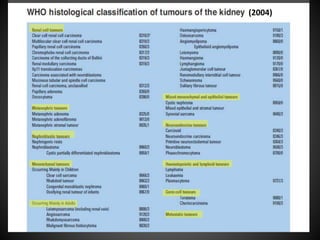
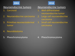
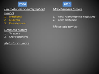
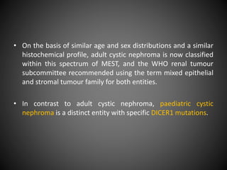
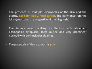
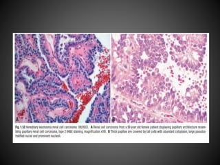
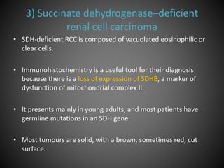
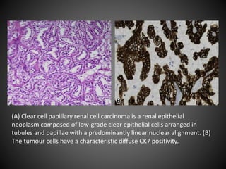
The document summarizes updates to the 2016 WHO classification of renal cell carcinoma compared to the 2004 classification. Key changes include recognizing RCC as distinct subtypes based on histology, architecture, location, associated diseases and molecular alterations. The 2016 classification adds 9 new renal tumor entities, separates some subtypes, and groups familial and sporadic forms of RCC together. It provides greater specificity in RCC diagnosis and classification based on recent advances in understanding RCC pathogenesis and genetics.
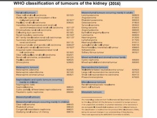

![• The new 2016 WHO classification refers to subtypes
that have been named on the basis of predominant
Cytoplasmic features (eg, clear cell and chromophobe renal
cell carcinomas [RCCs]),
Architectural features (eg, papillary RCC),
Anatomic location of tumours (eg, collecting duct and renal
medullary carcinomas), and
Correlation with a specific renal disease background (eg,
acquired cystic disease– associated RCCs)](https://image.slidesharecdn.com/whoclassification2016renalcellcarcinoma-231018134845-0540942b/85/WHO-CLASSIFICATION-2016-RENAL-CELL-CARCINOMA-pptx-27-320.jpg)
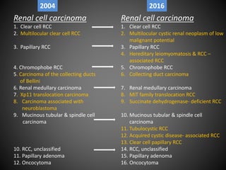
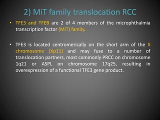
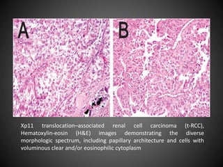
![ As well as molecular alterations pathognomonic for RCC
subtypes (eg, MiT family translocation carcinomas and
succinate dehydrogenase [SDH]–deficient renal carcinomas)
or
Familial predisposition syndromes (eg, hereditary
leiomyomatosis and RCC [HLRCC] syndrome– associated RCC).](https://image.slidesharecdn.com/whoclassification2016renalcellcarcinoma-231018134845-0540942b/85/WHO-CLASSIFICATION-2016-RENAL-CELL-CARCINOMA-pptx-28-320.jpg)
![• In contrast to the 2004 WHO classification, familial forms of
RCC, which also occur in sporadic form (eg, clear cell RCC
[ccRCC] in patients with von Hippel-Lindau [VHL] syndrome or
chromophobe RCC in patients with Birt- Hogg-Dube´
syndrome) are now discussed with the corresponding
sporadic tumour types in joint chapters.](https://image.slidesharecdn.com/whoclassification2016renalcellcarcinoma-231018134845-0540942b/85/WHO-CLASSIFICATION-2016-RENAL-CELL-CARCINOMA-pptx-29-320.jpg)


![PERI-PROSTHETIC FRACTURE NAIL-PLATE CONSTRUCT [NPC].pptx](https://cdn.slidesharecdn.com/ss_thumbnails/drarunkumardrmohamedashrafperiprostheticfrasturenail-plateconstructnpc-260209164459-7e9d15a1-thumbnail.jpg?width=640&height=640&fit=bounds)


