Download to read offline

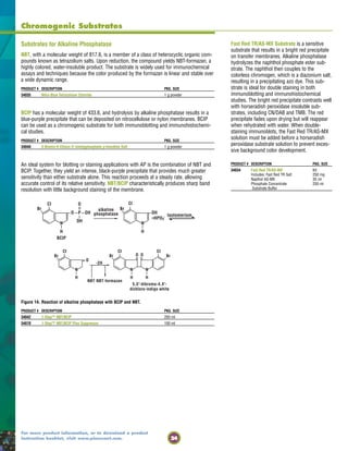
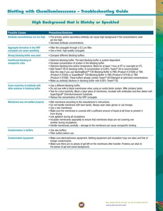
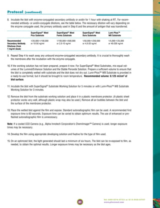
![Formulate Wash Buffers
Add detergent to blocking/wash
buffers to reduce nonspecific binding.
[Skip this step if you use StartingBlock™ T20 Blocking Buffer
in PBS (Product # 37539) or TBS (Product # 37543) or
SuperBlock® T20 Blocking Buffer in PBS (Product #
37516) or TBS (Product # 37536). These buffers already
contain Tween®-20 Detergent at optimized concentrations.]
Surfact-Amps®
Brand Detergents containing:
• Tween®
-20 (Product # 28320)
and Tween®
-80 (Product # 28328)
• Triton®
X-100 (Product # 28314) and Triton®
X-114 (Product # 28332)
• Nonidet P-40 (Product # 28324)
• Brij®
-35 (Product # 28316) and Brij®
-58 (Product # 28336)
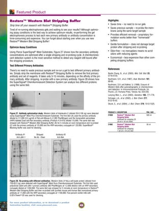
Stripping Buffer
Reprobe the blot if necessary.
• Restore™ Western Blot Stripping Buffer
(Product # 21059)
• IgG Elution Buffer (Product #s 21004
and 21009)
Film
Expose the membrane to X-ray film.
• CL-XPosure™ Film
5" x 7" sheets, (Product #s 34090 and 34092);
8" x 10" sheets, (Product #s 34091 and 34093)
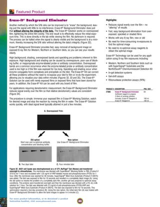
• Erase-It® Background Eliminator Kit (Product # 21065)
Enzyme Substrates
Add the detection reagent.
Chemiluminescent Substrates:
• SuperSignal®
West Pico Chemiluminescent
Substrate (Product #s 34077 and 34080)
• SuperSignal®
West Femto Maximum Sensitivity
Substrate (Product #s 34096
and 34095)
• SuperSignal®
West Dura
Extended Duration Substrate (Product #s 34076 and 34075)
• Lumi-Phos™ WB Substrate (Product # 34150)
Colorimetric Substrates:
• 1-Step™ Chloronaphthol (Product # 34012)
• TMB-Blotting (Product # 34018)
• NBT/BCIP (Product # 34042)
• Metal Enhanced DAB (Product # 34065)
HRP
SuperSignal®
Substrate
Primary and Secondary Detection Reagents
Incubate the membrane with antibody.
For a complete list, visit the antibody selection guide
on our web site (www.piercenet.com) accessible
under the Products tab.
For direct detection methods we offer:
• Monoclonal Antibodies
• Fluorescent Probes and Labeling Kits
• Enzyme Labeling Kits
For indirect detection methods we offer:
• Biotinylation Kits
• Protein A, Protein G and Protein L labeled with fluorescein,
rhodamine, HRP, AP or biotin
• Avidin, Streptavidin and NeutrAvidin™ Biotin-Binding Protein
labeled with fluorescein, rhodamine, HRP or AP
• Secondary antibodies labeled with fluorescein, rhodamine,
HRP, AP or biotin
HRP
Ag
For convenience and economy, Pierce also offers complete Western
blotting Kits that include chemiluminescent substrates, enzyme-
conjugated antibodies, blocking buffers and standard buffers.
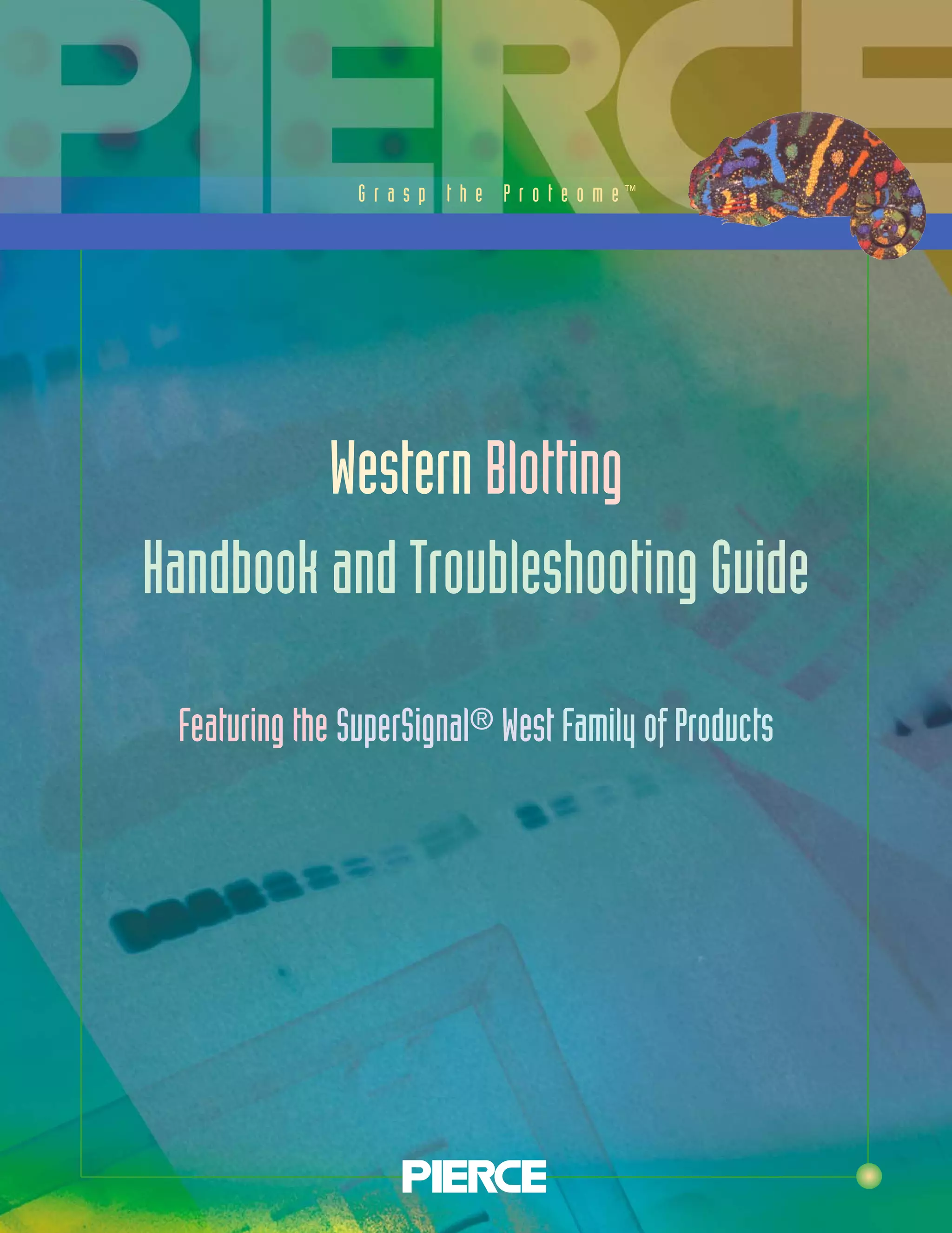
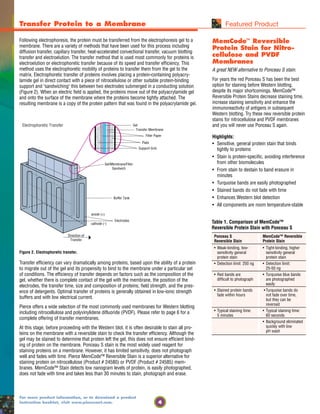
Western Blotting
the Pierce Way
STEP 4B
STEP 5
STEP 6
STEP 7
STEP 8
3](https://image.slidesharecdn.com/wb1600990-150315200940-conversion-gate01/85/Wb1600990-5-320.jpg)
![13
Tel: 800-874-3723 or 815-968-0747
www.piercenet.com
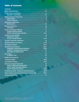
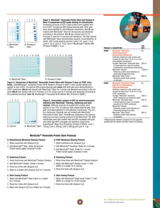
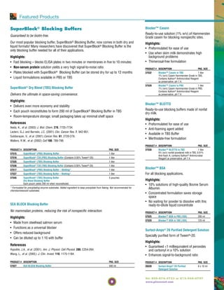
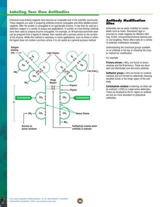
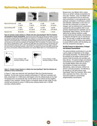
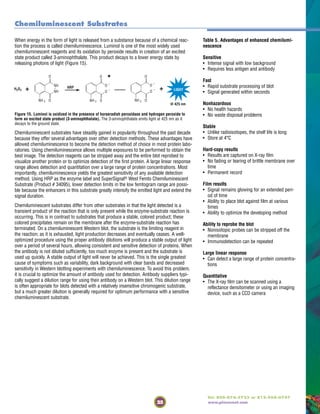
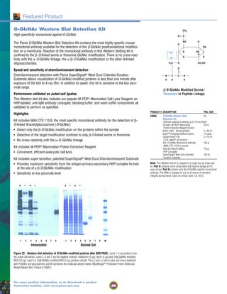
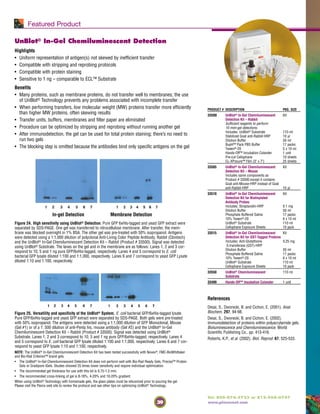
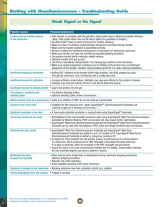
Affinity-Purified Secondary Antibodies
*See Table 3 on page 12 for the Key to Abbreviations.
Product #
Pkg. Size
Specificity Description Host Unconj. Biotin-LC Fluorescein Rhodamine Peroxidase Alk. Phos.
Anti-BOVINE Bovine IgG (H+L) Goat 31100 31710
2 mg 2 ml
Bovine IgG (H+L) Rabbit 31103 31712
2 mg 1.5 ml
Anti-CHICKEN Chicken IgY (H+L) Rabbit 31104 31720 31501 31401
2 mg 1.5 ml 1.5 mg 1.5 ml
Anti-GOAT Goat IgG (H+L) Donkey 31108
1.5 mg
Goat IgG (H+L) Mouse 31107 31730 31512 31400
(min x HnMsRb Sr Prot)* 1.5 mg 1 ml 1 mg 1 ml
Goat IgG (H+L) Rabbit 31105 31732 31509 31650 31402 31300
2 mg 1.5 mg 1.5 mg 1.5 mg 1.5 ml 1 ml
Goat IgG [F(ab')2] Rabbit 31153 31753 31553 31403 31405
2 mg 1.5 ml 1.5 mg 1.5 ml 1 ml
Goat IgG (Fc) Rabbit 31133 31733 31533 31433 31337
2 mg 1.5 ml 1.5 mg 1.5 ml 1 ml
Anti-GOAT F(ab')2 Goat IgG (H+L) Rabbit 31109 31302
Fragment of (min x Hn Sr Prot)* 0.5 mg 0.5 ml
Host Antibody
Anti-GUINEA PIG Guinea Pig IgG (H+L) Goat 31114
2 mg
Anti-HAMSTER Hamster IgG (H+L) Goat 31115 31750
1.5 mg 1.5 mg
Hamster IgG (H+L) Rabbit 31120 31587 31652
2 mg 1.5 mg 1.5 mg
Anti-HORSE Horse IgG (H+L) Goat 31116 31760
2 mg 1.5 mg
Anti-HUMAN Human IgG (H+L) Goat 31130 31770 31529 31656 31410 31310
2 mg 1.5 mg 2 mg 2 mg 2 ml 1 ml
Human IgG Goat 31118
Gamma Chain Specific 0.5 mg
Human IgG (H+L) Goat 31119 31774 31531 31412
(min x BvHsMs Sr Prot)* 1.5 mg 1.5 ml 1.5 mg 1.5 ml
Human IgG [F(ab')2] Goat 31122 31312
2 mg 1 ml
Human IgG [F(ab')2] Goat 31132 31414
(min x BvHsMs Sr Prot)* 1.5 mg 1.5 ml
Human IgG (Fc) Goat 31123 31416
(min x BvHsMs Sr Prot)* 1.5 mg 1.5 ml
Human IgM (Fc5µ) Goat 31136 31575 31415
2 mg 2 mg 2 ml
Human IgM (µ) Goat 31124 31778
0.5 mg 0.5 mg
Human IgM (Fc5µ) Goat 31138
(min x Bv Sr Prot)* 1.5 mg
Human IgA (α) Goat 31140 31577 31417 31314
2 mg 2 mg 2 ml 1 ml
Human IgG + IgM Goat 31134 31776
(H+L) 2 mg 2 ml
Human IgA + IgG Goat 31128 31782 31418 31316
+ IgM (H+L) 2 mg 2 ml 2 ml 1 ml
Human Kappa Chain Goat 31129 31780
0.5 mg 0.5 mg
Human Lambda Chain Goat 31131
0.5 mg
Human IgG (H+L) Mouse 31135 31420
(min x Ms Sr Prot)* 2 mg 1.5 ml](https://image.slidesharecdn.com/wb1600990-150315200940-conversion-gate01/85/Wb1600990-15-320.jpg)
![14
For more product information, or to download a product
instruction booklet, visit www.piercenet.com.
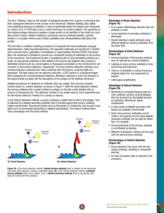
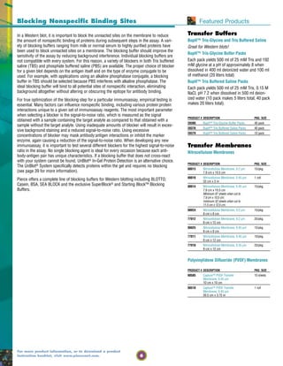
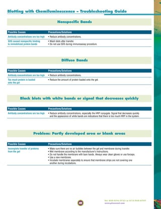
Affinity-Purified Secondary Antibodies
*See Table 3 on page 12 for the Key to Abbreviations.
Product #
Pkg. Size
Specificity Description Host Unconj. Biotin-LC Fluorescein Rhodamine Peroxidase Alk. Phos.
Anti-HUMAN Human IgG (H+L) Mouse 31137 31784
continued (min x BvHsMs Sr Prot)* 1.5 mg 1 ml
Human IgG (H+L) Rabbit 31143 31786
2 mg 1.5 ml
Human IgG (H+L) Rabbit 31147
(min x Ms Sr Prot)* 1.5 mg
Human IgG (Fc) Rabbit 31142 31789 31535 31423 31318
2 mg 1.5 ml 1.5 mg 1.5 ml 1 ml
Human IgM (Fc5µ) Rabbit 31149
2 mg
Anti-HUMAN Human IgG (Fc) Goat 31163
F(ab')2 Fragment 1 mg
of Host Antibody
Human IgG (H+L) Goat 31165
1 mg
Human IgA + IgG Goat 31539
+ IgM (H+L) 1 mg
Human IgG Mouse 31155
(min x MsBvHs Sr Prot)* 1.5 mg
Anti-MOUSE Mouse IgA (α) Goat 31169
(min x Hn Sr Prot)* 1 mg
Mouse IgA + IgG Goat 31171
+ IgM (H+L) 2 mg
Mouse IgG (H+L) Goat 31160 31800 31569 31660 31430 31320
2 mg 2 ml 2 mg 2 mg 2 ml 1 ml
Mouse IgG (H+L) Goat 31164 31802 31541 31661 31432 31322
(min x BvHnHs Sr Prot)* 1.5 mg 1.5 mg 1.5 mg 1.5 mg 1.5 ml 1 ml
Mouse IgG [F(ab')2] Goat 31166 31803 31543 31436 31324
2 mg 2 ml 2 mg 2 ml 1 ml
Mouse IgG (Fc) Goat 31168 31805 31547 31663 31437 31325
2 mg 2 ml 2 mg 2 mg 2 ml 1 ml
Mouse IgG (Fc) Goat 31170 31439 31327
(min x BvHnHs Sr Prot)* 1.5 mg 1.5 ml 1 ml
Mouse IgM (µ) Goat 31172 31804 31992 31662 31440 31326
2 mg 0.5 mg 2 mg 2 mg 2 ml 1 ml
Mouse IgM (µ) Goat 31176 31585 31664
(min x BvHnHs Sr Prot)* 1.5 mg 1.5 mg 1.5 mg
Mouse IgG + IgM Goat 31182 31807 31586 31444 31328
(H+L) 2 mg 2 ml 1.5 mg 2 ml 1 ml
Mouse IgG + IgM Goat 31184 31446 31330
(H+L) (min x BvHnHs Sr Prot)* 1.5 mg 1.5 ml 1 ml
Mouse IgG (H+L) Horse 31181 31806
1.5 mg 1.5 mg
Mouse IgG (H+L) Rabbit 31188 31810 31561 31665 31450 31329
2 mg 1.5 ml 1.5 mg 1.5 mg 1.5 ml 1 ml
Mouse IgG (H+L) Rabbit 31190 31812 31452 31334
(min x Hn Sr Prot)* 1.5 mg 1 ml 1 ml 0.5 ml
Mouse IgG [F(ab')2] Rabbit 31192 31811 31559 31666 31451 31331
2 mg 1.5 ml 1.5 mg 1.5 mg/ 1.5 ml 1 ml
Mouse IgG (Fc) Rabbit 31194 31813 31555 31455 31332
2 mg 1.5 ml 1.5 mg 1.5 ml 1 ml
Mouse IgM (µ) Rabbit 31196 31814 31557 31456 31333
2 mg 1.5 ml 1.5 mg 1.5 ml 1 ml
Mouse IgG + IgM Rabbit 31198 31815 31558 31457 31335
(H+L) 2 mg 1.5 ml 1.5 mg 1.5 ml 1 ml
Mouse IgG (H+L) Goat 31185 31565 31438
(min x BvHnHs Sr Prot)* 1 mg 1 mg 0.5 ml](https://image.slidesharecdn.com/wb1600990-150315200940-conversion-gate01/85/Wb1600990-16-320.jpg)
![15
Tel: 800-874-3723 or 815-968-0747
www.piercenet.com
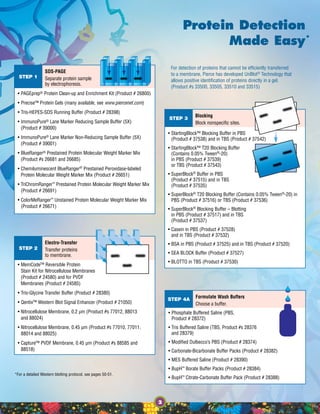
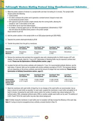
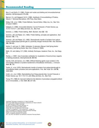
Affinity-Purified Secondary Antibodies
*See Table 3 on page 12 for the Key to Abbreviations.
Product #
Pkg. Size
Specificity Description Host Unconj. Biotin-LC Fluorescein Rhodamine Peroxidase Alk. Phos.
Anti-MOUSE Mouse IgM (µ) Goat 31178
F(ab')2 Fragment 1 mg
of Host Antibody
continued
Mouse IgM (µ) Goat 31186 31442
(min x BvHnHs Sr Prot) 1 mg 0.5 ml
Mouse IgG + IgM Goat 31448
(H+L) (min x BvHnHs Sr Prot) 0.5 ml
Mouse IgG (H+L) Rabbit 31189
1 mg
Anti-RABBIT Rabbit IgG (H+L) Donkey 31821 31568 31458 31345
(min x BvGtHnHsMsRtSh Sr Prot) 0.5 ml 0.5 mg 0.5 ml 0.5 ml
Rabbit IgG (H+L) Goat 31210 31820 31670 31460 31340
2 mg 1.5 mg 2 mg 2 ml 1 ml
Rabbit IgG (H+L) Goat 31212 31822 31583 31462 31342
(min x Hn Sr Prot) 1.5 mg 1.5 ml 1.5 mg 1.5 ml 1 ml
Rabbit IgG [F(ab')2] Goat 31234 31823 31573 31461 31343
2 mg 2 ml 2 mg 2 ml 1 ml
Rabbit IgG (Fc) Goat 31216 31463 31341
2 mg 2 ml 1 ml
Rabbit IgG (H+L) Mouse 31213 31824 31584 31674 31464
(min x GtHnMsSh Sr Prot) 1.5 mg 1 ml 1 mg 1 mg 1 ml
Anti-RABBIT Rabbit IgG (H+L) Goat 31214 31579
F(ab')2 Fragment 1 mg 1 mg
of Host Antibody
Rabbit IgG (H+L) Goat 31215
(min x Hn Sr Prot) 1 mg
Rabbit IgG (Fc) Goat 31217 31581
1 mg 1 mg
Anti-RAT Rat IgG (H+L) Goat 31220 31830 31629 31680 31470 31350
2 mg 2 ml 2 mg 2 mg 2 ml 1 ml
Rat IgG [F(ab')2] Goat 31474
2 ml
Rat IgG (Fc) Goat 31226 31833 31621 31475 31353
2 mg 2 ml 2 mg 2 ml 1 ml
Rat IgM (µ) Goat 31228 31832 31631 31476 31354
2 mg 2 ml 2 mg 2 ml 1 ml
Rat IgG (H+L) Rabbit 31218 31834
2 mg 1.5 mg
Rat IgG (H+L) Rabbit 31219 31836
(min x Ms Sr Prot) 0.5 mg 0.5 mg
Anti-RAT Rat IgG (H+L) Rabbit 31227
F(ab')2 Fragment 1 mg
of Host Antibody
Rat IgG + IgM Goat 31625
(H+L) 1 mg
Rat IgG (H+L) Mouse 31225 31633 31682
(min x Ms Sr Prot) 1 mg 0.5 mg 0.5 mg
Anti-SHEEP Sheep IgG (H+L) Rabbit 31240 31840 31627 31480 31360
2 mg 1.5 mg 1.5 mg 1.5 ml 1 ml
Sheep IgG (Fc) Rabbit 31241 31841 31441 31356
2 mg 1.5 ml 1.5 ml 1 ml
Sheep IgG [F(ab')2] Rabbit 31244 31844 31481 31344
2 mg 1.5 ml 1.5 ml 1 ml
Anti-SHEEP Sheep IgG (H+L) Rabbit 31229
F(ab')2 Fragment 1 mg
of Host Antibody
Anti-SWINE Swine IgG (H+L) Goat 31231
2 mg](https://image.slidesharecdn.com/wb1600990-150315200940-conversion-gate01/85/Wb1600990-17-320.jpg)


![35
Tel: 800-874-3723 or 815-968-0747
www.piercenet.com
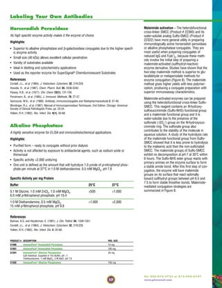
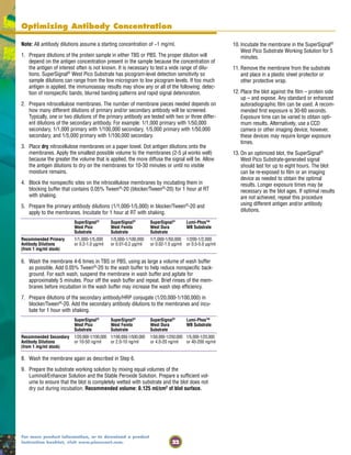
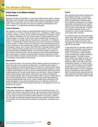
Studying Protein Interactions by Far-Western Blotting
Far-Western blotting was originally developed to screen protein expression libraries with
32P-labeled glutathione S-transferase (GST)-fusion protein. Far-Western blotting is now used
to identify protein:protein interactions. In recent years, far-Western blotting has been used
to determine receptor:ligand interactions and to screen libraries for interacting proteins. It is
possible to study the effect of post-translational modifications on protein:protein interac-
tions, examine interaction sequences using synthetic peptides as probes and identify
protein:protein interactions without using antigen-specific antibodies with this method of
analysis.
Far-Western blotting vs. Western blotting
The far-Western blotting technique is quite similar to Western blotting. In a Western blot,
an antibody is used to detect the corresponding antigen on a membrane. In a classical
far-Western analysis, a labeled or antibody-detectable “bait” protein is used to probe and
detect the target “prey” protein on the membrane. The sample (usually a lysate) contain-
ing the unknown prey protein is separated by sodium dodecyl sulfate-polyacrylamide gel
electrophoresis (SDS-PAGE) or native PAGE and then transferred to a membrane. When
attached to the surface of the membrane, the prey protein becomes accessible to probing.
After transfer, the membrane is blocked and then probed with a known bait protein, which
usually is applied in pure form. Following reaction of the bait protein with the prey pro-
tein, a detection system specific for the bait protein is used to identify the corresponding
band (Table 10).
Specialized Far-Western Analysis
By creative design of bait protein variants and other controls, the far-Western blotting
method can be adapted to yield very specific information about protein:protein interactions.
For example, Burgess, et al. used a modified far-Western blotting approach to determine
sites of contact among subunits of a multi-subunit complex. By an “ordered fragment lad-
der” far-Western analysis, they were able to identify the interaction domains of E. coli RNA
polymerase β´ subunit. The protein was expressed as a polyhistidine-tagged fusion, then
partially cleaved and purified using a Ni2+-chelate affinity column. The polyhistidine-tagged
fragments were separated by SDS-PAGE and transferred to a nitrocellulose membrane. The
fragment-localized interaction domain was identified using a 32P-labeled protein probe.
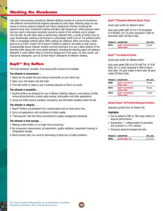
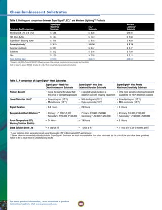
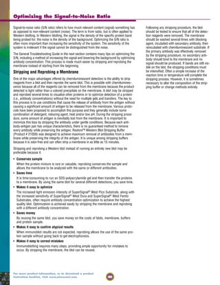
Table 10. Comparison of Western blotting and far-Western blotting methods
Step Western Blotting Far-Western Analysis
Gel Electrophoresis Native or denaturing (usually) Native (usually) or denaturing
Transfer System Optimal membrane and transfer system determined empirically Optimal membrane and transfer system determined empirically
Blocking Buffer Optimal blocking system determined empirically Optimal blocking system determined empirically
Detection Unlabeled primary antibody→ Unlabeled bait protein→
(several possible Enzyme-labeled secondary antibody→ Enzyme-labeled bait-specific antibody→
strategies)* Substrate reagent Substrate reagent
Enzyme-labeled primary antibody→ Radiolabeled bait protein→
[Arrows designate Substrate reagent Exposure to film
sequence of steps Biotinylated antibody→ Biotinylated bait protein→
in the detection Enzyme-labeled streptavidin→ Enzyme-labeled streptavidin→
strategy] Substrate reagent Substrate reagent
Fusion-tagged bait protein→
Tag-specific antibody→
Enzyme-labeled secondary antibody→
Substrate reagent
* Labeled antibodies generally are enzyme-labeled (either horseradish peroxidase or alkaline phosphatase). By contrast, bait proteins generally are not enzyme-labeled because a
large enzyme label is likely to sterically hinder unknown binding sites between bait and prey proteins. Other labeling and detection schemes are possible.
Far-Western Blotting
Importance of Native Prey Protein Structure in
Far-Western Analysis
Far-Western blotting procedures must be per-
formed with care and attention to preserving as
much as possible the native conformation and
interaction conditions for the proteins under
study. Denatured proteins may not be able to
interact, resulting in a failure to identify an inter-
action. Alternatively, proteins presented in
non-native conformations may interact in novel,
artificial ways, resulting in “false-positive” interac-
tions. The prey protein in particular is subjected
to preparative processing steps for far-Western
blotting that can have significant effects on detec-
tion of protein:protein interactions. This is not to
imply that identification of valid interactions is not
possible, but only to stress the importance of
appropriate validation and use of controls.](https://image.slidesharecdn.com/wb1600990-150315200940-conversion-gate01/85/Wb1600990-37-320.jpg)
![43
Tel: 800-874-3723 or 815-968-0747
www.piercenet.com
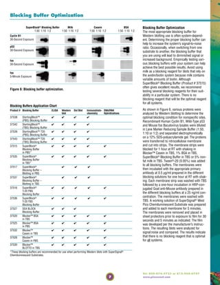
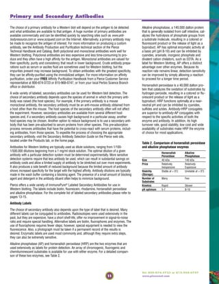
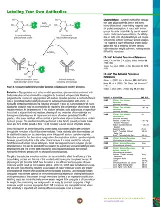
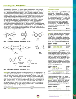
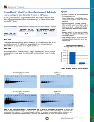
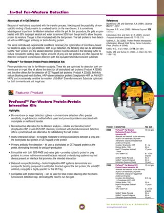
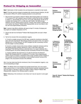
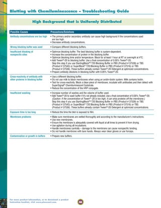
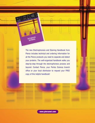
Increasing the Sensitivity of a Western Blot
1. Rinse membrane after
transfer with ultrapure water
2. Incubate membrane with
Reagent 1 for 2 minutes
on a shaker
3. Rinse membrane with
ultrapure water
(repeat 5 times)
1
Ultrapure
H2O
Ultrapure
H2O
4. Incubate membrane with
Reagent 2 for 10 minutes
on a shaker
2
5. Rinse membrane with
ultrapure water
(repeat 5 times)
Ultrapure
H2O Start your
detection
protocol.
Total time = 15 minutes
Figure 29. Enhanced chemiluminescent detection of identical serial dilutions of IL-6 before and after
treatment with Qentix™ Western Blot Signal Enhancer.
Qentix™ [ken´-tiks] Western Blot Signal Enhancer
It’s like intensifying screens in a bottle.
There are many ways to increase the sensitivity of a Western blot. Some methods are as
simple as switching substrates or blocking buffers, while others are more time-consuming
such as optimizing antibody titer or checking for proper protein transfer. Those solutions are
covered in the troubleshooting section of this handbook.
One of the more certain and easiest ways to increase the sensitivity of any Western blot is to
use the new Qentix™ Western Blot Signal Enhancer.
Qentix™ Western Blot Signal Enhancer does for enzyme-/substrate-based blotting what
intensifying screens do for radioactive blotting – it increases the signal up to 10-fold (or one
order of magnitude) in only 15 minutes.
The Qentix™ Western Blot Signal Enhancer membrane treatment is a simple, 15-minute pro-
cedure that can be added to your current Western blotting protocol. The result is an
increase in the intensity of target protein bands on the Western blot or detection of target
proteins at a level that could not previously be detected. Some protein targets have resulted
in a 10-fold increase in band intensity after treatment with the Western Blot Signal Enhancer
compared to the typical detection protocol without treatment.
Untreated blot Blot treated with Qentix™ Western Blot Enhancer
Figure 31. Qentix™ Western Blot Signal Enhancer Protocol – performed after transfer and before blocking.
Figure 30. Enhanced chromogenic detection of identical serial dilutions of IL-6 before and after treat-
ment with Qentix™ Western Blot Signal Enhancer.
Untreated blot Blot treated with Qentix™ Western Blot Enhancer
1 2 3 4 1 2 3 4
Highlights:
Enhances chemiluminescent, fluorescent and
colorimetric detection up to 10-fold
• Treatment with Western Blot Signal Enhancer can
boost the band intensity from three- to 10-fold,
regardless of what substrate is used
Enhances detection of targets transferred to
either nitrocellulose or PVDF*, independent of
membrane pore size
• Works with the most commonly used Western blot-
ting membranes
• Signal intensity has been increased with targets
such as mouse IL-6, p53, NF-κB, BRCA1 and EGF
Room temperature-stable, ready-to-use
reagents
• No thawing, formulating or diluting necessary
15-minute protocol
• Optimized to save time and improve detection capa-
bility of your specific analyte
* Signal enhancement of proteins on PVDF membrane has
been shown to be variable from no significant enhancement
for some proteins, to several-fold enhancement for others.
PRODUCT # DESCRIPTION PKG. SIZE
21050 Qentix™ Western Blot Kit
Signal Enhancer*
Sufficient reagent for ten
10 cm x 10 cm blots.
Includes: Enhancer Reagent 1 250 ml
Enhancer Reagent 2 250 ml](https://image.slidesharecdn.com/wb1600990-150315200940-conversion-gate01/85/Wb1600990-45-320.jpg)


This document provides guidance on performing and optimizing Western blot techniques. It discusses sample preparation, protein transfer, blocking, washing, primary and secondary antibody incubation, substrate selection, detection methods, troubleshooting, and related products. Key steps include separating proteins by electrophoresis, transferring them to a membrane, blocking non-specific binding, probing with primary and secondary antibodies, applying a substrate to produce a detectable signal, and capturing the results.


![StarBuzz-August 7th-2009-web[1]](https://cdn.slidesharecdn.com/ss_thumbnails/9952008-111030162352-phpapp02-thumbnail.jpg?width=640&height=640&fit=bounds)


