
Recommended
More Related Content
Similar to UV PPT.ppt
Similar to UV PPT.ppt (20)
Recently uploaded
Recently uploaded (20)
UV PPT.ppt
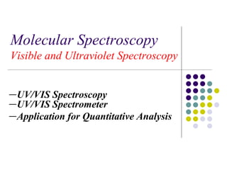
- 1. Molecular Spectroscopy Visible and Ultraviolet Spectroscopy -UV/VIS Spectroscopy -UV/VIS Spectrometer -Application for Quantitative Analysis
- 2. U V Spectrophotometer n 2014 Y2* p Y1 Y1 Y2 Y3 * Y4 *
- 3. Ultraviolet: 190~400nm Violet: 400 - 420 nm Indigo: 420 - 440 nm Blue: 440 - 490 nm Green: 490 - 570 nm Yellow: 570 - 585 nm Orange: 585 - 620 nm Red: 620 - 780 nm
- 4. Electronic Spectroscopy Ultraviolet (UV) and visible (VIS) spectroscopy This is the earliest method of molecular spectroscopy. A phenomenon of interaction of molecules with ultraviolet and visible lights. Absorption of photon results in electronic transition of a molecule, and electrons are promoted from ground state to higher electronic states.
- 5. Quantitative Analysis Beer’s Law A=ebc e: the molar absorptivity (L mol-1 cm-1) b: the path length of the sample c :the concentration of the compound in solution, expressed in mol L-1
- 7. UV Spectroscopy Instrumentation and Spectra A. Instrumentation : 1. The construction of a traditional UV-VIS spectrometer is very similar to an IR, as similar functions – sample handling, irradiation, detection and output are required 2. Here is a simple schematic that covers most modern UV spectrometers: sample reference detector I0 I0 I0 I log(I0/I) = A 200 700 l, nm monochromator/ beam splitter optics UV-VIS sources
- 8. Components of a Spectrophotometer Light Source Deuterium Lamps-a truly continuous spectrum in the ultraviolet region is produced by electrical excitation of deuterium at low pressure. (160nm~375nm) Tungsten Filament Lamps-the most common source of visible and near infrared radiation.
- 9. Components of a Spectrophotometer Monochromator Used as a filter: the monochromator will select a narrow portion of the spectrum (the bandpass) of a given source Used in analysis: the monochromator will sequentially select for the detector to record the different components (spectrum) of any source or sample emitting light.
- 11. Principle of Photomultiplier Detector The type is commonly used. The detector consists of a photo emissive cathode coupled with a series of electron- multiplying dynode stages, and usually called a photomultiplier. The primary electrons ejected from the photo- cathode are accelerated by an electric field so as to strike a small area on the first dynode.
- 12. Principle of Photomultiplier Detector The impinging electrons strike with enough energy to eject two to five secondary electrons, which are accelerated to the second dynode to eject still more electrons. A photomultiplier may have 9 to 16 stages, and overall gain of 106~109 electrons per incident photon.
- 13. Single and Double Beam Spectrometer Single-Beam: There is only one light beam or optical path from the source through to the detector. Double-Beam: The light from the source, after passing through the monochromator, is split into two separate beams-one for the sample and the other for the reference.
- 14. 14 Instrumentation – Sample Handling • Virtually all UV spectra are recorded solution-phase • Cells can be made of plastic, glass or quartz Only quartz is transparent in the full 200- 700 nm range; plastic and glass are only suitable for visible spectra. A typical sample cell (commonly called a cuvet):
- 15. Steps in carrying out a colorimetric analysis. Choose the wavelength of maximum absorbance. Prepare a calibration curve using known quantities of the complex measured at this wavelength. Measure the absorbance of your unknown sample. Calculate the concentration from the equation of the best fit line. UV / visible Spectroscopy
- 16. Standard Addition Method Standard addition must be used whenever the matrix of a sample changes the analytical sensitivity of the method. In other words, the slope of the working curve for standards made with distilled water is different from the same working curve.
- 17. Preparation of the Standards The concentration and volume of the stock solution added should be chosen to increase the concentration of the unknown by about 30% in each succeeding flask.
- 18. Preparation of the Sample Sampling Homogenization (By Grinding/ Blender) Digestion Wet Oxidation Dry Ashing Microwave Isolation Colour development by suitable reagent Dilution to different volume Preparation of Standard & read absorbance
- 19. External Standard and the Calibration Curve
- 20. Clarity of the solution The solution must be free of precipitates. Turbidity scatters and absorbs light. High sensitivity It is desirable that the colour reaction be highly sensitive. i.e. e is very large. UV / visible Spectroscopy
- 21. Advantages of colorimetric analysis. Better at low concentrations than titrimetric or gravimetric analysis. Can be applied under conditions where there are no satisfactory titrimetric or gravimetric procedures. Very rapid once a calibration curve as been obtained. UV / visible Spectroscopy
- 22. UV / visible Spectroscopy Six criteria for a successful analysis Specificity of the colour reaction Proportionality between colour and concentration Stability of the colour Reproducibility Clarity of the solution High sensitivity.
- 23. THANKING YOU
Editor's Notes
- 34
- 33
- 35
- 28