1. (12) United States Patent
Ferrari et al.
US007450779B2
US 7,450,779 B2
Nov. 11, 2008
(10) Patent N0.:
(45) Date of Patent:
(54) DE-NOISING DIGITAL RADIOLOGICAL
IMAGES
(75) Inventors: Ricardo J. Ferrari, Edmonton (CA);
Robin Winsor, Calgary (CA)
(73) Assignee: Imaging Dynamics Company Ltd.,
Calgary, Alberta (CA)
( * ) Notice: Subject to any disclaimer, the term ofthis
patent is extended or adjusted under 35
U.S.C. 154(b) by 479 days.
(21) Appl.No.: 11/131,286
(22) Filed: May 18, 2005
(65) Prior Publication Data
US 2005/0259889 A1 Nov. 24, 2005
Related US. Application Data
(60) Provisional application No. 60/573,287, ?led on May
21, 2004.
(51) Int. Cl.
G06K 9/40 (2006.01)
G06K 9/00 (2006.01)
(52) U.S.Cl. ..................................... .. 382/275; 382/128
(58) Field of Classi?cation Search ............... .. 382/275,
382/128-132, 254, 274; 600/310
See application ?le for complete search history.
(56) References Cited
U.S. PATENT DOCUMENTS
5,309,496 A 5/1994 Winsor
OTHER PUBLICATIONS
Ye, Zhen; Lu, Cheng-Chang; A Complex Wavelet Domain Markov
Model for Image Denoising; Sep. 14-17, 2003; IEEE; vol. 3; p.
365-368.*
Mascarenhas, Nelson D. A.; Furuie, Sergio S.; Portal, Angel L. S.;
Global Projection Estimation Methods for the Tomographic Recon
struction of Images With Poisson Noise; Dec. 1993; IEEE Transac
tions on Nuclear Science; vol. 40, No. 6; p. 2008-2013*
Bradley, A (2003) Shift-invariance in discrete Wavelet transform. In:
Sun et al Proceedings of the Seventh Digital Image Computing:
Techniques and Applications p. 29-38.
Crouse et al (1998) Wavelet-based statistical signal processing using
hidden Markov models. IEEE Transactions on Signal Processing
46:886-902.
Dippel et al (2002) Multiscale contrast enhancement for
radiographies: Laplacian pyramid versus fast Wavelet transform.
IEEE Transactions on Medical Imaging 21:343-353.
Donoho (1995) De-noising by soft-thresholding. IEEE Transactions
on Information Theory 41:613-627.
Donoho et al (1995) Adapting to unknown smoothness via Wavelet
shrinkage. Journal of American Statistical Association 90: 1200
1224.
(Continued)
Primary ExamineriAaron W Carter
(74) Attorney, Agent, or FirmiGoWling LaFleur Henderson
LLP; D. Doak Horne; BrianY. Lee
(57) ABSTRACT
This invention relates to a method for de-noising digital
radiographic images based upon a Wavelet-domain Hidden
Markov Tree (HMT) model. The method uses the
Anscombe’s transformation to adjust the original image to a
Gaussian noise model. The image is then decomposed in
different sub-bands of frequency and orientation responses
using a dual-tree complex Wavelet transform, and the HMT is
used to model the marginal distribution of the Wavelet coef
?cients. TWo different methods Were used to denoise the
Wavelet coef?cients. Finally, the modi?ed Wavelet coef?
cients are transformed back into the original domain to get the
de-noised image.
14 Claims, 14 Drawing Sheets
2. US 7,450,779 B2
Page 2
OTHER PUBLICATIONS
Durand et al(200l) Artifact free signal de-noising With Wavelets. In:
International Conference in Acoustics, Speech and Signal Process
ing. p. 3685-3688.
Kingsbury (1999) Image processing With complex Wavelets. Philo
sophical Transactions of the Royal Society of London 35712543
2560.
Laine et al (1994) Mammographic feature enhancement by
multiscale analysis. IEEE Transactions on Medical Imaging 13:725
740.
Romberg et al (2001) Bayesian tree-structured image modeling using
Wavelet-domain hidden Markov models. IEEE Transactions on
Image Processing 10:1056-1068.
* cited by examiner
7. US. Patent Nov. 11,2008 Sheet 5 0f 14 US 7,450,779 B2
Radiation dose versus denoising
perfomance
I lmage1
I lmage2
I lrnage3
El lmage4__
6543222222Am312?“
Image used in the HMT-training
8. US. Patent Nov. 11,2008 Sheet 6 0f 14 US 7,450,779 B2
0.03 T l l ‘l I
0.025 -
1:
2 — Large state
5 2 *—-— Small state _
rg 0'0 ——><— Densities summation
"J; I: Histogram of wavelet coef?cients
‘o
5" 0.015 —
1:
OJ
3 1
U‘ i
‘2 0.01 ' -
u
.
0.005 -
0.‘ 1 1.5 2 2.5
Magnitude of the complex wavelet coef?cients
Figure 6(a)
9. US. Patent Nov. 11,2008 Sheet 7 0f 14 US 7,450,779 B2
0.015
—— Large state
-—-— Small state
' —-*<— Densities summation
_ :1 Histogram of wavelet coef?cients
Frequencydistribution
0.2 0.4 0.6 ' 0.8 ‘ 1 1.2 1.4 1.6 1.8 2
Magnitude of the complex wavelet coef?clents
Figure 6(b)
10. US. Patent Nov. 11,2008 Sheet 8 0f 14 US 7,450,779 B2
—— Large state
~—-— Small state
—><— Densities summation
|:] Histogram of wavelet coef?cients
5.525%55:62“
Magnitude of the complex wavelet coef?cients
Figure 6(0)
11. US. Patent Nov. 11,2008 Sheet 9 0f 14 US 7,450,779 B2
— Large state
-—-— Small state
+ Densities summation
:l Histogram of wavelet coef?cients
I 1
Magnitude of the complex wavelet coef?cients
0.03
520.
00
0.035
0 O2
.015
0 01
0.005
C2525?5533i
2520155
Figure 6(d)
12. U S. Patent N v. 11,2008 Sheet 10 0f 14 US 7,450,779 B2
ig. 7(a)
Fig. 7(b)
Fig. 7(c)
13. US. Patent N v. 11,2008 Sheet 11 0f 14 US 7,450,779 B2
igure 8(a)
Figure 8(c)
14. US. Patent Nov. 11,2008 Sheet 12 0f 14 US 7,450,779 B2
Analysis of Noise Reduction
0, 1o -
o
s’ 8
.éo- 6 ~
0
‘6.n 4 ‘
E: 2 -
Z
0 _
Gaussian
Algorithm I tissue type
Figure 9 (a)
Analysis of Lack of Artifacts
14 -
8 12 -
g, -.
E
‘'6 a '
3 6 -
.0
E 4 -
3
z 2 _
0
Soft I Bone Soft I Bone Sofl Bone
Gaussian 2 Levels 4 Levels
Algorithm Itlssue
Figure 9(b)
15. US. Patent Nov. 11,2008 Sheet 13 0f 14 US 7,450,779 B2
Quality of Details
10
9
8..
7..
6
4
3
2..
1_
0
Soft I Bone
NumberofImages 0!
Gaussian 2 Levels 4 Levels
Algorithm Itlssue
Figure 9(c)
Analysis of bone sharpness
m 10 ~
Q
9 8 -
.5.
‘6 6 '
3
a “ '
:l 2 —
z
0 _
Gaussian 2 Levels 4 Levels
Algorithm
I Excellent I Good I Average I Poor [3 Not Acceptable‘!
Figure 9(d)
17. US 7,450,779 B2
1
DE-NOISING DIGITAL RADIOLOGICAL
IMAGES
RELATED APPLICATION
Priority is claimed from US. provisional application
60/573,287 ?led May 21, 2004, entitled DE-NOISING DIGI
TAL RADIOLOGICAL, listing Ricardo J. Ferrari and Robin
Winsor as inventors, such provisional application incorpo
rated herein by reference.
FIELD OF THE INVENTION
This invention relates generally to de-noising digital radio
logical images.
BACKGROUND OF THE INVENTION
General image de-noising techniques based upon the tra
ditional (orthogonal, maximally-decimated) discrete Wave
let-transform (DWT) have proved to provide the state-of-the
art in de-noising performance, in terms of peak signal-to
noise ratio (PSNR), according to many papers presented in
the literature, e.g. Crouse M, NoWak R, Baraniuk R (1998)
Wavelet-based statistical signal processing using hidden
Markov models. IEEE Transactions on Signal Processing
46:886-902, Donoho D (1995) De-noising by soft-threshold
ing; IEEE Transactions on Information Theory 41 :613-627,
and Romberg J, Choi H, Baraniuk R (2001) Bayesian tree
structured image modeling using Wavelet-domain hidden
Markov models; and, IEEE Transactions on Image Process
ing 10: 1056-1068. The basic idea behind this image-de-nois
ing approach is to decompose the noisy image by using a
Wavelet transform, to shrink or keep (by applying a soft or
hard thresholding technique) Wavelet coe?icients Which are
signi?cant relative to a speci?c threshold value or the noise
variance and to eliminate or suppress insigni?cant coef?
cients, as they are more likely related to the noise. The modi
?ed coef?cients are then transformed back into the original
domain in order to get the denoised image.
Despite the high PSNR values, most of these techniques
havetheirvisual performance degradedbythe introduction of
noticeable artifacts Which may limit their use in de-noising of
medical images. The common cause of artifacts in the tradi
tional Wavelet-based de-noising techniques is due to the
pseudo-Gibbs phenomenon Which is caused by the lack of
translation invariance of the Wavelet method. Shift variance
results from the use of critical sub-sampling (decimation) in
the DWT. Consequently, the Wavelet coef?cients are highly
dependent on their location in the sub-sampling lattice Which
directly affects the discrimination of large/small Wavelet
coe?icients, likely related to singularities/non-singularities,
respectively. Although this problem can be avoided by using
an undecimated DWT, it is too computationally expensive.
SUMMARY OF THE INVENTION
It is an object of the invention to provide a digital image
de-noising method that improves upon currently available
methods. One particular object of the invention is to provide
aneffective de-noisingmethodfor digital radiological images
that is computationally tractable.
According to one aspect ofthe invention, there is provided
a method of de-noising a digital radiological image. The
method comprises a training stage comprising:
(a) obtaining a digital radiological image ofa phantom;
(b) applying a direct Wavelet transformation to the phan
tom image to obtain Wavelet coe?icients ofthe phantom
image;
01
20
25
30
35
40
50
55
60
65
2
(c) estimating parameters of a noise and background dis
tribution and a singularity distribution that together ?ts
an observed overall distribution of the Wavelet coef?
cients, then,
(d) saving the estimated parameters.
After training, the de-noising method is applied to a digital
radiological image ofa subject, as folloWs: ?rst, apply a direct
Wavelet transformation to the subject image to obtain Wavelet
coef?cients of the subject image; then, use the saved esti
mated parameters to shrink or threshold the Wavelet coef?
cients of the subject image to reduce noise components;
?nally, apply an inverse Wavelet transformation to the shrunk
or thresholded Wavelet coef?cients to obtain a de-noised sub
ject image.
A dual-tree complex Wavelet transform can be used to
execute the direct Wavelet transformation of both the phan
tom image and the subject image. A Hidden Markov Tree
(HMT) model can be used to estimate the parameters of a
noise and background distribution and a singularity distribu
tion that together ?ts the observed overall distribution ofthe
Wavelet coe?icients. Where a HMT model is used, the subject
image canbe ?rst adjusted into a GaussianWhite noise model.
This adjustment can be performed by an Anscombe’s Trans
formation.
When training the de-noising method, multiple images of
the phantom can be taken, Wherein tWo or more ofthe phan
tom images have different signal to noise ratios. In such case,
the direct Wavelet transformation is applied to each phantom
image to obtain a set ofWavelet coef?cients for each phantom
image. Then, estimated parameters ofthe noise/background
and the singularity distributions are obtained for each set of
Wavelet coe?icients, and the parameters for an ef?cacious set
ofWavelet coef?cients are saved for later use in de-noising a
subject image. This process can be repeated for phantoms
representing different body parts, such that a database of
estimated parameters for different body parts is constructed;
this database can be used to de-noise images of different
subject body parts.
According to another aspect ofthe invention, there is pro
vided a computer readable memory having recorded thereon
statements and instructions for carrying out the de-noising
method described above. Alternatively, an apparatus for de
noising a digital radiological image canbe provided compris
ing means for carrying out each step in the above described
method. In particular, the apparatus can be a digital radiog
raphy system comprising a scintillator, a digital camera opti
cally coupled to the scintillator, and a computer in commu
nication With the camera and programmed With statements
and instructions for carrying out the de-noising method
described above on digital images captured by the camera.
BRIEF DESCRIPTION OF THE FIGURES
FIG. 1 is a How chart of a method for de-noising digital
radiographic images according to an embodiment of the
invention.
FIG. 2(a) is a photograph ofa phantom hand from Nuclear
Associates; FIG. 2(b) is a radiographic image obtained from
the hand phantom With 60 kVp, 3 .2 mAs, SID:100 cm, small
focal spot; and FIG. 2(c) is a clinical radiographic image
denoised by the de-noising method shoWn in FIG. 1. The
selected box in Figure (c) indicates the region area that Will be
Zoomed in for sake ofbetter visualiZation ofthe details ofthe
denoised images.
FIG. 3 is a schematic vieW of a dual-tree complex Wavelet
transform.
FIG. 4 is a ID-tree structure graph for the dependencies of
a Hidden Markov tree (HMT) model. Three levels are illus
trated. The trees for the tWo internal Wavelet coef?cients in
the level J+1 are not shoWn for the sake ofbetter visualiZation.
18. US 7,450,779 B2
3
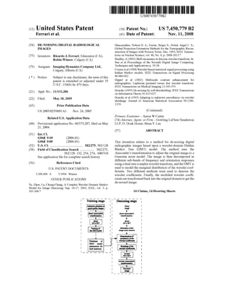
FIG. 5 is a graph of PSNR values resulting from the pro
cessing of four phantom images acquired using different
exposure levels. Each image Was used in turn to train a HMT
model. Following, the estimated HMT models Were used in
the de-noising algorithm. The PSNR average values from
column 1 to 4 in the attached table are 25.59, 22.64, 22.59,
and 22.65, respectively.
FIGS. 6(a) to (d) are examples oftWo-state Rayleigh mix
ture marginal distributions used to model the Wavelet coef?
cients. The densities summation and the histograms of the
Wavelet coef?cients are also shoWn. Plots Were obtained for
the ?rst four levels (a-d); subbands With orientation 0°.
FIGS. 7(a) to (0) are radiographic hand images of the
image shoWn in FIG. 2(0) denoisedby the de-noising method
With different levels: (a) 2 levels, (b) 3 levels, and (c) 4 levels.
FIGS. 8(a) to (0) are radiographic hand images of the
image shoWn in FIG. 2(0) denoised by a Gaussian ?lter With
different kernel sizes: FIGS. 8(a)-(0) kernel sizes equal to 2,
3, and 4 pixels, respectively.
FIGS. 9(a) to (d) are graphs of average results of a quali
tative assessment of the de-noising method. The graphs also
provide a comparison With de-noising using the Gaussian
?lter. The assessment included: analysis of noise reduction
(FIG. 9(a)), analysis ofartifacts (FIG. 9(b)), quality ofdetails
(FIG. 9(0)), and analysis ofbone sharpness (FIG. 9(d)).
FIG. 10 is a schematic illustration ofa digital radiographic
system used With a preferred embodiment of the present
invention.
DETAILED DESCRIPTION OF EMBODIMENTS
OF THE INVENTION
According to one embodiment of the invention and refer
ring to FIG. 1, a method for de-noising radiographic images
starts by pre-processing an original image usingAnscombe’s
variance stabilizing transformation, Which acts as if the data
arose from a Gaussian White noise model. The image is then
decomposed in different sub-bands of frequency and orien
tation responses using an overcomplete dual-tree complex
Wavelet transform (DT-CWT). By using the DT-CWT, visual
artifacts usually present in an image transformed by the tra
ditional DWT are signi?cantly minimized, With the advan
tage ofhaving a task that is still tractable in terms of compu
tation time. A Hidden Markov Tree (HMT) model is used to
describe the correlation among the Wavelet coef?cients by
modeling their marginal distribution and thus improving the
discrimination betWeen noisy and singularity pixels in an
image. Finally, the modi?ed Wavelet coef?cients are trans
formed back into the original domain in order to get the
de-noised image. The e?icacy of our method Was demon
strated on both phantom and real digital radiographic images
using quantitative and qualitative evaluation.
Digital Radiographic (DR) System: The DR systemusedin
our tests (referred as XplorerTM system) is an optically
coupled CCD based digital radiography unit. A schematic
illustration of such a unit is shoWn in FIG. 10. Referring to
FIG. 10, the digital radiography unit 12 comprises a CsI
scintillator 14 as the primary x-ray conversion layer and
couples the resulting light output to a CCD 20 by a mirror 16
and a lens 18 system. The 4K><4K CCD 20 is cooled to 2630
K resulting in a dark current rate ofless than one electron per
pixel per second. Images are digitized at 14 bits and subse
quently reduced for display to 12 bits. The Nyquist resolution
is 4.61 p/mm. The CCD 20 is coupledto a computer 22, Which
receives and processes the images detected by the CCD 20.
The Wavelet-based de-noising method is encoded in a pro
gram stored on computer readable medium in the computer
22.
System DQE is very high at loW frequencies but falls offat
higher frequencies, requiring the use of sharpening algo
rithms. This inevitably boosts noise Which can mask some
20
25
30
35
40
45
50
55
60
65
4
features. The Wavelet-based de-noising method is effective to
reduce the noise in the images, as discussed in detail beloW.
Hand Phantom and Image Dataset: A hand phantom from
NuclearAssociates as illustrated in FIG. 2(a) is comprised of
human skeletal parts embedded in anatomically accurate,
tissue-equivalent material. The materials have the same
absorption and secondary radiation-emitting characteristics
as living tissue. According to Nuclear Associates, all bone
marroW has been simulated With tissue-equivalent material,
Which permits critical detail study of bone structure and
sharpness comparisons using x-rays. In this Work, the phan
tom Was used to determine the characteristics of the image
noise variance and the appropriate image set to be used in the
training stage ofthe HMT model.
A total of ?fteen radiographic images of loWer and upper
extremities (hands, feet, Wrists and heels) Were analyzed. All
images Were acquired using the same type of digital radio
graphic system, described above in the section “Digital
Radiographic System”, With 108 un sampling interval and
l2-bits gray-level quantization. The images used in this Work
Were selected to characterize the best and Worse quality
images in terms of noise level.
Protocol for the Evaluation ofResults: The proposed algo
rithm Was evaluated quantitatively measuring the PSNR
using digital radiographic images from the phantom illus
trated in FIG. 2(a) and qualitatively using a set of ?fteen
clinical images.
The PSNR measure is de?ned as
Where Ii’,- and Ii’,- are the original and denoised images, respec
tively. xiJ is the pixel value in the spatial location (i,j) ofthe
original image, and N is the total number of pixels in the
image.
The qualitative analysis Was assessed according to the
opinion of tWo expert imaging specialists using a ranking
table. The images Were visually inspected on a computer 21"
monitor. Image intensity histogram-equalization and image
enhancement, using a standard unsharp-mask technique,
Were used for the sake ofbettervisualization ofthe de-noising
results. In addition, each processed image Was visually com
pared to the same original image ?ltered using the Gaussian
?lter. The kernel size ofthe Gaussian Was changed during the
analysis to provide the best tradeoffbetWeen sharpness ofthe
bone details and noise reduction. Table 1 Was ?lled out for all
?fteen images during the assessment ofthe algorithm.
TABLE 1
Example ofthe rank options and image characteristics analyzed
Which Were used by the tWo imaging specialists to assess
the results ofthe proposed de-noising algorithm
Image characteristics being assessed
Image # Noise Lack of Quality of
Anatomy reduction artifacts details Sharpness
Soft tissue i
Bone details
The images should be rated according to the following scores
1: excellent
2: good
3: average
4: poor
5: not acceptable
Noise Modeling andAnscombe’ s Transformation: In digi
tal radiographic systems there is a variety of imaging noise
19. US 7,450,779 B2
5
sources, Which originate from the different stages and ele
ments ofthe system, such as x-ray source, scattered radiation,
imaging screen, CCD camera, and electronic circuits among
others. The dominant cause of noise, hoWever, is due to the
quantum ?uctuations in the x-ray beam. In the present
method, a preprocessing stage is applied to the acquired
images to correct for the impulse noise, CCD dark current
noise and pixel nonuniforrnity.
It is Well knoWn that the Poisson distribution can be used to
model the arrival ofphotons and their expression by electron
counts on CCD detectors. Unlike Gaussian noise, Poisson
noise is proportional to the underlying signal intensity, Which
makes separating signal from noise a very dif?cult task.
Besides, Well established methods for image de-noising,
including the HMT model[2], are based upon the additive
White Gaussian noise model. Therefore, in order to overcome
this limitation, a variance stabilization (Anscombe’s) trans
formation[l3], described by
is applied to the original noise image. I(x,y) and IA(x,y) indi
cate the original and transformed images, respectively. The
Anscombe’s transformation acts as if the image data arose
from a Gaussian White noise model. More precisely, as the
number of photon counts increases, the noise variance in a
square-root image tends to a constant, independent of the
signal intensity. The inverse Anscombe’s transformation is
easily obtained by manipulating the above equation. In order
to have a more tractable problem, in this method We are
considering that the images are corrupted only by additive
Poisson noise. Other sources of noise, including electronic
noise normally present in digital radiographic systems, Were
not taken into account.
Dual Tree Complex Wavelet: Differently to the DWT, the
dual-tree complex Wavelet transform is a very attractive tech
nique for medical image de-noising since it performs as Well
as the undecimated DWT, in the context of shift invariance,
and With signi?cantly loWer computational cost.
The nearly shift invariant property is obtained With a real
biorthogonal transform having double the sampling rate at
each scale and by computing parallel Wavelet trees as illus
trated in FIG. 3, Which are differently subsampled. The DT
CWT presents perfect shift invariance at level 1, and approxi
mate shift invariance, beyond this level. The DT-CWT also
presents limitcd redundancy in the representation (4:1 for the
2D caseiindependent ofthe number of scales), good direc
tional selectivity (six oriented subbands: 115°, 145°, 175°),
and it permits perfect image reconstruction.
Hidden Markov Tree Model in the Wavelet Domain: The
HMT model, applied in the Wavelet context, is a statistical
model that can be used to capture statistical correlations
betWeen the magnitudes of Wavelet coel?cients across con
secutive scales of resolution. The HMT Works by modeling
the folloWing three important properties ofthe Wavelet coef
?cients:
Non-Gaussian distribution: The marginal distribution of
the magnitude of the complex Wavelet coef?cients can
be Well modeled by using a mixture of tWo-state Ray
leigh distributions. The choice for using the Rayleigh
mixture model instead of the Gaussian mixture model
Was based upon the fact that the real and imaginary parts
ofthe complex Wavelet coef?cients may be slightly cor
20
25
30
35
40
45
50
55
60
65
6
related, and therefore only the magnitudes of the com
plex Wavelet coef?cients Will present a nearly shift-in
variant property, but not the phase.
Persistency: Large/small Wavelet coef?cients related to
pixels in the image tend to propagate through scales of
the quad-trees. Therefore, a state variable is de?ned for
each Wavelet coef?cient Which associates the coef?cient
With one of the tWo-state Rayleigh marginal distribu
tions (one With small(S) and the other With large(L)
variance). The HMT model is then constructed by con
necting the state variables (L and S) across scales using
the Expectation-MaximiZation (EM) algorithm. FIG. 4
shoWs the ID-structure of the Hidden Markov tree
model.
Clustering: Adjacent Wavelet coel?cients of a particular
large/small coel?cient are very likely to share the same
state (large/small).
The HMT model is parameteriZedby the conditional prob
ability stating that the variable S]. is in state m given SP0.) is in
state n, or, ej,P(]-)’"’”:p(S]-:m|SP0-):n) m, n:l, . . . , 2. The state
probability of the root J is indicated by pSJ(m):p(S]-:m) and
the Rayleigh mixture parameters as p17,," and ojamz. The value
ofp17,," is set to Zero because the real and imaginary parts ofthe
complex Wavelet coef?cients must have Zero means (Wavelets
have Zero gain at dc). 0L,"2 is the variance. The parameters,
grouped into a vector 6:{pS](m),ej,PU)’"’”, OLMZ}, are deter
mined by the EM algorithm proposed in [2]. Herein, We
assume that the complex Wavelet coef?cients Wj folloW one of
the tWo-state Rayleigh distributions as
(3)
In order to have a more reliable and robust (not biased)
parameter estimation, the HMT model Was simpli?ed by
assuming that all the Wavelet coef?cients and state variables
Within a particular level of a subband have identical parent
childrelationships. Therefore, eachofthe six image subbands
obtained by using the DT-CWT Was trained independently
and hence presents its oWn set ofparameters. The magnitude
of the complex Wavelet coef?cients for each subband Were
modeled by the resulting mixture model
To take into account the dependencies among the Wavelets
coef?cients of different scales, a tree-graph representing a
parent-child relationship is used (see FIG. 4). The transition
of a speci?c Wavelet coel?cient j betWeen tWo consecutives
levels in the tree is speci?ed by the conditional probability
ejapwm’”. The algorithm fortraining the HMT model is knoWn
in the art, and for example, described in Crouse et. al. (1998)
“Wavelet-based statistical signal processing using hidden
Markov models”, IEEE Transactions on Signal Processing
46:886-902.
Training the HMT Model: The main goal of the training
stage is to ?nd the correlation among the Wavelet coef?cients
through the scales. Based upon experimental analysis and
also in a practical laboratory experimentusing the handphan
tom object, We have veri?ed that the best set of images to be
used in the training stage ofthe HMT model should have the
loWest level of noise and present enough image structure.
20. US 7,450,779 B2
7
To validate the above statement, the hand phantom Was
imaged With different radiation levels, according to the
parameters kVp, mAs as indicated in Table 2, given a set of
?ve images With different SNR values. The images Were used
in turn to train the ?ve models. The images Were then pro
cessed andthe PSNRWas recorded for further evaluation. The
results of the experiment are described beloW in a section
titled “Results”.
Selection of the clinical radiographic images used in the
training of the HMT model Was conducted by using a set of
representative images (outside of the testing image-set) of
each anatomy being studied (hand, foot, Wrist and heel). A
HMT model Was estimated for each speci?c anatomy. The
images Were visually chosen based on the level of noise and
amount ofbone details. Images With loWer level ofnoise and
richer in bone details Were given preference.
Noise Variance Estimation: Estimation of the noise vari
ance is an important step in our image de-noising algorithm
since it is used directly, along With the HMT parameters, in
our Wavelet-based ?ltering procedure. In the present Work,
the noise variance Was estimated as
2, 2 2
on ’ Oreal Xoimagz'nary (5)
Where Urea]2 and ol-magmary2 are, respectively, the noise vari
ance of the real and imaginary parts of the Wavelet coef?
cients computed by using the median absolute deviation
(MAD, [5]) algorithm.
TABLE 2
Parameters ofthe x-ray tube used in the experiment With the hand
phantom shown in FIG. 2. In this experiment, the SID Was set to
100 cm and the small focal spot Was used. Except for the ?rst set of
parameters the others are default values used in clinical application.
Image kVp mAs Type ofpatient usually applicable
1 60 2.5 pediatric
2 60 3.2 normal/medium
3 60 4.0 large
4 60 20 very high dose — NOT applicable
De-noising Usingthe HMT. The de-noisingprocedure pro
posed in this Work is composed oftWo shrinkage procedures:
one is used for the levels 1 and 2, and the other for the
subsequent levels. The rationality ofthis strategy is related to
the fact that the DT-CWT provides perfect shift-invariance
only at level 1, and approximate shift-invariance for the other
levels. Because of that, the capture of the inter-scale depen
dencies among the Wavelet coef?cients using the HMT model
starts to become unreliable beyond level 2 or 3, due to the
considerable image energy variation.
For the ?rst tWo levels of decomposition, the conditional
mean estimation of the noise-free Wavelet coe?icient Was
obtained using
2 (6)
Where p(S]-:m|W]-,0) is the probability of state m given the
noise Wavelet coe?icient Wj and the model parameters 0 com
putedby the EM algorithm. 0,42 is the variance ofthe additive
White Gaussian noise and E[ ] is the expectation operator.
Since the estimation of the subband variances 0L,"2 in the
HMT model is performed using noise Wavelet coe?icients,
20
25
30
35
40
45
50
55
60
65
8
their values are biased and should be corrected. The corrected
estimation is then obtained by
0, otherwise
After level 2, a modi?ed version ofthe soft-threshold proce
dure proposed in [10] Was used to ?nd the shrinkage factor
Which is applied to the real and imaginary parts of the com
plex Wavelet coe?icient Wj. In the above equation,
is the sigma function, S is an enhancement factor, andT:on/[3
is a thresholdvalue. [3 is considered as a smoothing parameter.
In the present Work the default values of S and [3 Were set to
1.3 and 0.9, respectively.
Results and Discussions: All the images illustrated in this
section Were post-processed using an image histogram equal
iZation, and unsharp mask technique, for the sake of better
visualiZation of the details. FIG. 5 shoWs the results of the
experiment carried out to determine the relation betWeen the
radiation dose and the algorithm performance, in terms of
PSNR The results Were used to con?rm that a high quality
image (the one obtained With a high x-ray dose, 60 kVp and
20 mAs) is in fact the best option to be used in the training of
the HMT model. By analyZing the average PSNR values We
noticed that Image 3 (obtained With 60 kVp and 4.0 mAs)
provides the second best average result. The Worse choice
Would be Image 1, acquired With 60 kVp and 2.5 mAs.
Despite the difference in the average values shoWn in FIG. 5,
and except for Image 4, the PSNR values obtained by using
different training images Were very similar. The x-ray tube
parameters used in the experiment are shoWn in Table 2.
FIG. 6 shoWs the results ofthe tWo-state Rayleigh mixture
model ?tting the marginal distribution of the Wavelet coef?
cients for the ?rst four consecutive levels (1 to 4) ofthe image
in FIG. 2(c). Visual inspection indicates the good curve
?tting provided by the Rayleigh function. Due to the high
image energy concentration around magnitude 0.25 in FIG.
6(a)-(b), application of a threshold technique to differentiate
large/small values Wavelet coe?icients Will not produce good
results. Indeed, HMT-based de-noising algorithms usually
outperform standard thresholding techniques because the
degree ofcoef?cient shrinkage is determined based not only
upon the value ofthe coef?cient but also upon its relationship
With its neighbors across scales (quad-tree relationship).
For the sake ofcomparison, FIGS. 7 and 8 shoW examples
of the radiographic hand image in FIG. 2(c) de-noised by
using the proposed technique With different levels of de
noising and the Gaussian ?lter With different kernel siZes. In
FIGS. 7(a) and 8(a), the granular appearance ofthe images is
typical ofimages corruptedby quantum noise. In these cases,
the Gaussian ?lter and the proposed algorithm using 2 levels
ofde-noising Were not e?icient in removing the noise. A huge