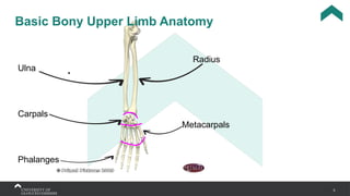
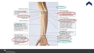
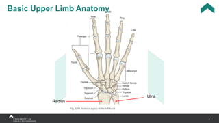
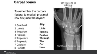
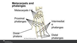
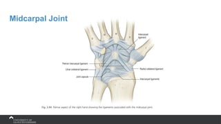
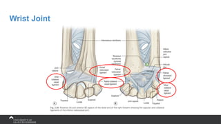
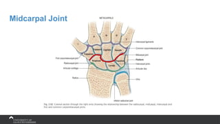
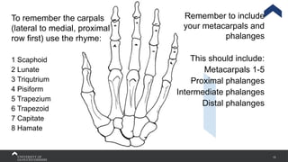
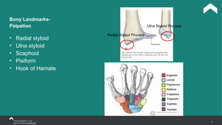
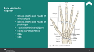
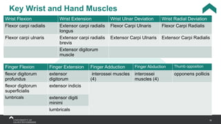
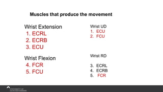
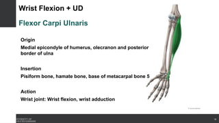
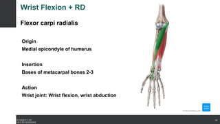
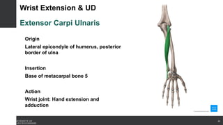
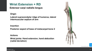
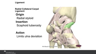
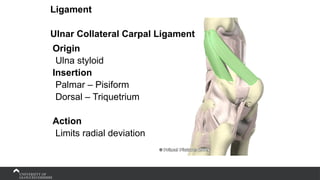
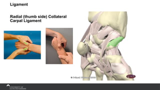
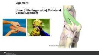
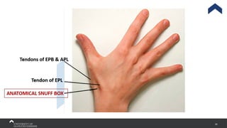
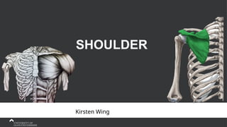
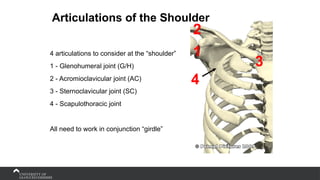
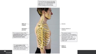
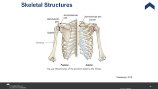
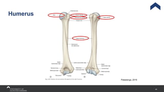
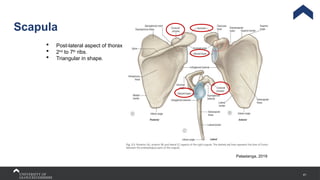
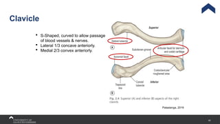
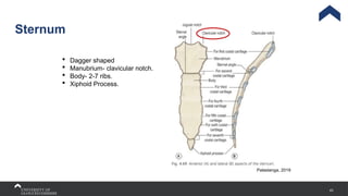
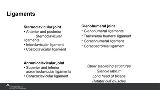
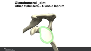
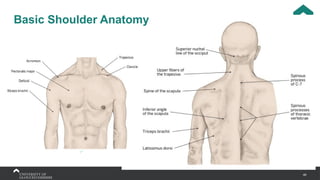
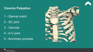
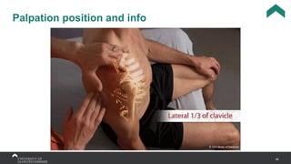
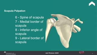
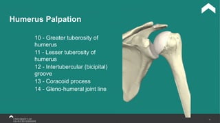
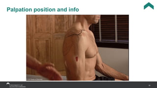
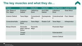
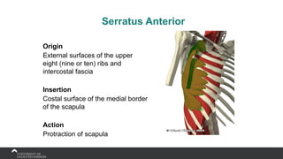
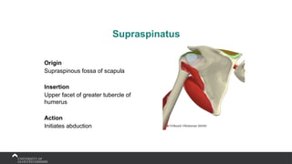
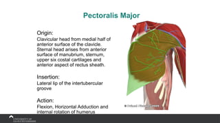
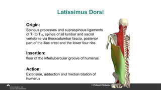
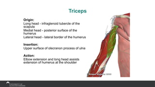
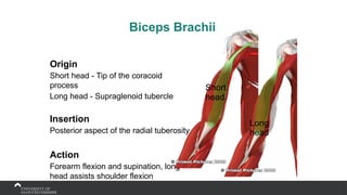
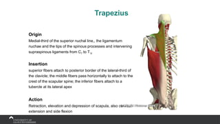
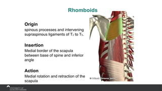
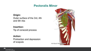
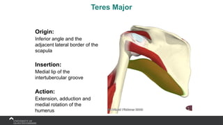
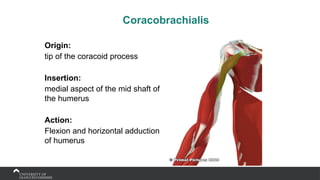
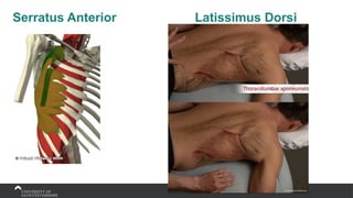
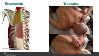
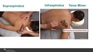
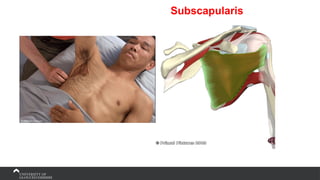
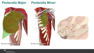
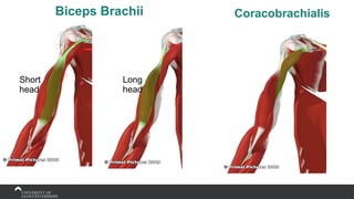
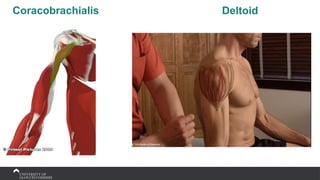
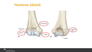
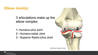
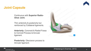
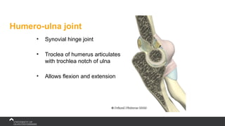
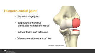
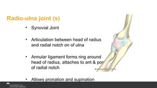
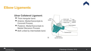
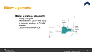
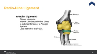
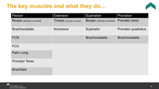
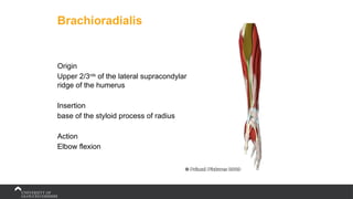
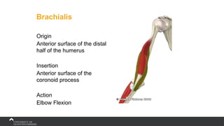
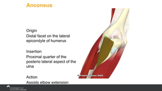
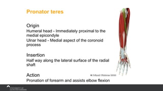
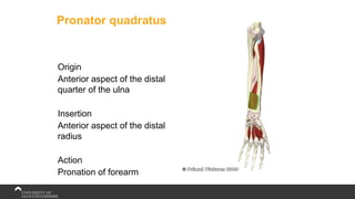
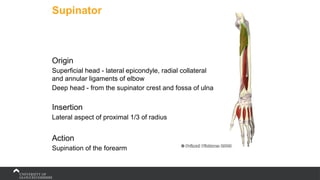
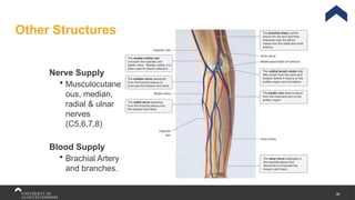
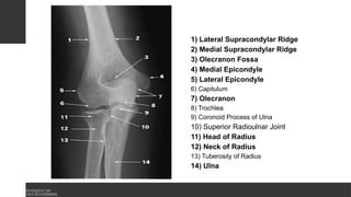
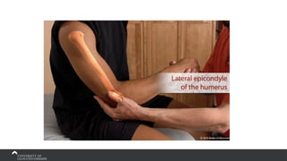
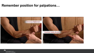
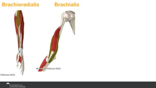
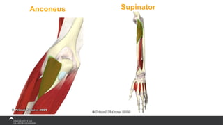
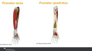
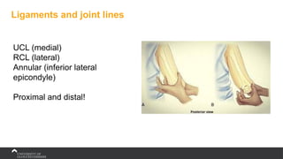
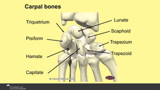
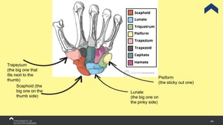
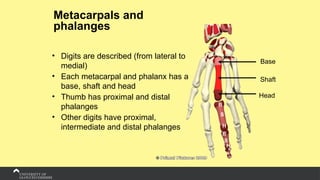
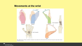
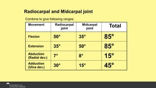
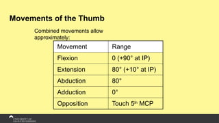
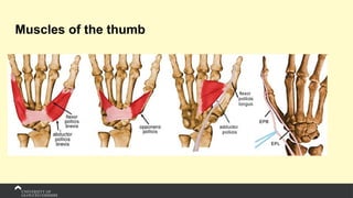
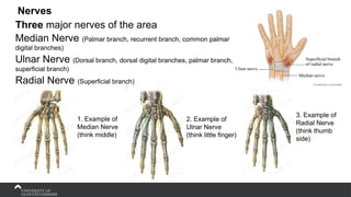
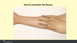
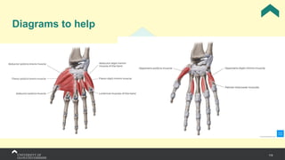
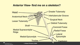
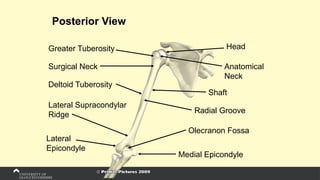
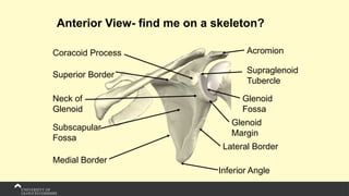
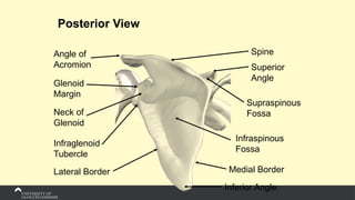
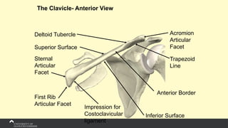
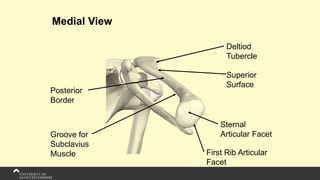
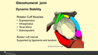
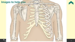
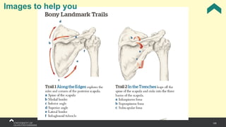
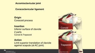
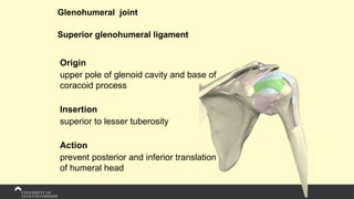
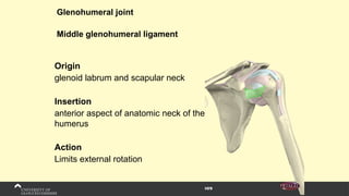
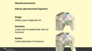
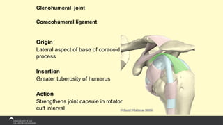
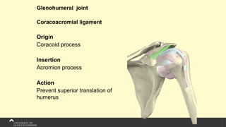
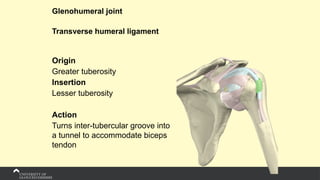
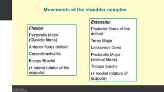
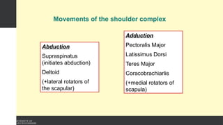
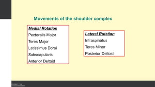
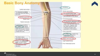
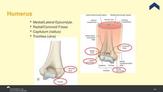
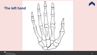
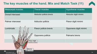
Anatomy Block
The Upper Limb
1. Wrist & Hand
2. Elbow
3. Shoulder Complex
SP7057
Kirsten Wing Sem 1, Week 2
2 Housekeeping reminder
In Person
Breaks
*Scheduled brain break after each joint
Kit (activity)
Please wear the appropriate clothing. You will be asked to leave/change if you do
not. So have a look around the room….
Questions
Yes great! Either raise your hand, ask me in a break or e-mail me after a session. Do not
disrupt others by interrupting. General, non personal questions must go into the discussion
forum.
Protocols
Please be ontime. You must do the attendance register.
3
Schedule
Elbow
1. Bone
2. Soft tissue
Shoulder
1. Bone
2. Soft
Tissue
Wrist &
Hand
1. Bone
2. Soft
Tissue
1st
9.15am
2nd
10.30
3rd
13.15
break lunch
Bring all
together
Wrist and Hand
Kirsten Wing
5
Basic Bony Upper Limb Anatomy
Ulna
Radius
Carpals
Phalanges
Metacarpals
6
7
Basic Upper Limb Anatomy
Radius
Ulna
Carpal bones
To remember the carpals
(lateral to medial, proximal
row first) use the rhyme:
1 Scaphoid
2 Lunate
3 Triqutrium
4 Pisiform
5 Trapezium
6 Trapezoid
7 Capitate
8 Hamate
1
2
3
4
5
6
7
8
Right hand: Posterior view
Silly
Little
Tommy
Pushes
Through
The
Car
Hole
Can you come up
with your
own???
Metacarpals and
phalanges
Metacarpals I-V
Proximal
phalanges Intermediat
e
phalanges
Distal
phalanges
Distal
phalanx
Midcarpal Joint
Wrist Joint
Midcarpal Joint
13
To remember the carpals
(lateral to medial, proximal
row first) use the rhyme:
1 Scaphoid
2 Lunate
3 Triqutrium
4 Pisiform
5 Trapezium
6 Trapezoid
7 Capitate
8 Hamate
Remember to include
your metacarpals and
phalanges
This should include:
Metacarpals 1-5
Proximal phalanges
Intermediate phalanges
Distal phalanges
14
Bony Landmarks-
Palpation
• Radial styloid
• Ulna styloid
• Scaphoid
• Pisiform
• Hook of Hamate
Radial Styloid Process
Ulna Styloid Process
15
Bony Landmarks-
Palpation
• Bases, shafts and heads of
metacarpals
• Bases, shafts and heads of
phalanges
• 1st carpometacarpal joint
• Radio-carpal joint line
• RCL
• UCL
16
Key Wrist and Hand Muscles
Wrist Flexion Wrist Extension Wrist Ulnar Deviation Wrist Radial Deviation
Flexor carpi radialis Extensor carpi radialis
longus
Flexor Carpi Ulnaris Flexor Carpi Radialis
Flexor carpi ulnaris Extensor carpi radialis
brevis
Extensor Carpi Ulnaris Extensor Carpi Radialis
Extensor digitorum
muscle
Finger Flexion Finger Extension Finger Adduction Finger Abduction Thumb opposition
flexor digitorum
profundus
extensor
digitorum
interossei muscles
(4)
interossei
muscles (4)
opponens pollicis
flexor digitorum
superficialis
extensor indicis
lumbricals extensor digiti
minimi
lumbricals
Wrist Extension
1. ECRL
2. ECRB
3. ECU
Wrist Flexion
4. FCR
5. FCU
Wrist UD
1. ECU
2. FCU
Wrist RD
3. ECRL
4. ECRB
5. FCR
Muscles that produce the movement
Wrist Flexion + UD
Flexor Carpi Ulnaris
Origin
Medial epicondyle of humerus, olecranon and posterior
border of ulna
Insertion
Pisiform bone, hamate bone, base of metacarpal bone 5
Action
Wrist joint: Wrist flexion, wrist adduction
18
Wrist Flexion + RD
Flexor carpi radialis
Origin
Medial epicondyle of humerus
Insertion
Bases of metacarpal bones 2-3
Action
Wrist joint: Wrist flexion, wrist abduction
19
Wrist Extension & UD
Extensor Carpi Ulnaris
Origin
Lateral epicondyle of humerus, posterior
border of ulna
Insertion
Base of metacarpal bone 5
Action
Wrist joint: Hand extension and
adduction
20
Wrist Extension + RD
Extensor carpi radialis longus
Origin
Lateral supracondylar ridge of humerus, lateral
intermuscular septum of arm
Insertion
Posterior aspect of base of metacarpal bone 2
Actions
Wrist joints: Hand extension, hand abduction
(radial deviation)
21
Wrist Extension + RD
Extensor carpi radialis brevis
Origin
Lateral epicondyle of humerus (common extensor tendon)
Insertion
Posterior aspect of base of metacarpal bone 3
Action
Wrist joints: Hand extension, hand abduction (radial
deviation)
22
Wrist Extension
Extensor digitorum muscle
Origin
Lateral epicondyle of humerus (common extensor
tendon)
Insertion
Extensor expansions of digits 2-5
Action
Metacarpophalangeal / Interphalangeal joints 2-5:
Finger extension
23
24
Remember… in order to palpate you
need to…
1. Effectively observe
2. Move the muscle if needed
3. Know where the muscle starts and ends
(theory bit)
4. Only then can you be hands on!
Wrist Flexion + UD
Flexor Carpi Ulnaris
25
Flex the
wrist to
expose the
tendons
Wrist Flexion + RD
Flexor carpi radialis
26
Flex the
wrist to
expose the
tendons
Wrist Extension & UD
Extensor Carpi Ulnaris
27
Wrist Extension + RD
Extensor carpi radialis longus and brevis
28
Wrist Extension
Extensor digitorum muscle
29
Ligament
Radial Collateral Carpal
Ligament
Origin
Radial styloid
Insertion
Scaphoid tuberosity
Action
Limits ulna deviation
Origin
Ulna styloid
Insertion
Palmar – Pisiform
Dorsal – Triquetrium
Action
Limits radial deviation
Ligament
Ulnar Collateral Carpal Ligament
Ligament
Radial (thumb side) Collateral
Carpal Ligament
Ligament
Ulnar (little finger side) Collateral
Carpal Ligament
Nerves
Three major nerves of the area
Median Nerve (Palmar branch, recurrent branch, common palmar
digital branches)
Ulnar Nerve (Dorsal branch, dorsal digital branches, palmar branch,
superficial branch)
Radial Nerve (Superficial branch)
1. Example of
Median Nerve
(think middle)
2. Example of
Ulnar Nerve
(think little finger)
3. Example of
Radial Nerve
(think thumb
side)
35
SHOULDER
Kirsten Wing
Articulations of the Shoulder
4 articulations to consider at the “shoulder”
1 - Glenohumeral joint (G/H)
2 - Acromioclavicular joint (AC)
3 - Sternoclavicular joint (SC)
4 - Scapulothoracic joint
All need to work in conjunction “girdle”
1
2
3
4
Bones
39
Skeletal Structures
Palastanga, 2019
Humerus
40
Humerus
Palastanga, 2019
41
Scapula
Palastanga, 2019
 Post-lateral aspect of thorax
 2nd
to 7th
ribs.
 Triangular in shape.
42
Clavicle
Palastanga, 2019
 S-Shaped, curved to allow passage
of blood vessels & nerves.
 Lateral 1/3 concave anteriorly.
 Medial 2/3 convex anteriorly.
43
Sternum
Palastanga, 2019
 Dagger shaped
 Manubrium- clavicular notch.
 Body- 2-7 ribs.
 Xiphoid Process.
Ligaments
Sternoclavicular joint
• Anterior and posterior
Sternoclavicular
ligaments
• Interclavicular ligament
• Costoclavicular ligament
Acromioclavicular joint
• Superior and inferior
acromioclavicular ligaments
• Coracoclavicular ligament
Glenohumeral joint
• Glenohumeral ligaments
• Transverse humeral ligament
• Coracohumeral ligament
• Coracoacromial ligament
Other stabilising structures
Glenoid labrum
Long head of biceps
Rotator cuff muscles
Glenohumeral joint
Other stabilisers – Glenoid labrum
46
Basic Shoulder Anatomy
47
Clavicle Palpation
1 - Sternal notch
2 - SC joint
3 - Clavicle
4 - A-C joint
5 - Acromian process
48
Palpation position and info
49
Scapula Palpation
6 - Spine of scapula
7 - Medial border of
scapula
8 - Inferior angle of
scapula
9 - Lateral border of
scapula
50
Palpation position and info
51
Humerus Palpation
10 - Greater tuberosity of
humerus
11 - Lesser tuberosity of
humerus
12 - Intertubercular (bicipital)
groove
13 - Coracoid process
14 - Gleno-humeral joint line
52
Palpation position and info
53
The key muscles and what they do…
Flexion Extension Adduction Abduction Medial Rotation Lateral Rotation
Clav. fibres of
Pec. Major.
Posterior
Deltoid
Latissimus
Dorsi
Ant & Mid
Deltoid
Latissimus
Dorsi
Teres Minor
Anterior Deltoid Teres Major Coracobrachi
alis
Supraspinatu
s
Coracobrachiali
s
Post. Deltoid
Coracobrachialis Latissimus
Dorsi
Teres Major Rotator Cuff Teres Major Infraspinatus
Biceps Brachii Pectoralis
Major (sternal
fibres)
Pectoralis
Major
Pectoralis Major
Triceps
brachii
Subscapularis
Anterior Deltoid
Serratus Anterior
Origin
External surfaces of the upper
eight (nine or ten) ribs and
intercostal fascia
Insertion
Costal surface of the medial border
of the scapula
Action
Protraction of scapula
Supraspinatus
Origin
Supraspinous fossa of scapula
Insertion
Upper facet of greater tubercle of
humerus
Action
Initiates abduction
Infraspinatus
Origin
Medial 2/3rds
of infraspinous
fossa of scapula
Insertion
Middle facet of greater tubercle
of humerus
Action
Lateral rotation of humerus
Teres Minor
Origin
Upper 2/3rds
of lateral border of the
scapula
Insertion
Lower facet of greater tubercle of
humerus
Action
Lateral rotation and adduction of humerus
Subscapularis
Origin
Medial 2/3rds
of subscapular fossa
Insertion
Lesser tubercle of humerus
Action
Medial rotation, and assists adduction, of
humerus
Deltoid
Origin:
Anterior border of lateral third of clavicle, the
lateral border of acromion and the lower lip of
scapular spine
Insertion:
Deltoid tuberosity, ½ way down the lateral
aspect of the shaft of humerus
Action:
Abduction of humerus, also anterior fibres aid
flexion and posterior fibres aid extension
Pectoralis Major
Origin:
Clavicular head from medial half of
anterior surface of the clavicle.
Sternal head arises from anterior
surface of manubrium, sternum,
upper six costal cartilages and
anterior aspect of rectus sheath.
Insertion:
Lateral lip of the intertubercular
groove
Action:
Flexion, Horizontal Adduction and
internal rotation of humerus
Latissimus Dorsi
Origin:
Spinous processes and supraspinous ligaments
of T7 to T12, spines of all lumbar and sacral
vertebrae via thoracolumbar fascia, posterior
part of the iliac crest and the lower four ribs
Insertion:
floor of the intertubercular groove of humerus
Action:
Extension, adduction and medial rotation of
humerus
Triceps
Origin:
Long head - infraglenoid tubercle of the
scapula
Medial head - posterior surface of the
humerus
Lateral head - lateral border of the humerus
Insertion:
Upper surface of olecranon process of ulna
Action:
Elbow extension and long head assists
extension of humerus at the shoulder
Biceps Brachii
Origin
Short head - Tip of the coracoid
process
Long head - Supraglenoid tubercle
Insertion
Posterior aspect of the radial tuberosity
Action
Forearm flexion and supination, long
head assists shoulder flexion
Short
head
Long
head
Trapezius
Origin
Medial-third of the superior nuchal line,, the ligamentum
nuchae and the tips of the spinous processes and intervening
supraspinous ligaments from C7 to T12
Insertion
superior fibers attach to posterior border of the lateral-third of
the clavicle; the middle fibers pass horizontally to attach to the
crest of the scapular spine; the inferior fibers attach to a
tubercle at its lateral apex
Action
Retraction, elevation and depression of scapula, also cervical
extension and side flexion
Rhomboids
Origin
spinous processes and intervening
supraspinous ligaments of T2 to T5.
Insertion
Medial border of the scapula
between base of spine and inferior
angle
Action
Medial rotation and retraction of the
scapula
Pectoralis Minor
Origin:
Outer surface of the 3rd, 4th
and 5th ribs
Insertion:
Tip of coracoid process
Action:
Protraction and depression
of scapula
Teres Major
Origin:
Inferior angle and the
adjacent lateral border of the
scapula
Insertion:
Medial lip of the
intertubercular groove
Action:
Extension, adduction and
medial rotation of the
humerus
Coracobrachialis
Origin:
tip of the coracoid process
Insertion:
medial aspect of the mid shaft of
the humerus
Action:
Flexion and horizontal adduction
of humerus
Serratus Anterior Latissimus Dorsi
Rhomboids Trapezius
Supraspinatus Infraspinatus Teres Minor
Subscapularis
Pectoralis Major Pectoralis Minor
Biceps Brachii
Short
head
Long
head
Coracobrachialis
Coracobrachialis Deltoid
Anatomy
of the Elbow
Kirsten Wing
Radius and Ulna (antero-
medial view)
Head
Radial
Tuberosity
RADIUS
Ulna Tuberosity
Coronoid Process
Olecranon Process
Anterior Border
Anterior Border
Trochlea Notch Neck
ULNA
Interosseous Border
Styloid process Styloid process
Head
Radius and Ulna (postero-
lateral view)
Supinator Crest
Posterior
Border
Ulna Notch
Posterior Border
Head
RADIUS
Olecranon Process
Neck
ULNA
Interosseous Borders
Styloid process Styloid process
79
Radius and Ulna
Head
Radial
Tuberosity
RADIUS
Ulna Tuberosity
Coronoid Process
Olecranon Process
Trochlea Notch
ULNA
Humerus (distal)
Elbow Joint(s)
3 articulations make up the
elbow complex
1- Humero-ulna Joint
2 - Humero-radial Joint
3 - Superior Radio-Ulna Joint
1 2
3
82
Joint Capsule
Continuous with Superior Radio-
Ulnar Joint.
Thin anteriorly & posteriorly but
reinforced by Collateral ligaments.
Anteriorly: Coronoid & Radial Fossa
to Cornoid Process & Annular
ligament.
Posteriorly: Olecranon process to
Annular ligament.
(Palastanga & Soames, 2012)
Humero-ulna joint
• Synovial hinge joint
• Troclea of humerus articulates
with trochlea notch of ulna
• Allows flexion and extension
Humero-radial joint
• Synovial hinge joint
• Capitulum of humerus
articulates with head of radius
• Allows flexion and extension
• Often not considered a “true” joint
Radio-ulna joint (s)
• Synovial Joint
• Articulation between head of radius
and radial notch on of ulna
• Annular ligament forms ring around
head of radius, attaches to ant & post
of radial notch
• Allows pronation and supination
86
Elbow Ligaments
Ulnar Collateral Ligament:
 Thick triangular band.
 Anterior, Medial Epicondyle to
Coronoid Process.
 Posterior, Medial Epicondyle to
Medial Olecranon Process.
 Both united by intermediate band.
(Palastanga & Soames, 2012)
87
Elbow Ligaments
(Palastanga & Soames, 2012)
Radial Collateral Ligament:
Strong, triangular.
Inferior Lateral Epicondyle (deep
to extensor tendons) to Annular
ligament.
Less distinctive than UCL.
88
Radio-Ulna Ligament
Annular Ligament:
Strong, triangular.
Inferior Lateral Epicondyle (deep
to extensor tendons) to Annular
ligament.
Less distinctive than UCL.
89
The key muscles and what they do…
Flexion Extension Supination Pronation
Biceps (already covered) Triceps (already covered) Biceps (already covered) Pronator teres
Brachioradialis Anconeus Supinator Pronator quadratus
FCR Brachioradialis Brachioradialis
FCU
Palm Long
Pronator Teres
Brachilais
Brachioradialis
Origin
Upper 2/3rds
of the lateral supracondylar
ridge of the humerus
Insertion
base of the styloid process of radius
Action
Elbow flexion
Brachialis
Origin
Anterior surface of the distal
half of the humerus
Insertion
Anterior surface of the
coronoid process
Action
Elbow Flexion
Anconeus
Origin
Distal facet on the lateral
epicondyle of humerus
Insertion
Proximal quarter of the
posterio lateral aspect of the
ulna
Action
Assists elbow extension
Pronator teres
Origin
Humeral head - Immediately proximal to the
medial epicondyle
Ulnar head - Medial aspect of the coronoid
process
Insertion
Half way along the lateral surface of the radial
shaft
Action
Pronation of forearm and assists elbow flexion
Pronator quadratus
Origin
Anterior aspect of the distal
quarter of the ulna
Insertion
Anterior aspect of the distal
radius
Action
Pronation of forearm
Supinator
Origin
Superficial head - lateral epicondyle, radial collateral
and annular ligaments of elbow
Deep head - from the supinator crest and fossa of ulna
Insertion
Lateral aspect of proximal 1/3 of radius
Action
Supination of the forearm
96
Other Structures
Nerve Supply
 Musculocutane
ous, median,
radial & ulnar
nerves
(C5,6,7,8)
Blood Supply
 Brachial Artery
and branches.
1) Lateral Supracondylar Ridge
2) Medial Supracondylar Ridge
3) Olecranon Fossa
4) Medial Epicondyle
5) Lateral Epicondyle
6) Capitulum
7) Olecranon
8) Trochlea
9) Coronoid Process of Ulna
10) Superior Radioulnar Joint
11) Head of Radius
12) Neck of Radius
13) Tuberosity of Radius
14) Ulna
Remember position for palpations…
Brachioradialis Brachialis
Anconeus Supinator
Pronator teres Pronator quadratus
Ligaments and joint lines
UCL (medial)
RCL (lateral)
Annular (inferior lateral
epicondyle)
Proximal and distal!
Extra
Resources
Carpal bones
Triquetrium
Pisiform
Hamate
Capitate
Lunate
Scaphoid
Trapezium
Trapezoid
106
Lunate
(the big one on
the pinky side)
Scaphoid (the
big one on the
thumb side)
Trapezium
(the big one that
i9s next to the
thumb)
Pisiform
(the sticky out one)
Metacarpals and
phalanges
• Digits are described (from lateral to
medial)
• Each metacarpal and phalanx has a
base, shaft and head
• Thumb has proximal and distal
phalanges
• Other digits have proximal,
intermediate and distal phalanges
Base
Shaft
Head
B
S
H
B
S
H
B
S
H
Movements at the wrist
Movements of the Midcarpal Joint
Radiocarpal and Midcarpal joint
Combine to give following ranges:
Movement Radiocarpal
joint
Midcarpal
joint
Total
Flexion 50° 35° 85°
Extension 35° 50° 85°
Abduction
(Radial dev.) 7° 8° 15°
Adduction
(Ulna dev.) 30° 15° 45°
Movements of the Thumb
Combined movements allow
approximately:
Movement Range
Flexion 0 (+90° at IP)
Extension 80° (+10° at IP)
Abduction 80°
Adduction 0°
Opposition Touch 5th
MCP
Muscles of the thumb
Nerves
Three major nerves of the area
Median Nerve (Palmar branch, recurrent branch, common palmar
digital branches)
Ulnar Nerve (Dorsal branch, dorsal digital branches, palmar branch,
superficial branch)
Radial Nerve (Superficial branch)
1. Example of
Median Nerve
(think middle)
2. Example of
Ulnar Nerve
(think little finger)
3. Example of
Radial Nerve
(think thumb
side)
How to remember the flexors
115
Diagrams to help
References
Cael, C. (2010)
Functional Anatomy
Philadelphia: Lippincott Williams & Wilkins
Drake, R.L, Vogl, W. and Mitchell, A.W.M. (2005)
Gray’s Anatomy for Students
Philadelphia; Elsevier
Primal Pictures CD-Rom (2003)
Palastanga, N. and Soames, R. (2012)
Anatomy and Human Movement (6th
Ed)
Edinburgh; Butterworth Heinemann.
Anterior View- find me on a skeleton?
Head
Anatomical Neck
Lesser Tuberosity
Shaft
Medial Supracondylar
Ridge
Medial Epicondyle
Greater Tuberosity
Intertubercular Groove
Surgical Neck
Deltoid Tuberosity
Radial Fossa
Lateral
Epicondyle
Capitulum
Coronoid Fossa
Troclea
Posterior View
Head
Anatomical
Neck
Shaft
Lateral Supracondylar
Ridge
Medial Epicondyle
Greater Tuberosity
Surgical Neck
Deltoid Tuberosity
Radial Groove
Lateral
Epicondyle
Olecranon Fossa
Anterior View- find me on a skeleton?
Acromion
Glenoid
Fossa
Glenoid
Margin
Subscapular
Fossa
Coracoid Process
Superior Border
Lateral Border
Medial Border
Inferior Angle
Neck of
Glenoid
Supraglenoid
Tubercle
Posterior View
Spine
Superior
Angle
Supraspinous
Fossa
Lateral Border Medial Border
Angle of
Acromion
Neck of
Glenoid
Infraglenoid
Tubercle
Inferior Angle
Infraspinous
Fossa
Glenoid
Margin
The Clavicle- Anterior View
Acromion
Articular
Facet
Trapezoid
Line
First Rib
Articular Facet
Deltoid Tubercle
Anterior Border
Sternal
Articular
Facet
Inferior Surface
Superior Surface
Impression for
Costoclavicular
ligament
Medial View
Deltiod
Tubercle
Superior
Surface
Posterior
Border
First Rib Articular
Facet
Sternal
Articular Facet
Groove for
Subclavius
Muscle
Glenohumeral joint
Dynamic Stability
Rotator Cuff Muscles
• Supraspinatus
• Infraspinatus
• Teres Minor
• Subscapularis
Rotator cuff interval
Supported by ligaments and tendons
124
Images to help you
125
Images to help you
126
Images to help you
Movement Pure ROM
(approx)
ROM
(approx)
Muscles
Flexion 90o-100o 180o Clav. fibres of Pec. Major.
Anterior Deltoid, Coracobrachialis
Extension 70o 90o Posterior Deltoid
Teres Major, Latissimus Dorsi
Abduction 120o 180o Ant & Mid Deltoid
Supraspinatus, Rotator Cuff
Adduction 50o-75o Latissimus Dorsi
Coracobrachialis, Teres Major
Medial Rot.
Lateral Rot.
60o-100o
80o-90o
Subscapularis
Teres Major, Pectoralis Major
Latissimus Dorsi
Infraspinatus
Teres Minor, Post. Deltoid
Muscles acting over the shoulder
Scapula Stabilisers
- Serratus Anterior
- Trapezius (upper, middle
and lower fibres)
- Rhomboids
Rotator cuff
- Supraspinatus
- Infraspinatus
- Teres minor
- Subscapularis
Other Key Movers
Deltoids (anterior, posterior and
middle)
Pectoralis major (sternal and
clavicular fibres)
Latissimus dorsi
Teres Major
Triceps (working over the
shoulder)
Biceps (Long head)
Coracobrachialis
Pec Minor
Sternoclavicular joint
Costoclavicular ligament
Origin
Inferior surface of proximal end of
clavicle
Insertion
Superior surface of 1st
rib and costal
cartilage
Action
Limits elevation of clavicle
Supports inferior joint capsule
Acromioclavicular joint
Superior & inferior acromioclavicular ligaments
Origin
Superior and inferior surfaces of
distal clavicle
Insertion
Superior and inferior surfaces of
medial acromion
Action
Strengthen joint capsule
Acromioclavicular joint
Coracoclavicular ligament
Origin
Coracoid process
Insertion
Inferior surface of clavicle
2 parts
Conoid & Trapezoid
Action
Limit superior translation of clavicle
against scapula (at AC joint)
Conoid
Trapezoid
Glenohumeral joint
Superior glenohumeral ligament
Origin
upper pole of glenoid cavity and base of
coracoid process
Insertion
superior to lesser tuberosity
Action
prevent posterior and inferior translation
of humeral head
Glenohumeral joint
Middle glenohumeral ligament
Origin
glenoid labrum and scapular neck
Insertion
anterior aspect of anatomic neck of the
humerus
Action
Limits external rotation
Glenohumeral joint
Inferior glenohumeral ligament
Origin
Inferior part of glenoid rim
Insertion
Lower part of anatomical neck of
humerus
Action
Limits abduction of humerus
Glenohumeral joint
Coracohumeral ligament
Origin
Lateral aspect of base of coracoid
process
Insertion
Greater tuberosity of humerus
Action
Strengthens joint capsule in rotator
cuff interval
Glenohumeral joint
Coracoacromial ligament
Origin
Coracoid process
Insertion
Acromion process
Action
Prevent superior translation of
humerus
Glenohumeral joint
Transverse humeral ligament
Origin
Greater tuberosity
Insertion
Lesser tuberosity
Action
Turns inter-tubercular groove into
a tunnel to accommodate biceps
tendon
Movements of the shoulder complex
Flexion
Pectoralis Major
(Clavicle fibres)
Anterior fibres deltoid
Coracobrachiarlis
Biceps Brachii
(+ lateral rotator of the
scapula)
Extension
Posterior fibres of the
deltoid
Teres Major
Latissimus Dorsi
Pectoralis Major
(sternal fibres)
Triceps brachii
(+ medial rotators of
scapula)
Movements of the shoulder complex
Abduction
Supraspinatus
(initiates abduction)
Deltoid
(+lateral rotators of
the scapular)
Adduction
Pectoralis Major
Latissimus Dorsi
Teres Major
Coracobrachiarlis
(+medial rotators of
scapula)
Movements of the shoulder complex
Medial Rotation
Pectoralis Major
Teres Major
Latissimus Dorsi
Subscapularis
Anterior Deltoid
Lateral Rotation
Infraspinatus
Teres Minor
Posterior Deltoid
141
Basic Bony Anatomy
142
Humerus
 Medial/Lateral Epicondyle.
 Radial/Coronoid Fossa
 Capitulum (radius)
 Trochlea (ulna)
143
Skeletal Structures: Posterior View
144
Cael, 2010
Ligaments of the Elbow
Region: Anterior View
145
Ligaments of the Elbow Region:
Posterior View
Cael, 2010
146
Bursae
(Cael, 2010)
1) Lateral Supracondylar Ridge
2) Medial Supracondylar Ridge
3) Olecranon Fossa
4) Medial Epicondyle
5) Lateral Epicondyle
6) Capitulum
7) Olecranon
8) Trochlea
9) Coronoid Process of Ulna
10) Superior Radioulnar Joint
11) Head of Radius
12) Neck of Radius
13) Tuberosity of Radius
14) Ulna
148
Extra Images
(Cael, 2010)
149
Carrying Angle
Line of Humerus & Forearm are not
the same.
Forearm abducted at angle, why?
 Medial edge of Trochlea
projects approx. 6mm
below the lateral edge.
 Typically Females have
larger angles.
Masked in full flexion & in pronation
of the extended forearm.
References
Cael, C. (2010)
Functional Anatomy
Philadelphia: Lippincott Williams & Wilkins
Drake, R.L, Vogl, W. and Mitchell, A.W.M. (2005)
Gray’s Anatomy for Students
Philadelphia; Elsevier
Primal Pictures CD-Rom (2003)
Palastanga, N. and Soames, R. (2012)
Anatomy and Human Movement (6th
Ed)
Edinburgh; Butterworth Heinemann.
151
Extra Images
152
Extra Images
Tasks
Please complete the following two diagrams
and bring a permanent marker (something that
will stick to a rubber glove)
All the bones
of the hand remember
your marker
154
Anatomy
155
The right hand
156
The left hand
157
The key muscles of the hand. Mix and Match Task (11)
Metacarpal muscles Thenar muscles Hypothenar muscles
158
The key muscles of the hand. Mix and Match Task (11)
Metacarpal muscles Thenar muscles Hypothenar muscles
Dorsal interossei Abductor pollicis brevis Abductor digiti minimi
Palmar interossei Adductor pollicis Flexor digiti minimi
Lumbricals Flexor pollicis brevis Opponens digiti minimi
Opponens pollicis Palmaris brevis
159
The ligaments…
Ligament Attachments Function Notes
Glenohumeral ligaments
Coracoclavicular ligament
Coracoacromial ligament
160
The key muscles and what they do…
Flexion Extension Adduction Abduction Medial Rotation Lateral Rotation
Clav. fibres of
Pec. Major.
Posterior
Deltoid
Latissimus
Dorsi
Ant & Mid
Deltoid
Latissimus
Dorsi
Teres Minor
Anterior Deltoid Teres Major Coracobrachi
alis
Supraspinatu
s
Coracobrachiali
s
Post. Deltoid
Coracobrachialis Latissimus
Dorsi
Teres Major Rotator Cuff Teres Major Infraspinatus
Biceps Brachii Pectoralis
Major (sternal
fibres)
Pectoralis
Major
Pectoralis Major
Triceps
brachii
Subscapularis
Anterior Deltoid

Upper Limb Anatomy Slides forearm , arm,

  • 1.
    Anatomy Block The UpperLimb 1. Wrist & Hand 2. Elbow 3. Shoulder Complex SP7057 Kirsten Wing Sem 1, Week 2
  • 2.
    2 Housekeeping reminder InPerson Breaks *Scheduled brain break after each joint Kit (activity) Please wear the appropriate clothing. You will be asked to leave/change if you do not. So have a look around the room…. Questions Yes great! Either raise your hand, ask me in a break or e-mail me after a session. Do not disrupt others by interrupting. General, non personal questions must go into the discussion forum. Protocols Please be ontime. You must do the attendance register.
  • 3.
    3 Schedule Elbow 1. Bone 2. Softtissue Shoulder 1. Bone 2. Soft Tissue Wrist & Hand 1. Bone 2. Soft Tissue 1st 9.15am 2nd 10.30 3rd 13.15 break lunch Bring all together
  • 4.
  • 5.
    5 Basic Bony UpperLimb Anatomy Ulna Radius Carpals Phalanges Metacarpals
  • 6.
  • 7.
    7 Basic Upper LimbAnatomy Radius Ulna
  • 8.
    Carpal bones To rememberthe carpals (lateral to medial, proximal row first) use the rhyme: 1 Scaphoid 2 Lunate 3 Triqutrium 4 Pisiform 5 Trapezium 6 Trapezoid 7 Capitate 8 Hamate 1 2 3 4 5 6 7 8 Right hand: Posterior view Silly Little Tommy Pushes Through The Car Hole Can you come up with your own???
  • 9.
    Metacarpals and phalanges Metacarpals I-V Proximal phalangesIntermediat e phalanges Distal phalanges Distal phalanx
  • 10.
  • 11.
  • 12.
  • 13.
    13 To remember thecarpals (lateral to medial, proximal row first) use the rhyme: 1 Scaphoid 2 Lunate 3 Triqutrium 4 Pisiform 5 Trapezium 6 Trapezoid 7 Capitate 8 Hamate Remember to include your metacarpals and phalanges This should include: Metacarpals 1-5 Proximal phalanges Intermediate phalanges Distal phalanges
  • 14.
    14 Bony Landmarks- Palpation • Radialstyloid • Ulna styloid • Scaphoid • Pisiform • Hook of Hamate Radial Styloid Process Ulna Styloid Process
  • 15.
    15 Bony Landmarks- Palpation • Bases,shafts and heads of metacarpals • Bases, shafts and heads of phalanges • 1st carpometacarpal joint • Radio-carpal joint line • RCL • UCL
  • 16.
    16 Key Wrist andHand Muscles Wrist Flexion Wrist Extension Wrist Ulnar Deviation Wrist Radial Deviation Flexor carpi radialis Extensor carpi radialis longus Flexor Carpi Ulnaris Flexor Carpi Radialis Flexor carpi ulnaris Extensor carpi radialis brevis Extensor Carpi Ulnaris Extensor Carpi Radialis Extensor digitorum muscle Finger Flexion Finger Extension Finger Adduction Finger Abduction Thumb opposition flexor digitorum profundus extensor digitorum interossei muscles (4) interossei muscles (4) opponens pollicis flexor digitorum superficialis extensor indicis lumbricals extensor digiti minimi lumbricals
  • 17.
    Wrist Extension 1. ECRL 2.ECRB 3. ECU Wrist Flexion 4. FCR 5. FCU Wrist UD 1. ECU 2. FCU Wrist RD 3. ECRL 4. ECRB 5. FCR Muscles that produce the movement
  • 18.
    Wrist Flexion +UD Flexor Carpi Ulnaris Origin Medial epicondyle of humerus, olecranon and posterior border of ulna Insertion Pisiform bone, hamate bone, base of metacarpal bone 5 Action Wrist joint: Wrist flexion, wrist adduction 18
  • 19.
    Wrist Flexion +RD Flexor carpi radialis Origin Medial epicondyle of humerus Insertion Bases of metacarpal bones 2-3 Action Wrist joint: Wrist flexion, wrist abduction 19
  • 20.
    Wrist Extension &UD Extensor Carpi Ulnaris Origin Lateral epicondyle of humerus, posterior border of ulna Insertion Base of metacarpal bone 5 Action Wrist joint: Hand extension and adduction 20
  • 21.
    Wrist Extension +RD Extensor carpi radialis longus Origin Lateral supracondylar ridge of humerus, lateral intermuscular septum of arm Insertion Posterior aspect of base of metacarpal bone 2 Actions Wrist joints: Hand extension, hand abduction (radial deviation) 21
  • 22.
    Wrist Extension +RD Extensor carpi radialis brevis Origin Lateral epicondyle of humerus (common extensor tendon) Insertion Posterior aspect of base of metacarpal bone 3 Action Wrist joints: Hand extension, hand abduction (radial deviation) 22
  • 23.
    Wrist Extension Extensor digitorummuscle Origin Lateral epicondyle of humerus (common extensor tendon) Insertion Extensor expansions of digits 2-5 Action Metacarpophalangeal / Interphalangeal joints 2-5: Finger extension 23
  • 24.
    24 Remember… in orderto palpate you need to… 1. Effectively observe 2. Move the muscle if needed 3. Know where the muscle starts and ends (theory bit) 4. Only then can you be hands on!
  • 25.
    Wrist Flexion +UD Flexor Carpi Ulnaris 25 Flex the wrist to expose the tendons
  • 26.
    Wrist Flexion +RD Flexor carpi radialis 26 Flex the wrist to expose the tendons
  • 27.
    Wrist Extension &UD Extensor Carpi Ulnaris 27
  • 28.
    Wrist Extension +RD Extensor carpi radialis longus and brevis 28
  • 29.
  • 30.
    Ligament Radial Collateral Carpal Ligament Origin Radialstyloid Insertion Scaphoid tuberosity Action Limits ulna deviation
  • 31.
    Origin Ulna styloid Insertion Palmar –Pisiform Dorsal – Triquetrium Action Limits radial deviation Ligament Ulnar Collateral Carpal Ligament
  • 32.
    Ligament Radial (thumb side)Collateral Carpal Ligament
  • 33.
    Ligament Ulnar (little fingerside) Collateral Carpal Ligament
  • 34.
    Nerves Three major nervesof the area Median Nerve (Palmar branch, recurrent branch, common palmar digital branches) Ulnar Nerve (Dorsal branch, dorsal digital branches, palmar branch, superficial branch) Radial Nerve (Superficial branch) 1. Example of Median Nerve (think middle) 2. Example of Ulnar Nerve (think little finger) 3. Example of Radial Nerve (think thumb side)
  • 35.
  • 36.
  • 37.
    Articulations of theShoulder 4 articulations to consider at the “shoulder” 1 - Glenohumeral joint (G/H) 2 - Acromioclavicular joint (AC) 3 - Sternoclavicular joint (SC) 4 - Scapulothoracic joint All need to work in conjunction “girdle” 1 2 3 4
  • 38.
  • 39.
  • 40.
  • 41.
    41 Scapula Palastanga, 2019  Post-lateralaspect of thorax  2nd to 7th ribs.  Triangular in shape.
  • 42.
    42 Clavicle Palastanga, 2019  S-Shaped,curved to allow passage of blood vessels & nerves.  Lateral 1/3 concave anteriorly.  Medial 2/3 convex anteriorly.
  • 43.
    43 Sternum Palastanga, 2019  Daggershaped  Manubrium- clavicular notch.  Body- 2-7 ribs.  Xiphoid Process.
  • 44.
    Ligaments Sternoclavicular joint • Anteriorand posterior Sternoclavicular ligaments • Interclavicular ligament • Costoclavicular ligament Acromioclavicular joint • Superior and inferior acromioclavicular ligaments • Coracoclavicular ligament Glenohumeral joint • Glenohumeral ligaments • Transverse humeral ligament • Coracohumeral ligament • Coracoacromial ligament Other stabilising structures Glenoid labrum Long head of biceps Rotator cuff muscles
  • 45.
  • 46.
  • 47.
    47 Clavicle Palpation 1 -Sternal notch 2 - SC joint 3 - Clavicle 4 - A-C joint 5 - Acromian process
  • 48.
  • 49.
    49 Scapula Palpation 6 -Spine of scapula 7 - Medial border of scapula 8 - Inferior angle of scapula 9 - Lateral border of scapula
  • 50.
  • 51.
    51 Humerus Palpation 10 -Greater tuberosity of humerus 11 - Lesser tuberosity of humerus 12 - Intertubercular (bicipital) groove 13 - Coracoid process 14 - Gleno-humeral joint line
  • 52.
  • 53.
    53 The key musclesand what they do… Flexion Extension Adduction Abduction Medial Rotation Lateral Rotation Clav. fibres of Pec. Major. Posterior Deltoid Latissimus Dorsi Ant & Mid Deltoid Latissimus Dorsi Teres Minor Anterior Deltoid Teres Major Coracobrachi alis Supraspinatu s Coracobrachiali s Post. Deltoid Coracobrachialis Latissimus Dorsi Teres Major Rotator Cuff Teres Major Infraspinatus Biceps Brachii Pectoralis Major (sternal fibres) Pectoralis Major Pectoralis Major Triceps brachii Subscapularis Anterior Deltoid
  • 54.
    Serratus Anterior Origin External surfacesof the upper eight (nine or ten) ribs and intercostal fascia Insertion Costal surface of the medial border of the scapula Action Protraction of scapula
  • 55.
    Supraspinatus Origin Supraspinous fossa ofscapula Insertion Upper facet of greater tubercle of humerus Action Initiates abduction
  • 56.
    Infraspinatus Origin Medial 2/3rds of infraspinous fossaof scapula Insertion Middle facet of greater tubercle of humerus Action Lateral rotation of humerus
  • 57.
    Teres Minor Origin Upper 2/3rds oflateral border of the scapula Insertion Lower facet of greater tubercle of humerus Action Lateral rotation and adduction of humerus
  • 58.
    Subscapularis Origin Medial 2/3rds of subscapularfossa Insertion Lesser tubercle of humerus Action Medial rotation, and assists adduction, of humerus
  • 59.
    Deltoid Origin: Anterior border oflateral third of clavicle, the lateral border of acromion and the lower lip of scapular spine Insertion: Deltoid tuberosity, ½ way down the lateral aspect of the shaft of humerus Action: Abduction of humerus, also anterior fibres aid flexion and posterior fibres aid extension
  • 60.
    Pectoralis Major Origin: Clavicular headfrom medial half of anterior surface of the clavicle. Sternal head arises from anterior surface of manubrium, sternum, upper six costal cartilages and anterior aspect of rectus sheath. Insertion: Lateral lip of the intertubercular groove Action: Flexion, Horizontal Adduction and internal rotation of humerus
  • 61.
    Latissimus Dorsi Origin: Spinous processesand supraspinous ligaments of T7 to T12, spines of all lumbar and sacral vertebrae via thoracolumbar fascia, posterior part of the iliac crest and the lower four ribs Insertion: floor of the intertubercular groove of humerus Action: Extension, adduction and medial rotation of humerus
  • 62.
    Triceps Origin: Long head -infraglenoid tubercle of the scapula Medial head - posterior surface of the humerus Lateral head - lateral border of the humerus Insertion: Upper surface of olecranon process of ulna Action: Elbow extension and long head assists extension of humerus at the shoulder
  • 63.
    Biceps Brachii Origin Short head- Tip of the coracoid process Long head - Supraglenoid tubercle Insertion Posterior aspect of the radial tuberosity Action Forearm flexion and supination, long head assists shoulder flexion Short head Long head
  • 64.
    Trapezius Origin Medial-third of thesuperior nuchal line,, the ligamentum nuchae and the tips of the spinous processes and intervening supraspinous ligaments from C7 to T12 Insertion superior fibers attach to posterior border of the lateral-third of the clavicle; the middle fibers pass horizontally to attach to the crest of the scapular spine; the inferior fibers attach to a tubercle at its lateral apex Action Retraction, elevation and depression of scapula, also cervical extension and side flexion
  • 65.
    Rhomboids Origin spinous processes andintervening supraspinous ligaments of T2 to T5. Insertion Medial border of the scapula between base of spine and inferior angle Action Medial rotation and retraction of the scapula
  • 66.
    Pectoralis Minor Origin: Outer surfaceof the 3rd, 4th and 5th ribs Insertion: Tip of coracoid process Action: Protraction and depression of scapula
  • 67.
    Teres Major Origin: Inferior angleand the adjacent lateral border of the scapula Insertion: Medial lip of the intertubercular groove Action: Extension, adduction and medial rotation of the humerus
  • 68.
    Coracobrachialis Origin: tip of thecoracoid process Insertion: medial aspect of the mid shaft of the humerus Action: Flexion and horizontal adduction of humerus
  • 69.
  • 70.
  • 71.
  • 72.
  • 73.
  • 74.
  • 75.
  • 76.
  • 77.
    Radius and Ulna(antero- medial view) Head Radial Tuberosity RADIUS Ulna Tuberosity Coronoid Process Olecranon Process Anterior Border Anterior Border Trochlea Notch Neck ULNA Interosseous Border Styloid process Styloid process Head
  • 78.
    Radius and Ulna(postero- lateral view) Supinator Crest Posterior Border Ulna Notch Posterior Border Head RADIUS Olecranon Process Neck ULNA Interosseous Borders Styloid process Styloid process
  • 79.
    79 Radius and Ulna Head Radial Tuberosity RADIUS UlnaTuberosity Coronoid Process Olecranon Process Trochlea Notch ULNA
  • 80.
  • 81.
    Elbow Joint(s) 3 articulationsmake up the elbow complex 1- Humero-ulna Joint 2 - Humero-radial Joint 3 - Superior Radio-Ulna Joint 1 2 3
  • 82.
    82 Joint Capsule Continuous withSuperior Radio- Ulnar Joint. Thin anteriorly & posteriorly but reinforced by Collateral ligaments. Anteriorly: Coronoid & Radial Fossa to Cornoid Process & Annular ligament. Posteriorly: Olecranon process to Annular ligament. (Palastanga & Soames, 2012)
  • 83.
    Humero-ulna joint • Synovialhinge joint • Troclea of humerus articulates with trochlea notch of ulna • Allows flexion and extension
  • 84.
    Humero-radial joint • Synovialhinge joint • Capitulum of humerus articulates with head of radius • Allows flexion and extension • Often not considered a “true” joint
  • 85.
    Radio-ulna joint (s) •Synovial Joint • Articulation between head of radius and radial notch on of ulna • Annular ligament forms ring around head of radius, attaches to ant & post of radial notch • Allows pronation and supination
  • 86.
    86 Elbow Ligaments Ulnar CollateralLigament:  Thick triangular band.  Anterior, Medial Epicondyle to Coronoid Process.  Posterior, Medial Epicondyle to Medial Olecranon Process.  Both united by intermediate band. (Palastanga & Soames, 2012)
  • 87.
    87 Elbow Ligaments (Palastanga &Soames, 2012) Radial Collateral Ligament: Strong, triangular. Inferior Lateral Epicondyle (deep to extensor tendons) to Annular ligament. Less distinctive than UCL.
  • 88.
    88 Radio-Ulna Ligament Annular Ligament: Strong,triangular. Inferior Lateral Epicondyle (deep to extensor tendons) to Annular ligament. Less distinctive than UCL.
  • 89.
    89 The key musclesand what they do… Flexion Extension Supination Pronation Biceps (already covered) Triceps (already covered) Biceps (already covered) Pronator teres Brachioradialis Anconeus Supinator Pronator quadratus FCR Brachioradialis Brachioradialis FCU Palm Long Pronator Teres Brachilais
  • 90.
    Brachioradialis Origin Upper 2/3rds of thelateral supracondylar ridge of the humerus Insertion base of the styloid process of radius Action Elbow flexion
  • 91.
    Brachialis Origin Anterior surface ofthe distal half of the humerus Insertion Anterior surface of the coronoid process Action Elbow Flexion
  • 92.
    Anconeus Origin Distal facet onthe lateral epicondyle of humerus Insertion Proximal quarter of the posterio lateral aspect of the ulna Action Assists elbow extension
  • 93.
    Pronator teres Origin Humeral head- Immediately proximal to the medial epicondyle Ulnar head - Medial aspect of the coronoid process Insertion Half way along the lateral surface of the radial shaft Action Pronation of forearm and assists elbow flexion
  • 94.
    Pronator quadratus Origin Anterior aspectof the distal quarter of the ulna Insertion Anterior aspect of the distal radius Action Pronation of forearm
  • 95.
    Supinator Origin Superficial head -lateral epicondyle, radial collateral and annular ligaments of elbow Deep head - from the supinator crest and fossa of ulna Insertion Lateral aspect of proximal 1/3 of radius Action Supination of the forearm
  • 96.
    96 Other Structures Nerve Supply Musculocutane ous, median, radial & ulnar nerves (C5,6,7,8) Blood Supply  Brachial Artery and branches.
  • 97.
    1) Lateral SupracondylarRidge 2) Medial Supracondylar Ridge 3) Olecranon Fossa 4) Medial Epicondyle 5) Lateral Epicondyle 6) Capitulum 7) Olecranon 8) Trochlea 9) Coronoid Process of Ulna 10) Superior Radioulnar Joint 11) Head of Radius 12) Neck of Radius 13) Tuberosity of Radius 14) Ulna
  • 99.
    Remember position forpalpations…
  • 100.
  • 101.
  • 102.
  • 103.
    Ligaments and jointlines UCL (medial) RCL (lateral) Annular (inferior lateral epicondyle) Proximal and distal!
  • 104.
  • 105.
  • 106.
    106 Lunate (the big oneon the pinky side) Scaphoid (the big one on the thumb side) Trapezium (the big one that i9s next to the thumb) Pisiform (the sticky out one)
  • 107.
    Metacarpals and phalanges • Digitsare described (from lateral to medial) • Each metacarpal and phalanx has a base, shaft and head • Thumb has proximal and distal phalanges • Other digits have proximal, intermediate and distal phalanges Base Shaft Head B S H B S H B S H
  • 108.
  • 109.
    Movements of theMidcarpal Joint
  • 110.
    Radiocarpal and Midcarpaljoint Combine to give following ranges: Movement Radiocarpal joint Midcarpal joint Total Flexion 50° 35° 85° Extension 35° 50° 85° Abduction (Radial dev.) 7° 8° 15° Adduction (Ulna dev.) 30° 15° 45°
  • 111.
    Movements of theThumb Combined movements allow approximately: Movement Range Flexion 0 (+90° at IP) Extension 80° (+10° at IP) Abduction 80° Adduction 0° Opposition Touch 5th MCP
  • 112.
  • 113.
    Nerves Three major nervesof the area Median Nerve (Palmar branch, recurrent branch, common palmar digital branches) Ulnar Nerve (Dorsal branch, dorsal digital branches, palmar branch, superficial branch) Radial Nerve (Superficial branch) 1. Example of Median Nerve (think middle) 2. Example of Ulnar Nerve (think little finger) 3. Example of Radial Nerve (think thumb side)
  • 114.
    How to rememberthe flexors
  • 115.
  • 116.
    References Cael, C. (2010) FunctionalAnatomy Philadelphia: Lippincott Williams & Wilkins Drake, R.L, Vogl, W. and Mitchell, A.W.M. (2005) Gray’s Anatomy for Students Philadelphia; Elsevier Primal Pictures CD-Rom (2003) Palastanga, N. and Soames, R. (2012) Anatomy and Human Movement (6th Ed) Edinburgh; Butterworth Heinemann.
  • 117.
    Anterior View- findme on a skeleton? Head Anatomical Neck Lesser Tuberosity Shaft Medial Supracondylar Ridge Medial Epicondyle Greater Tuberosity Intertubercular Groove Surgical Neck Deltoid Tuberosity Radial Fossa Lateral Epicondyle Capitulum Coronoid Fossa Troclea
  • 118.
    Posterior View Head Anatomical Neck Shaft Lateral Supracondylar Ridge MedialEpicondyle Greater Tuberosity Surgical Neck Deltoid Tuberosity Radial Groove Lateral Epicondyle Olecranon Fossa
  • 119.
    Anterior View- findme on a skeleton? Acromion Glenoid Fossa Glenoid Margin Subscapular Fossa Coracoid Process Superior Border Lateral Border Medial Border Inferior Angle Neck of Glenoid Supraglenoid Tubercle
  • 120.
    Posterior View Spine Superior Angle Supraspinous Fossa Lateral BorderMedial Border Angle of Acromion Neck of Glenoid Infraglenoid Tubercle Inferior Angle Infraspinous Fossa Glenoid Margin
  • 121.
    The Clavicle- AnteriorView Acromion Articular Facet Trapezoid Line First Rib Articular Facet Deltoid Tubercle Anterior Border Sternal Articular Facet Inferior Surface Superior Surface Impression for Costoclavicular ligament
  • 122.
    Medial View Deltiod Tubercle Superior Surface Posterior Border First RibArticular Facet Sternal Articular Facet Groove for Subclavius Muscle
  • 123.
    Glenohumeral joint Dynamic Stability RotatorCuff Muscles • Supraspinatus • Infraspinatus • Teres Minor • Subscapularis Rotator cuff interval Supported by ligaments and tendons
  • 124.
  • 125.
  • 126.
  • 127.
    Movement Pure ROM (approx) ROM (approx) Muscles Flexion90o-100o 180o Clav. fibres of Pec. Major. Anterior Deltoid, Coracobrachialis Extension 70o 90o Posterior Deltoid Teres Major, Latissimus Dorsi Abduction 120o 180o Ant & Mid Deltoid Supraspinatus, Rotator Cuff Adduction 50o-75o Latissimus Dorsi Coracobrachialis, Teres Major Medial Rot. Lateral Rot. 60o-100o 80o-90o Subscapularis Teres Major, Pectoralis Major Latissimus Dorsi Infraspinatus Teres Minor, Post. Deltoid
  • 128.
    Muscles acting overthe shoulder Scapula Stabilisers - Serratus Anterior - Trapezius (upper, middle and lower fibres) - Rhomboids Rotator cuff - Supraspinatus - Infraspinatus - Teres minor - Subscapularis Other Key Movers Deltoids (anterior, posterior and middle) Pectoralis major (sternal and clavicular fibres) Latissimus dorsi Teres Major Triceps (working over the shoulder) Biceps (Long head) Coracobrachialis Pec Minor
  • 129.
    Sternoclavicular joint Costoclavicular ligament Origin Inferiorsurface of proximal end of clavicle Insertion Superior surface of 1st rib and costal cartilage Action Limits elevation of clavicle Supports inferior joint capsule
  • 130.
    Acromioclavicular joint Superior &inferior acromioclavicular ligaments Origin Superior and inferior surfaces of distal clavicle Insertion Superior and inferior surfaces of medial acromion Action Strengthen joint capsule
  • 131.
    Acromioclavicular joint Coracoclavicular ligament Origin Coracoidprocess Insertion Inferior surface of clavicle 2 parts Conoid & Trapezoid Action Limit superior translation of clavicle against scapula (at AC joint) Conoid Trapezoid
  • 132.
    Glenohumeral joint Superior glenohumeralligament Origin upper pole of glenoid cavity and base of coracoid process Insertion superior to lesser tuberosity Action prevent posterior and inferior translation of humeral head
  • 133.
    Glenohumeral joint Middle glenohumeralligament Origin glenoid labrum and scapular neck Insertion anterior aspect of anatomic neck of the humerus Action Limits external rotation
  • 134.
    Glenohumeral joint Inferior glenohumeralligament Origin Inferior part of glenoid rim Insertion Lower part of anatomical neck of humerus Action Limits abduction of humerus
  • 135.
    Glenohumeral joint Coracohumeral ligament Origin Lateralaspect of base of coracoid process Insertion Greater tuberosity of humerus Action Strengthens joint capsule in rotator cuff interval
  • 136.
    Glenohumeral joint Coracoacromial ligament Origin Coracoidprocess Insertion Acromion process Action Prevent superior translation of humerus
  • 137.
    Glenohumeral joint Transverse humeralligament Origin Greater tuberosity Insertion Lesser tuberosity Action Turns inter-tubercular groove into a tunnel to accommodate biceps tendon
  • 138.
    Movements of theshoulder complex Flexion Pectoralis Major (Clavicle fibres) Anterior fibres deltoid Coracobrachiarlis Biceps Brachii (+ lateral rotator of the scapula) Extension Posterior fibres of the deltoid Teres Major Latissimus Dorsi Pectoralis Major (sternal fibres) Triceps brachii (+ medial rotators of scapula)
  • 139.
    Movements of theshoulder complex Abduction Supraspinatus (initiates abduction) Deltoid (+lateral rotators of the scapular) Adduction Pectoralis Major Latissimus Dorsi Teres Major Coracobrachiarlis (+medial rotators of scapula)
  • 140.
    Movements of theshoulder complex Medial Rotation Pectoralis Major Teres Major Latissimus Dorsi Subscapularis Anterior Deltoid Lateral Rotation Infraspinatus Teres Minor Posterior Deltoid
  • 141.
  • 142.
    142 Humerus  Medial/Lateral Epicondyle. Radial/Coronoid Fossa  Capitulum (radius)  Trochlea (ulna)
  • 143.
  • 144.
    144 Cael, 2010 Ligaments ofthe Elbow Region: Anterior View
  • 145.
    145 Ligaments of theElbow Region: Posterior View Cael, 2010
  • 146.
  • 147.
    1) Lateral SupracondylarRidge 2) Medial Supracondylar Ridge 3) Olecranon Fossa 4) Medial Epicondyle 5) Lateral Epicondyle 6) Capitulum 7) Olecranon 8) Trochlea 9) Coronoid Process of Ulna 10) Superior Radioulnar Joint 11) Head of Radius 12) Neck of Radius 13) Tuberosity of Radius 14) Ulna
  • 148.
  • 149.
    149 Carrying Angle Line ofHumerus & Forearm are not the same. Forearm abducted at angle, why?  Medial edge of Trochlea projects approx. 6mm below the lateral edge.  Typically Females have larger angles. Masked in full flexion & in pronation of the extended forearm.
  • 150.
    References Cael, C. (2010) FunctionalAnatomy Philadelphia: Lippincott Williams & Wilkins Drake, R.L, Vogl, W. and Mitchell, A.W.M. (2005) Gray’s Anatomy for Students Philadelphia; Elsevier Primal Pictures CD-Rom (2003) Palastanga, N. and Soames, R. (2012) Anatomy and Human Movement (6th Ed) Edinburgh; Butterworth Heinemann.
  • 151.
  • 152.
  • 153.
  • 154.
    Please complete thefollowing two diagrams and bring a permanent marker (something that will stick to a rubber glove) All the bones of the hand remember your marker 154 Anatomy
  • 155.
  • 156.
  • 157.
    157 The key musclesof the hand. Mix and Match Task (11) Metacarpal muscles Thenar muscles Hypothenar muscles
  • 158.
    158 The key musclesof the hand. Mix and Match Task (11) Metacarpal muscles Thenar muscles Hypothenar muscles Dorsal interossei Abductor pollicis brevis Abductor digiti minimi Palmar interossei Adductor pollicis Flexor digiti minimi Lumbricals Flexor pollicis brevis Opponens digiti minimi Opponens pollicis Palmaris brevis
  • 159.
    159 The ligaments… Ligament AttachmentsFunction Notes Glenohumeral ligaments Coracoclavicular ligament Coracoacromial ligament
  • 160.
    160 The key musclesand what they do… Flexion Extension Adduction Abduction Medial Rotation Lateral Rotation Clav. fibres of Pec. Major. Posterior Deltoid Latissimus Dorsi Ant & Mid Deltoid Latissimus Dorsi Teres Minor Anterior Deltoid Teres Major Coracobrachi alis Supraspinatu s Coracobrachiali s Post. Deltoid Coracobrachialis Latissimus Dorsi Teres Major Rotator Cuff Teres Major Infraspinatus Biceps Brachii Pectoralis Major (sternal fibres) Pectoralis Major Pectoralis Major Triceps brachii Subscapularis Anterior Deltoid

Editor's Notes

  • #25 https://www.youtube.com/watch?v=LfTb0MP7e-8
  • #26 https://www.youtube.com/watch?v=LfTb0MP7e-8
  • #28 https://learnmuscles.com/blog/2016/12/06/causes-tennis-elbow/
  • #42 S-Shaped, curved to allow passage of blood vessels & nerves. Lateral 1/3 concave anteriorly. Medial 2/3 convex anteriorly. Inferiorly: Costoclavicular Ligament. Subclavian Groove Coracoclavicular Ligament
  • #43 Dagger shaped Manubrium- clavicular notch. Body- 2-7 ribs. Xiphoid Process.
  • #72 We can’t directly palpate subscap
  • #82 Collateral= outside...medial and lateral ANTERIORLY: blends with some fibres of brachialis (flexes elbow POSTERIORLY: reinforced by RCL, encircles head of radius, allows radius to rotate (pro/sup)
  • #85 Proximal and distal
  • #86 UCL- fans out from medial epicondyle reinforces med/lat aspects of the joint capsule
  • #89 Ant. View: Medial Epicondyle  common flexor tendon origin  important muscles are FCR + FCU + FDS (Palmaris longus absent in about 14% of population – hook hand and wrist/grip – medial epicondyle to distal half flexor retinaculum and palmar aponeurosis) FCU – two heads humeral attachment at medial epicondyle of the humerus and ulnar head at olecranon and posterior border of proximal 3rd of ulna  pisiform, hook of hamate and base of 5th metacarpal FCR – Medial epicondyle of humerus  base of 2nd metacarpal FDS – two heads: humeroulnar head from medial epicondyle of humerus, UCL and coronoid process + radial head from superior half of ant. Radius to bodies of 2nd-4th phalanges (THESE ACT OVER THE WRIST) Over the elbow: Biceps Brachii – Two heads – Long head from superior glenoid tubercle and short head from tip coracoid process  radial tuberosity. Brachialis  ant. Distal half of humerus  anterior surface of coronoid process Brachioradialis  Upper 2/3rds of the lateral supracondylar ridge of the humerus  styloid process of distal radius Pronator Teres  Humeral head - Immediately proximal to the medial epicondyle ​+ Ulnar head - Medial aspect of the coronoid process  Half way along the lateral surface of the radial shaft Supinator  Lateral epicondyle of humerus RCL/AL + supinator fossa  lateral aspect of proximal 3rd of radius
  • #96 ROM of elbow flexion 150 degrees limiting factors bicep
  • #127 This will help with SP4041
  • #142 Medial epicondyle larger than lateral Trochlea- medial aspect/more extensive Capitulum- rounded surface Fossas on the olecranon and cornoid
  • #149 5-15 degrees
  • #151 Sternal and clavicular heads of sternomastoid Clavicular and acromial attachments of trapezius Sternal and clavicular heads of pectoralis major, and laterally to outer lip of bicipital groove. Clavicular and acromial attachments of deltoid: distally to midshaft humerus. Pectoralis minor Coracobrachialis: distally to medial humeral midshaft Short head of biceps Supraspinatus Subscapularis Long head of triceps Latissimus dorsi: rotates around teres major to floor of bicipital groove Teres major Brachioradialis Brachialis Extensor carpi radialis longus Common extensor origin Pronator teres: distally to lateral radial midshaft Common flexor origin Flexor digitorum superficialis: distal split onto middle phalanx Supinator: from lateral ulna around posterior aspect of upper radius Ulnar head of pronator teres Radial attachment of biceps Flexor digitorum profundus: to distal phalanx Flexor pollicis longus: to base of distal phalanx Pronator quadratus: between adjacent radius and ulna Flexor carpi ulnaris Abductor pollicis longus Tendons of extensor carpi radialis longus and brevis Extensor pollicis brevis Extensor pollicis longus
  • #152 Sternal and clavicular heads of sternomastoid Trapezius  Brachial plexus  Subclavian artery Sternal and clavicular heads of pectoralis major Deltoid Axillary artery Biceps, short head Biceps, long head Serratus anterior Median nerve Radial nerve Brachial artery Common flexor origin Brachioradialis Pronator teres Biceps tendon and aponeurosis Flexor carpi ulnaris Flexor carpi radialis Ulnar artery Radial artery Palmaris longus, overlying median nerve at wrist Ulnar nerve Thenar muscles Hypothenar mus
  • #154 Stickers session
  • #157 Match the words into the table Abductor pollicis brevis: Thumb abduction Adductor pollicis: Thumb adduction Abductor digiti minimi: Abduction, flexion and extension of little finger Flexor digiti minimi: Flexion, lateral rotation and opposition of little finger Opponens digiti minimi: Flexion, lateral rotation and opposition of little finger Palmaris brevis: Tightens palmar aponeurosis, tightens grip Dorsal interossei: flexion and extension of fingers 2-4 Palmar interossei: flexion and extension of fingers 2, 4 & 5 Lumbricals: flexion and extension of fingers 2-5 Flexor pollicis brevis: Thumb flexion Opponens pollicis: Thumb opposition
  • #158 Match the words into the table Abductor pollicis brevis: Thumb abduction Adductor pollicis: Thumb adduction Abductor digiti minimi: Abduction, flexion and extension of little finger Flexor digiti minimi: Flexion, lateral rotation and opposition of little finger Opponens digiti minimi: Flexion, lateral rotation and opposition of little finger Palmaris brevis: Tightens palmar aponeurosis, tightens grip Dorsal interossei: flexion and extension of fingers 2-4 Palmar interossei: flexion and extension of fingers 2, 4 & 5 Lumbricals: flexion and extension of fingers 2-5 Flexor pollicis brevis: Thumb flexion Opponens pollicis: Thumb opposition