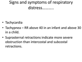
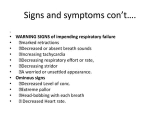
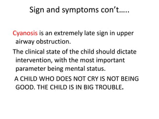
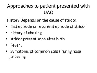
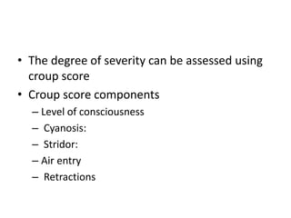
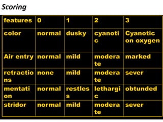
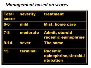
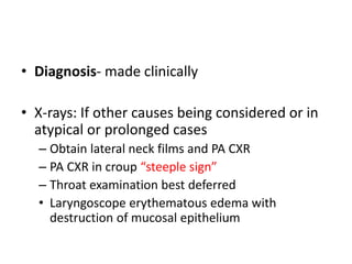
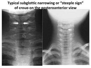
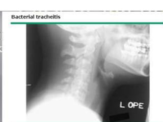
3. Introduction
• Acute upper airway obstruction from any
cause can be a life-threatening emergency.
• Complete obstruction will result in respiratory
failure followed by cardiac arrest, in a matter
of minutes.
• A child with a partial obstruction may have an
adequate airway initially. However, this
condition can deteriorate rapidly.
4. • The extrathoracic airways narrow during
inspiration and widen during expiration.
• Obstruction of the extrathoracic airway causes
intraluminal pressure distal to the obstruction
to fall during inspiration, which adds to the
obstruction.
5. CF
• Inspiratory stridor,:
• Stridor is a harsh, high-pitched, musical sound
produced by turbulent airflow through a
partially obstructed airway.)
• Although acute stridor usually is caused by
acute airway infections, other disorders may
be present and should be considered when
symptoms are severe, prolonged, or recurrent.
6. • hoarseness,
• and both suprasternal and intercostal
retractions are signs of a partial obstruction of
the larynx and extrathoracic trachea.
• If the obstruction is severe, then agitation,
cyanosis, acidosis, and respiratory failure
occur
7. • Compared to adults, infants and young
children have small airways and can quickly
develop clinically significant upper airway
obstruction
8. causes
• The most common cause of upper airway
obstruction in children is croup .
• Choking on a foreign body also occurs
frequently .
9. DDX of stridor
• Viral croup —Barking cough,Respiratory distress,Hoarse
voice,If due to measles, signs of measles .
• Retropharyngeal abscess —Soft tissue swelling ,Difficulty in
swallowing,Fever
• Foreign body —Sudden history of choking,Respiratory
distress
• Diphtheria —Bull neck appearance due to enlarged cervical
nodes and oedema,Red throat,Grey pharyngeal
membrane,Blood-stained nasal discharge,No evidence of
DTP vaccination
• Congenital anomaly —Stridor present since birth
10. Signs and symptoms of respiratory
distress………..
• Tachycardia
• Tachypnea – RR above 40 in an infant and above 30
in a child.
• Suprasternal retractions indicate more severe
obstruction than intercostal and subcostal
retractions.
11. Signs and symptoms con’t….
.
• WARNING SIGNS of impending respiratory failure
• marked retractions
• Decreased or absent breath sounds
• Increasing tachycardia
• Decreasing respiratory effort or rate,
• Decreasing stridor
• A worried or unsettled appearance.
• Ominous signs
• Decreased Level of conc.
• Extreme pallor
• Head-bobbing with each breath
• Decreased Heart rate.
12. Sign and symptoms con’t…..
Cyanosis is an extremely late sign in upper
airway obstruction.
The clinical state of the child should dictate
intervention, with the most important
parameter being mental status.
A CHILD WHO DOES NOT CRY IS NOT BEING
GOOD. THE CHILD IS IN BIG TROUBLE.
13. Approaches to patient presented with
UAO
History Depends on the cause of stridor:
• first episode or recurrent episode of stridor
• history of choking
• stridor present soon after birth.
• Fever ,
• Symptoms of common cold ( runny nose
,sneezing
14. Examination
• Audible stridor
• Hoarsness of the voice
• Barking type of cough
• Other findings specific to the cause (bull neck
appearance and grey pharyngeal membrane in
diphtheria, maculopapular skin rashes in measles
etc).
• Decreased or absent air entry, cyanosis, signs of
respiratory distress in severe cases
15. General Treatment
• Airway, airway, airway
• Supplemental oxygen
• Position of comfort
• REMEMBER – almost all children with an U.A.O.
can be bag-valve-mask ventilated, and this should
always be tried first in a respiratory failure
situation.
• Advanced airway management, IV, cardiac
monitor, Pulse ox, etc.
16. General Treatment con’t…..
• Always have a smaller ET tube readily
available because of the possibility of
significant airway edema.
• Watch for aspiration
17. • Remember…………… Failure to manage the
airway is the leading cause of preventable
deaths in children.
19. Etiology and Epidemiology
• Most common cause of stridor after neonatal
period
• Age: 3 months – 5years, peak is in the second
year of life
• Males are more frequently affected, Common in
winter
• Recurrence common till 3-6yrs and decreases
with age
• 15 %have strong family history of croup
20. ETIOLOGY
• Etiology-mostly viral
• Parainfluenza virus type I,II,III(75% of the cases)
• Incubation 2-6 days, virus shed for about 2 weeks
• Others
– Influenza A or B,
– RSV, rhinovirus, and measles
– Adenovirus
– Mycoplasma pneumoniae
– Diphteria_rarely
21. Clinical Manifestations
• Most common cause of URTO
• Pts usually have rhinorrhea, pharyngitis, mild cough
&low grade fever 1–3 days before the signs and
symptoms of upper airway obstruction become
apparent
• The child then develops the characteristic “barking”
cough, hoarseness, and inspiratory stridor
• As a disease progress patients will have signs of
respiratory distress
• Symptoms are worse at night, resolve with in a
week
• Other Family members may have mild resp. illness
22. • The degree of severity can be assessed using
croup score
• Croup score components
– Level of consciousness
– Cyanosis:
– Stridor:
– Air entry
– Retractions
25. • Diagnosis- made clinically
• X-rays: If other causes being considered or in
atypical or prolonged cases
– Obtain lateral neck films and PA CXR
– PA CXR in croup “steeple sign”
– Throat examination best deferred
• Laryngoscope erythematous edema with
destruction of mucosal epithelium
28. Treatment
–Supportive:
• Oxygen,
• IV fluid,
• Close monitoring
• Often improvement after child has been in cold
night air or moist air from shower
–Antipyretics if fever present
–Antibiotics not indicated
–IV fluid hydration only if necessary
29. –Stridor at rest or child in respiratory
distress- treat with epinephrine and
steroids
–Intubation if respiratory failure or pending
30. Treatment
• Racemic Epinephrine
• Recommended to watch patient for 3 hrs
before considering discharge
• Dexamethasone
– Steroids-used with moderate to severe episodes
of croup
– 0.3-0.6 mg/kg IM stat
• Tracheostomy: impending respiratory failure
20%
31. Croup: Discharge criteria
• After three to four hours of observation, children who remain
comfortable may be discharged home if they meet the following
criteria
• No stridor at rest
• Normal pulse oximetry
• Good air exchange
• Normal color
• Normal level of consciousness
• Demonstrated ability to tolerate fluids by mouth
• Caregivers understand the indications for return to care and would
be able to return if necessary
• Follow-up with the primary care provider within the next 24 hours.
32. Complications
• Seen in 15% of patients
• Otitis media
• Bacterial tracheatis
• Bacterial pneumonia
• Cervical lymphadenitis
33. SPASMODIC CROUP
• Spasmodic croup (ie, acute spasmodic laryngitis) is
characterized:
• by acute attacks of inspiratory stridor that tend to occur
suddenly during the evening or at night,
• last several hours, and then subside, only to recur during
the next several days.
• It usually is seen in children between 6 months and 3
years of age.
• Its etiology is uncertain, but it may represent recurrent
viral laryngotracheitis.
• The child with spasmodic croup usually awakens with a
barking, metallic cough and marked inspiratory stridor.
34. • The degree of inspiratory obstruction can be
striking, with retractions of the supraclavicular
and substernal areas.
• Fever is absent, and although the child may have
had a mild upper respiratory infection preceding
the attack,
• examination of the posterior pharynx reveals
only minimal, if any, signs of inflammation.
• Acute adductor spasm of the vocal cords also
may be a cause, possibly triggered by a mild viral
illness or allergy.
35. • Placing the child in a closed bathroom in which a hot
shower is running may bring relief in a few minutes.
• Paradoxically, exposure to the cool night air while en
route to a hospital frequently breaks the attack before
the hospital is reached.
• Aerosol treatment with racemic epinephrine usually
terminates the attack. If symptoms are atypical or
frequent, structural causes of recurrent stridor should
be considered.
• Occurrence in later childhood and adolescence
suggests psychogenic illness, especially if symptoms do
not occur during sleep
36. foreign bodies aspiration
• Infants and toddlers use their mouths to
explore their surroundings.
• Most victims of foreign body aspiration are
older infants and toddlers.
• Children <3 yr of age account for 73% of cases.
Preambulatory toddlers can aspirate objects
given to them by older siblings.
37. • One third of aspirated objects are nuts,
particularly peanuts. Fragments of raw carrot,
apple, dried beans, popcorn, and sunflower or
watermelon seeds are also aspirated, as are
small toys or toy parts.
38. Clinical Manifestations
Three stages
1 Initial event: Violent paroxysms of coughing,
choking, gagging, and possibly airway
obstruction occur immediately when the
foreign body is aspirated
2 Asymptomatic interval: The foreign body
becomes lodged, reflexes fatigue, and the
immediate irritating symptoms subside
39. 3 Complications: Obstruction, erosion, or
infection develops to direct attention again to
the presence of a foreign body.
complications include fever, cough,
hemoptysis, pneumonia, and atelectasis
40. Diagnosis
• A positive history must never be ignored. A
negative history may be misleading.
• Choking or coughing episodes accompanied
by wheezing are highly suggestive of an airway
foreign body .
• Most airway foreign bodies lodge in a
bronchus (right bronchus ∼58% of cases); the
location is the larynx or trachea in ∼10% of
cases
41. • The patient is asymptomatic and the
radiograph is normal in 15-30% of cases.
Opaque foreign bodies occur in only 10-25%
of cases.
• If there is a high index of suspicion,
bronchoscopy should be performed despite
negative imaging studies
42. • The treatment of choice for airway foreign
bodies is prompt endoscopic removal with
rigid instruments.
• Bronchoscopy is deferred only until
preoperative studies have been obtained
43. BACTERIAL TRACHEITIS
(PSEUDOMEMBRANOUS CROUP
• Bacterial tracheitis is a rare but important
cause of severe upper airway obstruction and
should be considered in older patients with
stridor or in infants who do not respond to
racemic epinephrine inhalation.
• Children with this condition initially have
symptoms that are similar to epiglottitis or
severe viral laryngotracheitis
44. • Lateral neck radiographs show subglottic narrowing and
foreign material in the tracheal lumen.
• Endoscopy reveals extensive sloughing of the respiratory
epithelium and large amounts of mucopurulent secretions
and debris blocking the trachea.
• These secretions may be difficult to remove and
progressively occlude the airway.
• Staphylococcus aureus is the most common organism that
is associated with this condition;
• nontypable H. influenzae, Branhamella catarrhalis, and
Streptococcus pneumoniae are less often the cause
45. • Treatment includes culture of secretions,
• appropriate intravenous antibiotics,
• oxygen,
• avoidance of fluid overload,
• and endotracheal intubation if there are signs
of progressive respiratory failure.
49. Epidemiology
• Age: 2-7 years affected
• Peak incidence-3&half yrs
• M:F=3:2
• No seasonal variation
50. ETIOLOGY
• Causative Organisms
–H. influenza type B almost all prior to
vaccination
• Rarely
–Streptococcus pyogenes
–Streptococcus pneumoniae
–Staphylococcus aureus
51. CLINICAL MANIFESTATIONS
• Classic symptoms- abrupt onset of high fever, sore
throat, dysphagia, and drooling of saliva, +/-
stridor.
• The barking cough typical of croup is rare.
• Usually, no other family members are ill with acute
respiratory symptoms.
• The child may assume the tripod position, sitting
upright and leaning forward with the chin up and
mouth open while bracing on the arms
52. • In a patient in whom the diagnosis is certain or
probable based on clinical grounds, laryngoscopy
should be performed expeditiously in a controlled
environment such as an operating room or
intensive care unit.
– “cherry red” swollen epiglottis by laryngoscopy.
• Anxiety-provoking interventions such as
phlebotomy, intravenous line placement, placing
the child supine, or direct inspection of the oral
cavity should be avoided until the airway is
secure.
53. • The otherwise healthy child suddenly develops a:
– sore throat &
– fever.
• Within a matter of hours,
– the patient appears toxic,
– swallowing is difficult &
– breathing is labored.
54. • Drooling is usually present
• Hyperextended neck in an attempt to maintain airway.
• The child may assume the tripod position,
– sitting upright &
– leaning forward
– with the chin up & mouth open
– while bracing on the arms.
56. MANAGEMENT
• The principles of management:
• Epiglottitis is a medical emergency and warrants
immediate treatment with an artificial airway
– Routine tracheostomy , intubation
• Oxygen
• Antibiotics
– Ceftriaxone or Chloramphenicol for 7-10 DAYS
57. Retropharyngeal and Para
pharyngeal abscess
• Neck contains deeply located LNs including retro &lateral pharyngeal
nodes w/c drain the upper air way &digestive tract
• Retropharyngeal space is located between the pharynx & the cervical
vertebrae extending down to superior mediastinum.
• Lateral pharyngeal space is bounded by pharynx medially carotid sheath
posteriorly & muscles of styloid process laterally.
• The two spaces communicate with each other.
• Infection usually extends from infection of oropharnyx
• Once infected, the nodes progress through 3 stages
cellulitis, phlegmon and abscess
58. Etiology and Epidemiology
• Usually polymicrobial
• Usual pathogens include group A strept., oropharyngeal
anaerobes and S.aures
• Hib, klebsela are other causes
• Common b/n 3-4yrs of age
• Males are affected more than females
• Rare after 5yrs b/c retropharyngeal nodes involute at this
age
59. CLINICAL MANIFESTATIONS
Retropharyngeal abscess
Fever, irritability, decreased oral intake and drooling of saliva
Neck stiffness, tortocolis & refusal to move the neck
Muffled voice, stridor and respiratory distress
Bulging of posterior pharyngeal wall
Cervical adenopathy may be present
Lateral pharyngeal abscess
Fever, dysphagia &prominent bulge on the lateral pharyngeal wall
Sometimes there is medial displacement of tonsils
60.
61. Investigations
Culture from the pus
CT
X-ray(wide retropharyngeal space >1/2 the thickness of
adjoining vertebrae)
DDX
epiglottis
Foreign body aspiration
Meningitis
Lymphoma
Hematoma
Vertebral osteomyelitis
62. Management
• IV Abcs with or without drainage
• 50% of Pts do not need drainage
• Indications for drainage
-obstruction
-failure to respond to IV Abcs
COMPLICATIONS
• Upper air way obstruction
• Rupture leading to aspiration pneumonia
• Mediastinitis
• Thrombophlebitis of internal jugular vein(Lemierre Ds)
• Erosion of carotid artery sheath
63. Diphtheria
• Diphtheria is a bacterial infection caused by
Corynebacterium diphtheria and it can be
prevented by immunization.
• Infection in the upper airway or nasopharynx
produces a grey membrane which, when
present in the larynx or trachea, can cause
stridor and obstruction
64. • Nasal involvement produces a bloody
discharge. Diphtheria toxin causes muscular
paralysis and myocarditis, which is associated
with increased mortality.
65. Diagnosis
• Examine the child’s nose and throat and look for
a grey, adherent membrane, which cannot be
wiped off with a swab.
• Great care is needed when examining the throat,
as this may precipitate complete obstruction of
the airway.
• A child with pharyngeal diphtheria may have an
obviously swollen neck, termed a ‘bull neck’
66. Treatment
• Antitoxin - give 40 000 units of diphtheria
antitoxin (IM or IV) immediately, because
delay can lead to increased mortality.
• Antibiotics -any child with suspected
diphtheria should be given procaine penicillin
(50, 000 units/kg IM) daily for 7 days.
68. Congenital causes of UAO
• Laryngomalacia
• Congenital subglottic stenosis
• Vocal cord paralysis
• Congenital laryngeal webs and atresia
• Congenital subglottic hemangioma
• Laryngoceles and sacular cyst
• Posterior laryngeal cleft and
laryngetracheoesophageal cleft
69. Laryngomalacia
• Laryngomalacia is the most common
congenital laryngeal anomaly and the most
common cause of stridor in infants and
children.
• Sixty percent of congenital laryngeal
anomalies in children with stridor are due to
laryngomalacia.
70. CLINICAL MANIFESTATIONS
• Stridor is inspiratory, low-pitched, and exacer-
bated by any exertion: crying, agitation, or
feeding.
• The stridor is caused, in part, by decreased
laryngeal tone leading to supraglottic collapse
during inspiration.
• Symptoms usually appear within the 1st 2 wk of
life and increase in severity for up to 6 mo,
although gradual improvement can begin at any
time.
71. • DIAGNOSIS
• The diagnosis is made primarily based on
symptoms.
• The diagnosis is confirmed by outpatient
flexible laryngoscopy .
• When the work of breathing is moderate to
severe, airway films and chest radio- graphs
are indicated
72. • TREATMENT
• Expectant observation is suitable for most infants
because most symptoms resolve spontaneously as the
child and airway grow.
• In 15-20% of patients symptoms are severe enough to
cause progressive respiratory distress, cyanosis, or
failure to thrive. In these patients surgical intervention
via supraglattoplasty is considered
• Supraglattoplasty is 90% successful in relieving upper
airway obstruction caused by laryngomalacia.
73. CongenitalSubglotticStenosis
• Congenital subglottic stenosis is the second
most common cause of stridor.
• The subglottis is the narrowest part of the
upper airway in a child.
• Subglottic stenosis is a narrowing of the
subglottic larynx, which is the space extending
from the undersurface of the true vocal cords
to the inferior margin of the cricoid cartilage.
74. CLINICAL MANIFESTATIONS
• It typically causes respiratory distress and
biphasic or primarily inspiratory stridor.
• It may be congenital or acquired.
• Symptoms often occur with a respiratory tract
infection as the edema and thickened
secretions of a common cold narrow an
already compromised airway leading to
recurrent or persistent croup like symptoms.
75. • Biphasic or primarily inspiratory stridor is the
typical presenting symptom for congenital
subglottic stenosis.
• Recurrent or persistent croup usually occurs in
these children at 6 mo of age or younger.
• The edema and thickened secretions of the
common cold further narrow an already
marginal airway that leads to croup-like
symptoms.
76. • DIAGNOSIS
• The diagnosis made by airway radiographs is
confirmed by direct laryngoscopy.
• During diagnostic laryngoscopy the subglottic
larynx is visualized directly and sized
objectively using endotracheal tubes
77. • The percentage of stenosis is determined by
comparing the size of the patients’ larynx to a
standard of laryngeal dimensions based on
age.
• Stenosis >50% is usually symptomatic and
often requires treatment.
78. • TREATMENT
• Anterior laryngotracheal decompression
(cricoid split) or laryngotra- cheal
reconstruction with cartilage grafting
79. Vocal Cord Paralysis
• Vocal cord paralysis is the third most common
congenital laryngeal anomaly that produces
stridor in infants and children.
• Unilateral vocal cord paralysis is most often
iatrogenic as a result of surgical treatment for
gastrointestinal (tracheoesophageal fistula)
and cardiovascular (patent ductus arteriosis
repair) anomalies.
80. CLINICAL MANIFESTATIONS
• Unilateral paralysis causes aspiration,
coughing, and choking; the cry is weak and
breathy, but stridor and other symptoms of
airway obstruc- tion are less common.
• . Bilateral vocal cord paralysis produces airway
obstruction manifested by high- pitched
inspiratory stridor: a phonatory sound or
inspiratory cry.
81. • DIAGNOSIS
• The diagnosis of vocal cord paralysis is made
by awake flexible laryn- goscopy. A thorough
investigation for the underlying primary cause
is indicated.
82. • TREATMENT
• Treatment is based on the severity of the symptoms.
Vocal cord paraly- sis in infants usually resolves
spontaneously within 6-12 mo.
• If it does not resolve within 2-3 yr, it is unlikely to do
so.
• Bilateral paralysis can require temporary tracheotomy.
Procedures that widen the posterior glottis to relieve
the obstruction include laryngotracheal reconstruc-
tion using an endoscopically placed posterior glottis
cartilage graft, or arytenoidectomy, or arytenoid
lateralization.