
Recommended
More Related Content
Similar to UL-fascial spaces in palm upper limb topic in anatomy (1).pdf
Similar to UL-fascial spaces in palm upper limb topic in anatomy (1).pdf (20)
Recently uploaded
Recently uploaded (20)
UL-fascial spaces in palm upper limb topic in anatomy (1).pdf
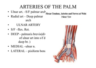
- 1. . ARTERIES OF THE PALM • Ulnar art. –S/F palmar arch • Radial art – Deep palmar arch • ULNAR ARTERY • S/F –flex. Ret. • DEEP - palmaris brevis(div of ulnar art into s/f & deep br. ) • MEDIAL –ulnar n. • LATERAL – pisiform bone
- 2. SUPERFIAL PALMAR ARCH • Formation – s/f ter. br. of ulnar art. Completed on lat. side by one of the following • a) s/f palmar br.of radial art • b)arteria princeps pollicis • c)arteria radialis indicis • d)arteria nervi mediana • Relations • Deep – palmar aponeurosis • Infront- long flex. Tendons, • lumbrical muscles & palmar dig. brs.of median n. Branches -4palmar dig. Art.(1med.-proper p.d. a.,3lat.- common p.d.a.) join by palmar metacarpal art.of deep palmar arch
- 3. DEEP PALMAR ARCH Arterial arcade formed between i –term. end of radial art. ii- deep br. of ulnar art. Radial art > two heads of 1st Dorsal intr. > between oblique & tr. Head of adductor pollicis >deep palmar arch |< Below hook of hamate(rad.art. Divides into arteria princeps pollicis &arteria radialis indicis ) | Between abductor digiti minimi & flexor digiti minimi | deep br. Of ulnar art. With deep br. Of ulnar n.( within its concavity)
- 4. DEEP PALMAR ARCH • Relations • Deep- oblique head of adductor pollicis,long flex. Tendons & lumbricals • Infront- bases of metacarpals & interossei • Branches- i) 3 palmar metacarpal arteries ii) 3 perforating arteries (passes between two heads of 2nd to 4th dorsal intros.,accompany veins drain the palm into dorsal v. arch) iii) Recurrent br.-anastomose with ant. Carpal arch
- 5. FASCIAL SPACES IN PALM • Surgical imortance • Potencial spaces limited by fibrous septa • - mid palmar • -Thenar • -Pulp CENTARL PALMAR SPACE between thenar & hypothenar eminences Infront – Palmar aponeourosis , long flex. tendons with synovial sheath and lumbrical mucles Deep – Dense fascia covering the interossie and the metacarpales Medial – Medial palmar septa (hypothenar eminence) Lateral – Lateral palmar septa (thenar eminence) • The C. palmar space divided by intermediate fiber septa into – Mid palmar space on ulnar side – Thenar space on radial side
- 7. Mid palmar space • Triangular in shape Boundaries – Infront – a) Fl. tendons of little, ring & middle finger with their synovial sheath b) Third and fourth lumbrical muscles Behind – Dense facia covering the interossei and metacarples of third and fourth sapce. Lateral – Intermediate palmar septa Medial – Hypothenar muscles separated by medial palmar septum Proximal- closed Distal- Extends as diverticula along fascial sheath of 3rd & 4th lumbrical muscles Applied- pus drained by splitting web betrween 3rd & 4th or 4th & 5th &exploring lumbical canals
- 8. Triangular in shape Thenar space Boundaries- Infront-a)muscles of thenar eminence b) flex. Tendons of index c) 1st & 2nd lumbrical muscles Behind- adductor polllicis Lateral- tendon of flex. Pollicis its sheath separated by lat. Palmar septum from thenar muscles Medial- Intermediate septum Proximal- closed Distal – 1st &2nd lumbricals 2nd lumbrical canal connect with mid palmar space Digital synovial sheath of 2nd ,3rd ,4th extend upto distal border of mid palmar & thenar space. Pus may burst into them
- 11. Pulp space • Location- between palmar skin & distal phalanges lies distal to fibrous flex. Sheath of flexor tendons • Tight compartments containing s/c fat & bl v. • Distal 4/5th distal phal. – gets digital arteries by penetrating fibrous septa • PROXIMAL 1/5TH WITHOUT TRAVERSING THE SEPTA • Applied – whitlow (infection) • Throbbing pain.> neglected> avascular necrosis