Download to read offline
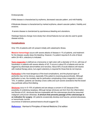
![Patients with proctitis usually pass fresh blood or blood-stained mucus, either mixed with stool
or streaked onto the surface of a normal or hard stool. They also have tenesmus, or urgency
with a feeling of incomplete evacuation, but rarely have abdominal pain. With proctitis or
proctosigmoiditis, proximal transit slows, which may account for the constipation commonly
seen in patients with distal disease.
When the disease extends beyond the rectum, blood is usually mixed with stool or grossly
bloody diarrhea may be noted. Colonic motility is altered by inflammation with rapid transit
through the inflamed intestine. When the disease is severe, patients pass a liquid stool
containing blood, pus, and fecal matter. Diarrhea is often nocturnal and/or postprandial.
Although severe pain is not a prominent symptom, some patients with active disease may
experience lower abdominal discomfort or mild central abdominal cramping. Severe cramping
and abdominal pain can occur with severe attacks of the disease. Other symptoms in moderate
to severe disease include anorexia, nausea, vomiting, fever, and weight loss.
Physical signs of proctitis include a tender anal canal and blood on rectal examination. With
more extensive disease, patients have tenderness to palpation directly over the colon. Patients
with toxic colitis have severe pain and bleeding, and those with megacolon have hepatic
tympany. Both may have signs of peritonitis if a perforation has
occurred.
Laboratory, Endoscopic, and Radiographic Features
Active disease can be associated with a rise in acute-phase reactants (C-reactive protein
[CRP]), platelet count, and erythrocyte sedimentation rate (ESR), and a decrease in
hemoglobin. Fecal lactoferrin, a glycoprotein present in activated neutrophils, is a highly
sensitive and specific marker for detecting intestinal inflammation. Fecal calprotectin is
present in neutrophils and monocytes and levels correlate well with histologic inflammation,
predict relapses, and detect pouchitis. Both fecal lactoferrin and calprotectin are becoming an
integral part of IBD management and are used frequently to rule out active inflammation versus
symptoms of irritable bowel or bacterial overgrowth. In severely ill patients, the serum albumin
level will fall rather quickly. Leukocytosis may be present but is not a specific indicator of
disease activity. Proctitis or proctosigmoiditis rarely causes a rise in CRP.
Diagnosis relies on the
● patient’s history;
● clinical symptoms;
● negative stool examination for bacteria, C. difficile toxin, and ova and parasites;
● sigmoidoscopic appearance ; and
● histology of rectal or colonic biopsy specimens.
Sigmoidoscopy is used to assess disease activity and is usually performed before treatment. If
the patient is not having an acute flare, colonoscopy is used to assess disease extent and
activity.](https://image.slidesharecdn.com/ulcerativecolitisuc-241019035515-73f98d2c/85/ULCERATIVE-COLITIS-UC-Pathology-Clinical-Features-Complications-pdf-3-320.jpg)

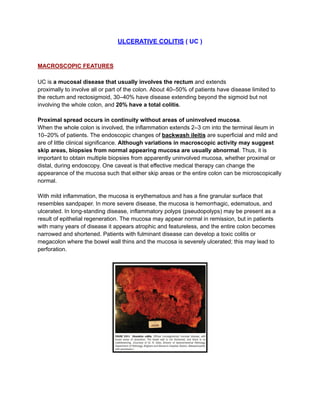
Ulcerative colitis (UC) is a mucosal disease primarily affecting the colon, characterized by variable extents of inflammation and symptoms such as diarrhea, rectal bleeding, and abdominal pain. Diagnosis is based on clinical history, symptom evaluation, and endoscopic findings, while complications may include toxic megacolon and perforation. Management relies on effective medical therapy, which can alter mucosal appearance, and is crucial to prevent severe outcomes.


