well-fitting shoes are essential
Corns under the metatarsals can be helped by soft spongy soles, but sometimes need orthopaedic surgery to alter weight bearing.
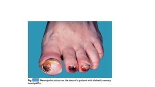
Special care is needed with corns on ischaemic or diabetic feet, which are at greater risk of infection and ulceration