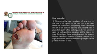
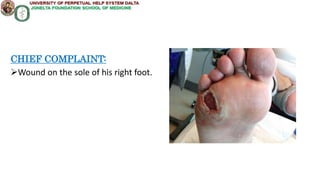
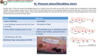
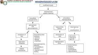
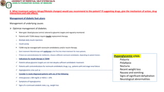
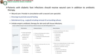
ENDOCRINOLOGY-
DERMATOLOGY
SUBGROUP –1, BLOCK 11-12
 SUN STEFFANY
 SWAIN JAYADEV
 THIRUNAMPALLI NETHIBOTTU SHASHI KUMAR
 THOMPSON ROBERT NELSON JENCY
Case scenario:
A 58-year-old farmer complains of a wound on
the sole of his right foot. He says that it has been
there for six months, progressing from a sore the
size of a ten-centavo coin to the present size. It
gives him pain while walking, and even when at
rest. He feels pricking sensation at the tips of his
toes. Sometimes there is numbness of the soles of
his feet. He also complains of having to wake up at
night to urinate. He has been losing weight for the
past six months as well.
IDENTIFYING DATA:
Name: Not given in case
Age: 58 Years old
Gender: Male
Occupation: Farmer
CHIEF COMPLAINT:
Wound on the sole of his right foot.
HISTORY OF PRESENT ILLNESS:
 He complains of a wound on the sole of his right
foot. He says that it has been there for six months,
progressing from a sore the size of a ten-centavo coin
to the present size. It gives him pain while walking,
and even when at rest.
Pricking sensation at the tips of his toes
Numbness of soles of feet sometimes
He also complains of having to wake up at night to
urinate
Weight loss for last 6 months.
Medication History:
Not Given In Case
Family History:
Not Given In Case
Personal and Social History:
Not Given In Case
Review of Systems:
General: (+) weight loss, (-) fatigue, (-) fever
skin : (+) Ulcer on foot
HEENT: (-) Headaches, (-) blurring of vision, (-)
earaches, (-) sinus trouble, (-) hoarseness
Neck: (-) Swollen glands, (-) goiter
Respiratory: (-) cough, (-) asthma
Cardiovascular: (-) palpitations, (-) chest pain, discomfort
Gastrointestinal: (-) constipation, (-) diarrhea
Peripheral Vascular: (-) swelling, tenderness (-) colour
changes
Urinary: (-) pain, (-) urgency, (+) frequency
Musculoskeletal: (+)pain in the wound area while walking and rest ,
(-) muscle pain, (-) sitffness, (-) joint pain,
Psychiatric: (-) memory changes, (-) depression
Neurologic: (+) Pricking sensation on toes , (+) numbness of soles of
feet, (-) Paralysis, (-) vertigo, (-) changes in speech
Hematologic: (-) easy bruising, bleeding
Endocrine: (-) heat or cold intolerance, (-) excessive sweating,
(-) excessive hunger, thirst.
Salient Features:
 58 years old, male
 non-healing wound on feet (6 months)
 Single, large ulcer on lateral side of sole of right foot close to toes
 Pain while walking and at rest because of ulcer
 Pricking sensation at tips of toes (Paresthesia)
 Numbness on the soles of feet sometimes (Peripheral neuropathy)
 Wakes up at night to urinate (Nocturia)
 Losing weight (6 months)
1. Describe the skin findings.
⮚The skin findings in the patient's foot include single deep ulcer with
regular border surrounded by both pale and erythematous thickened skin
located below 5th Metatarsal head on the plantar surface of right foot.
2. What may be causing the patient’s skin condition? (Primary diagnosis)
Explain.
Primary diagnosis: Foot ulcer probable secondary to uncontrolled DM
The patient's skin condition is most likely caused by a foot ulcer, which is a
type of open sore or wound that develops on the foot. Foot ulcers are
commonly associated with underlying medical conditions, such as long-term
effects of diabetes, which can cause damage to the nerves and blood vessels in
the feet, leading to poor circulation and reduced sensation. This can result in
minor injuries, such as blisters or cuts, becoming infected and developing into
foot ulcers.
Rule In Rule out
58 yrs. Old Male
non-healing wound on feet for last 6
months
Ulcer at plantar aspect of right foot under
5thmetatarsal
Pain while walking and at rest
Wound Surrounded by thickened skin
Paresthesia
Peripheral neuropathy
Nocturia
Weight loss for past 6 months
3. Give at least five differential diagnoses (other causes of the skin condition)
I. ISCHEMIC ARTERIAL ULCERS
II. VENOUS ULCERS
III. Pressure ulcers/Decubitus ulcers
IV. Pyoderma gangrenosum
V. Cellulitis
Rule In Rule out
+ 58 yrs. Old Male - loss of hair
+ non-healing wound on feet for last 6
months
- Cold skin
+ Pain while walking and at rest - Wound surrounded by thinned skin
+ Small size wound at early stage
I. ISCHEMIC ARTERIAL ULCERS
Arterial ulcers are often found between or on the tips of the toes, on the heels, on the outer ankle or tibia (around lateral malleolus), other pressure points
caused by poor blood flow to the foot, or atherosclerosis. They may appear as small, deep wounds with a "punched out" appearance, and may be surrounded
by shiny, hairless skin. Underlying conditions for the arterial ulcers are diabetes, hypertension, smoking, previous vascular disease to which it’s significant with
the patient’s history. The arterial ulcer characteristics are also very similar.
Rule In Rule out
+ 58 yrs. Old Male - painless, edema
+ non-healing wound on feet for last 6
months
- Irregular shaped wound
- Wound surrounded by shiny skin
II. VENOUS ULCERS
Venous ulcers: These are caused by problems with the veins in the inner lower legs, gaiter area and may appear as shallow, irregularly shaped wounds with
uneven edges. They are typically located around the ankle and may be accompanied by swelling and varicose veins.
Rule In Rule out
+ 58 yrs. Old Male - Immobility
+ non-healing wound on feet for last
6 months
- Perception of pain
+ Pain while walking and at rest - Skin perfusion (e.g., in peripheral artery
disease, heart failure, vasoconstriction,
or shock)
+ Full-thickness skin loss -
Risk factor- Sign and symptoms of DM
III. Pressure ulcers/Decubitus ulcers
Pressure ulcers: These are caused by prolonged pressure on a specific area of the foot and are often seen in people who are bedridden or have limited
mobility. They may appear as open sores or areas of skin that have broken down, and are typically located on bony prominences, such as the sacrum, heel,
greater trochanter, lateral malleolus, elbows. These injuries typically develop over bony prominences when local pressure-induced hypoperfusion and necrosis
can lead to the loss of several or all skin layers.
Rule In Rule out
+ 58 yrs. Old Male - Irregular, undermined, violaceous border
+ non-healing wound on feet for last 6
months
- Granulation tissue
+ Pain while walking and at rest - purulent exudate
+ Risk factor- Sign and symptoms of DM
IV. Pyoderma gangrenosum
Pyoderma gangrenosum is an uncommon, ulcerative cutaneous condition, non infectious, neutrophilic dermatosis commonly associated with
underlying disease
• Classic/Ulcerative PG variant usually presents with painful ulcers ,commonly on legs (70%) •
Clinical features: presents as solitory or multiple small, tender, red-blue papules, plaques or pustules that evolve into painful ulcers with
characteristic violaceous undermined edges
There may be granulation tissue, necrosis or purulent exudate at the ulcer base
Pathergy occurs in 25%
Healing usually occurs with atrophic cribriform scar
Rule In Rule out
+ 58 yrs. Old Male - Bullae
+ non-healing wound on feet for last 6
months
- Purulent exudate
+ Pain while walking and at rest - Lymphadenitis
+ Risk factor- Sign and symptoms of DM - erythema
V. Cellulitis
Cellulitis is an acute inflammatory condition of the deep dermis and subcutaneous tissue usually found complicating a wound, ulcer or dermatosis.
Spreading and pyogenic in nature, it is characterized by localized pain, erythema, swelling and heat. The involved area, most commonly on the
leg, lacks sharp demarcation from uninvolved skin.
Cellulitis may be caused by indigenous flora colonizing the skin and appendages, like Staphylococcus aureus (S. aureus) and Streptococcus
pyogenes (S. pyogenes), or by a wide variety of exogenous bacteria.
PATHOPHYSIOLOGY of DM
PATHOPHYSIOLOGY of DFU
4. What laboratory examinations/diagnostic procedures are recommended for the patient?
What are your expected results?
Laboratory investigations
Blood glucose test: FBS, PPBS, HbA1c, RBG
Laboratory investigations
 Routine studies-Perform in all patients as part of the initial diagnostic workup and reassess at least annually.
 Renal function - to rule out kidney damage- eGFR that is rapidly decreasing or < 30 mL/min/1.73 m2
 Spot urinary albumin-to-creatinine ratio: to detect microalbuminuria
 Lipid panel------------------------------------------------------------------------------------------------------
 Additional studies-
 C-peptide: can help differentiate between types of diabetes
 ↑ C-peptide levels may indicate insulin resistance and hyperinsulinemia → T2DM
 ↓ C-peptide levels indicate an absolute insulin deficiency → T1DM
 Urinalysis:
 Glucosuria may be present if the renal threshold for glucose is reached (nonspecific for diabetes mellitus).
 Ketone bodies: positive in acute metabolic decompensation (diabetic ketoacidosis)
 Spot urinary albumin-to-creatinine ratio: to detect microalbuminuria
Laboratory investigations
CBC, ESR: Elevated WBC, ESR may indicate skin and soft tissue infection, acute/chronic
osteomyelitis
CRP: Inflammation
Blood cultures: help determine if a patient has bacteremia/septicemia, a life-threatening
condition
Wound cultures Biopsy and culture of infected tissue :To determine the type of bacteria
causing the infection, and to guide antibiotic treatment.
Imaging tests, such as X-rays or MRI, to evaluate the extent of tissue damage and the presence
of any underlying bone or joint involvement.
4. What treatment options (drugs/lifestyle changes) would you recommend to the patient? If suggesting drugs, give the mechanism of action, drug
interactions and side effects.
Management of diabetic foot ulcers
Management of underlying causes
 Optimize management of diabetes.
 Main goal: blood glucose control, tailored to glycemic targets and regularly monitored
 Patients with T1DM always require insulin replacement therapy.
o Multiple daily insulin injections
o Insulin pump
 T2DM may be managed with noninsulin antidiabetics and/or insulin therapy.
o Start treatment Monotherapy with metformin is the first-line initial treatment for most patients.
o If there are contraindications for metformin, choose a different noninsulin antidiabetic, depending on patient factors.
o Indications for insulin therapy in T2DM
 Patients whose glycemic targets are not met despite sufficient antidiabetic treatment
 Patients with contraindications for noninsulin antidiabetic drugs, e.g., patients with end-stage renal failure
 Hyperglycemic crisis such as ------------------------------------------------------------------------------------------------------------------------------------------------
o Consider in newly diagnosed patients with any of the following:
 Initial glucose ≥ 300 mg/dL or HbA1c > 10%
 Symptoms of hyperglycemia
 Signs of a continued catabolic state, e.g., weight loss
Hyperglycemic crisis
Polyuria
Polydipsia
Nocturia
Recent weight loss
Nausea and vomiting
Signs of significant dehydration
Neurological abnormalities
Patients with diabetic foot infections should receive wound care in addition to antibiotic
therapy.
 Wound care: Provide in consultation with a wound care specialist.
o Dressings to promote wound healing
o Debridement (e.g., surgical) including removal of surrounding calluses
 Initiate empiric antibiotic therapy for skin and soft tissue infections.
 Pain management: As the patient is experiencing pain, medications like acetaminophen or NSAIDs may be prescribed. In some cases, opioids may
be used, but they can be addictive, so caution is necessary.
 Specialized footwear
 Provide mechanical offloading from pressure points
 Reduce progression and recurrence of foot ulcers
 Decrease calluses
 Lifestyle recommendations for patients with diabetes mellitus
 Balanced diet and nutrition------------------------------------------------
 Recommend smoking cessation.
 Prevention of Diabetic Foot Ulcer and Infection
 Patient education (classes, handouts)
 Self examination
 Footwear and orthoses
 Lotion and lamb's wool
 Nutrition and self-management of glycemic control
 Foot screening examinations
 Aggressive early evaluation and treatment of foot lesions
 Multidisciplinary clinic setting
 Antimicrobial prophylaxis for elective surgery
 A high-fiber diet
 Eating nonstarchy vegetables, whole foods
 Avoiding refined sugar and grains
DRUGS MOA DRUG INTERACTIONS SIDE EFFECTS
INSULIN Insulin lowers blood glucose levels. It regulates carbohydrate,
protein and fat metabolism by inhibiting hepatic glucose
production and lipolysis, and enhancing peripheral glucose
disposal.
Decreased hypoglycaemic effect with corticosteroids, danazol,
diazoxide, diuretics, glucagon, isoniazid, phenothiazine derivatives,
somatropin, sympathomimetic agents, thyroid hormones, oestrogens,
progestins (e.g. in oral contraceptives), protease inhibitors and
atypical antipsychotic (e.g. olanzapine and clozapine).
Hypoglycaemia, insulin resistance, lipoatrophy,
hypokalaemia, blurred vision.
METFORMIN Metformin is a biguanide antihyperglycemic agent which
improves glucose tolerance by lowering both basal and
postprandial plasma glucose. It exerts its effect by decreasing
hepatic glucose production through inhibition of
gluconeogenesis and glycogenolysis, delaying intestinal
absorption of glucose, and improving insulin sensitivity by
increasing peripheral glucose uptake and utilisation.
Increased risk of lactic acidosis with carbonic anhydrase inhibitors (e.g.
acetazolamide, dichlorphenamide, topiramate, zonisamide), NSAIDs,
including cyclo-oxygenase (COX) II inhibitors; ACE inhibitors,
angiotensin II receptor antagonists, and loop diuretics. Increased risk
of hypoglycaemia with other antidiabetic agents (e.g. sulfonylurea,
meglitinides, insulin).
Significant: Vitamin B12 deficiency (prolonged use).
Gastrointestinal disorders: Nausea, vomiting, abdominal
pain or distress, flatulence, diarrhoea, dyspepsia, taste
disturbance.
Metabolism and nutrition disorders: Loss of appetite.
Musculoskeletal and connective tissue disorders: Asthenia.
Potentially Fatal: Lactic acidosis.
DOXYCYCLINE Doxycycline, a tetracycline congener, has bacteriostatic activity
against a broad range of gram-positive and gram-negative
bacteria. It inhibits protein synthesis by binding to the 30S
ribosomal subunit of the bacteria. It may also cause alterations
in the cytoplasmic membrane.
Absorption may be impaired by concurrently administered antacids or
other drugs containing Al, Ca, or Mg; oral Zn, Fe salts, or bismuth
preparations. May interfere with the bactericidal action of penicillin.
May potentiate the effects of anticoagulants. May potentiate the
hypoglycaemic effect of sulfonylureas.
Gastrointestinal disorders: Nausea, vomiting, diarrhoea,
upper abdominal pain, dry mouth, dysphagia, glossitis, black
hairy tongue; periodontal abscess, tooth disorder,
toothache.
General disorders and administration site conditions: Pain.
Hepatobiliary disorders: Cholestatic hepatitis, fatty liver
degeneration.
VANCOMYCIN Vancomycin is a glycopeptide antibiotic which binds tightly to D-
alanyl-D-alanine portion of cell wall precursor, blocking
glycopeptide polymerisation leading to the inhibition of
bacterial cell wall synthesis. It also impairs bacterial-cell-
membrane permeability and RNA synthesis.
Increased frequency of erythema, histamine-like flushing, and
anaphylactic reactions with anaesthetic agents. aminoglycosides,
amphotericin B, polymyxin B, bacitracin, colistin,
piperacillin/tazobactam, cisplatin, loop diuretics, NSAIDs) may
potentiate the toxicity of vancomycin.
Blood and lymphatic system disorders: Neutropenia
(reversible), thrombocytopenia.
Gastrointestinal disorders: Nausea; abdominal pain,
diarrhoea, flatulence, vomiting (oral).
General disorders and administration site conditions: Fever,
chills, fatigue; peripheral oedema (oral).
Musculoskeletal and connective tissue disorders: Muscle
spasm (chest and back), back pain.
Nervous system disorders: Headache.
CEFTRIAXONE Ceftriaxone is a 3rd generation broad-spectrum cephalosporin
antibiotic. It binds to 1 or more penicillin-binding proteins
(PBPs) inhibiting the final transpeptidation step of
peptidoglycan synthesis in the bacterial cell walls, leading to
bacterial cell lysis and death.
May increase the risk of bleeding with vitamin K antagonists (e.g.
warfarin). May increase the nephrotoxicity of aminoglycosides.
Significant: Biliary lithiasis, gallbladder sludge,
pseudolithiasis, pancreatitis (possibly secondary to biliary
obstruction), renal lithiasis (reversible), urolithiasis, acute
renal failure; encephalopathy, fungal or bacterial
superinfection (prolonged use).
Gastrointestinal disorders: Nausea, vomiting, diarrhoea.
Skin and subcutaneous tissue disorders: Rash, pruritus,
diaphoresis.
THANK YOU

Endo Derma Subgroup 1.pptx

  • 1.
    ENDOCRINOLOGY- DERMATOLOGY SUBGROUP –1, BLOCK11-12  SUN STEFFANY  SWAIN JAYADEV  THIRUNAMPALLI NETHIBOTTU SHASHI KUMAR  THOMPSON ROBERT NELSON JENCY
  • 2.
    Case scenario: A 58-year-oldfarmer complains of a wound on the sole of his right foot. He says that it has been there for six months, progressing from a sore the size of a ten-centavo coin to the present size. It gives him pain while walking, and even when at rest. He feels pricking sensation at the tips of his toes. Sometimes there is numbness of the soles of his feet. He also complains of having to wake up at night to urinate. He has been losing weight for the past six months as well.
  • 3.
    IDENTIFYING DATA: Name: Notgiven in case Age: 58 Years old Gender: Male Occupation: Farmer
  • 4.
    CHIEF COMPLAINT: Wound onthe sole of his right foot.
  • 5.
    HISTORY OF PRESENTILLNESS:  He complains of a wound on the sole of his right foot. He says that it has been there for six months, progressing from a sore the size of a ten-centavo coin to the present size. It gives him pain while walking, and even when at rest. Pricking sensation at the tips of his toes Numbness of soles of feet sometimes He also complains of having to wake up at night to urinate Weight loss for last 6 months.
  • 6.
    Medication History: Not GivenIn Case Family History: Not Given In Case Personal and Social History: Not Given In Case
  • 7.
    Review of Systems: General:(+) weight loss, (-) fatigue, (-) fever skin : (+) Ulcer on foot HEENT: (-) Headaches, (-) blurring of vision, (-) earaches, (-) sinus trouble, (-) hoarseness Neck: (-) Swollen glands, (-) goiter
  • 8.
    Respiratory: (-) cough,(-) asthma Cardiovascular: (-) palpitations, (-) chest pain, discomfort Gastrointestinal: (-) constipation, (-) diarrhea Peripheral Vascular: (-) swelling, tenderness (-) colour changes Urinary: (-) pain, (-) urgency, (+) frequency
  • 9.
    Musculoskeletal: (+)pain inthe wound area while walking and rest , (-) muscle pain, (-) sitffness, (-) joint pain, Psychiatric: (-) memory changes, (-) depression Neurologic: (+) Pricking sensation on toes , (+) numbness of soles of feet, (-) Paralysis, (-) vertigo, (-) changes in speech Hematologic: (-) easy bruising, bleeding Endocrine: (-) heat or cold intolerance, (-) excessive sweating, (-) excessive hunger, thirst.
  • 10.
    Salient Features:  58years old, male  non-healing wound on feet (6 months)  Single, large ulcer on lateral side of sole of right foot close to toes  Pain while walking and at rest because of ulcer  Pricking sensation at tips of toes (Paresthesia)  Numbness on the soles of feet sometimes (Peripheral neuropathy)  Wakes up at night to urinate (Nocturia)  Losing weight (6 months)
  • 11.
    1. Describe theskin findings. ⮚The skin findings in the patient's foot include single deep ulcer with regular border surrounded by both pale and erythematous thickened skin located below 5th Metatarsal head on the plantar surface of right foot.
  • 12.
    2. What maybe causing the patient’s skin condition? (Primary diagnosis) Explain. Primary diagnosis: Foot ulcer probable secondary to uncontrolled DM The patient's skin condition is most likely caused by a foot ulcer, which is a type of open sore or wound that develops on the foot. Foot ulcers are commonly associated with underlying medical conditions, such as long-term effects of diabetes, which can cause damage to the nerves and blood vessels in the feet, leading to poor circulation and reduced sensation. This can result in minor injuries, such as blisters or cuts, becoming infected and developing into foot ulcers.
  • 13.
    Rule In Ruleout 58 yrs. Old Male non-healing wound on feet for last 6 months Ulcer at plantar aspect of right foot under 5thmetatarsal Pain while walking and at rest Wound Surrounded by thickened skin Paresthesia Peripheral neuropathy Nocturia Weight loss for past 6 months
  • 14.
    3. Give atleast five differential diagnoses (other causes of the skin condition) I. ISCHEMIC ARTERIAL ULCERS II. VENOUS ULCERS III. Pressure ulcers/Decubitus ulcers IV. Pyoderma gangrenosum V. Cellulitis
  • 15.
    Rule In Ruleout + 58 yrs. Old Male - loss of hair + non-healing wound on feet for last 6 months - Cold skin + Pain while walking and at rest - Wound surrounded by thinned skin + Small size wound at early stage I. ISCHEMIC ARTERIAL ULCERS Arterial ulcers are often found between or on the tips of the toes, on the heels, on the outer ankle or tibia (around lateral malleolus), other pressure points caused by poor blood flow to the foot, or atherosclerosis. They may appear as small, deep wounds with a "punched out" appearance, and may be surrounded by shiny, hairless skin. Underlying conditions for the arterial ulcers are diabetes, hypertension, smoking, previous vascular disease to which it’s significant with the patient’s history. The arterial ulcer characteristics are also very similar.
  • 16.
    Rule In Ruleout + 58 yrs. Old Male - painless, edema + non-healing wound on feet for last 6 months - Irregular shaped wound - Wound surrounded by shiny skin II. VENOUS ULCERS Venous ulcers: These are caused by problems with the veins in the inner lower legs, gaiter area and may appear as shallow, irregularly shaped wounds with uneven edges. They are typically located around the ankle and may be accompanied by swelling and varicose veins.
  • 17.
    Rule In Ruleout + 58 yrs. Old Male - Immobility + non-healing wound on feet for last 6 months - Perception of pain + Pain while walking and at rest - Skin perfusion (e.g., in peripheral artery disease, heart failure, vasoconstriction, or shock) + Full-thickness skin loss - Risk factor- Sign and symptoms of DM III. Pressure ulcers/Decubitus ulcers Pressure ulcers: These are caused by prolonged pressure on a specific area of the foot and are often seen in people who are bedridden or have limited mobility. They may appear as open sores or areas of skin that have broken down, and are typically located on bony prominences, such as the sacrum, heel, greater trochanter, lateral malleolus, elbows. These injuries typically develop over bony prominences when local pressure-induced hypoperfusion and necrosis can lead to the loss of several or all skin layers.
  • 18.
    Rule In Ruleout + 58 yrs. Old Male - Irregular, undermined, violaceous border + non-healing wound on feet for last 6 months - Granulation tissue + Pain while walking and at rest - purulent exudate + Risk factor- Sign and symptoms of DM IV. Pyoderma gangrenosum Pyoderma gangrenosum is an uncommon, ulcerative cutaneous condition, non infectious, neutrophilic dermatosis commonly associated with underlying disease • Classic/Ulcerative PG variant usually presents with painful ulcers ,commonly on legs (70%) • Clinical features: presents as solitory or multiple small, tender, red-blue papules, plaques or pustules that evolve into painful ulcers with characteristic violaceous undermined edges There may be granulation tissue, necrosis or purulent exudate at the ulcer base Pathergy occurs in 25% Healing usually occurs with atrophic cribriform scar
  • 19.
    Rule In Ruleout + 58 yrs. Old Male - Bullae + non-healing wound on feet for last 6 months - Purulent exudate + Pain while walking and at rest - Lymphadenitis + Risk factor- Sign and symptoms of DM - erythema V. Cellulitis Cellulitis is an acute inflammatory condition of the deep dermis and subcutaneous tissue usually found complicating a wound, ulcer or dermatosis. Spreading and pyogenic in nature, it is characterized by localized pain, erythema, swelling and heat. The involved area, most commonly on the leg, lacks sharp demarcation from uninvolved skin. Cellulitis may be caused by indigenous flora colonizing the skin and appendages, like Staphylococcus aureus (S. aureus) and Streptococcus pyogenes (S. pyogenes), or by a wide variety of exogenous bacteria.
  • 20.
  • 21.
  • 22.
    4. What laboratoryexaminations/diagnostic procedures are recommended for the patient? What are your expected results? Laboratory investigations Blood glucose test: FBS, PPBS, HbA1c, RBG
  • 23.
    Laboratory investigations  Routinestudies-Perform in all patients as part of the initial diagnostic workup and reassess at least annually.  Renal function - to rule out kidney damage- eGFR that is rapidly decreasing or < 30 mL/min/1.73 m2  Spot urinary albumin-to-creatinine ratio: to detect microalbuminuria  Lipid panel------------------------------------------------------------------------------------------------------  Additional studies-  C-peptide: can help differentiate between types of diabetes  ↑ C-peptide levels may indicate insulin resistance and hyperinsulinemia → T2DM  ↓ C-peptide levels indicate an absolute insulin deficiency → T1DM  Urinalysis:  Glucosuria may be present if the renal threshold for glucose is reached (nonspecific for diabetes mellitus).  Ketone bodies: positive in acute metabolic decompensation (diabetic ketoacidosis)  Spot urinary albumin-to-creatinine ratio: to detect microalbuminuria
  • 24.
    Laboratory investigations CBC, ESR:Elevated WBC, ESR may indicate skin and soft tissue infection, acute/chronic osteomyelitis CRP: Inflammation Blood cultures: help determine if a patient has bacteremia/septicemia, a life-threatening condition Wound cultures Biopsy and culture of infected tissue :To determine the type of bacteria causing the infection, and to guide antibiotic treatment. Imaging tests, such as X-rays or MRI, to evaluate the extent of tissue damage and the presence of any underlying bone or joint involvement.
  • 25.
    4. What treatmentoptions (drugs/lifestyle changes) would you recommend to the patient? If suggesting drugs, give the mechanism of action, drug interactions and side effects. Management of diabetic foot ulcers Management of underlying causes  Optimize management of diabetes.  Main goal: blood glucose control, tailored to glycemic targets and regularly monitored  Patients with T1DM always require insulin replacement therapy. o Multiple daily insulin injections o Insulin pump  T2DM may be managed with noninsulin antidiabetics and/or insulin therapy. o Start treatment Monotherapy with metformin is the first-line initial treatment for most patients. o If there are contraindications for metformin, choose a different noninsulin antidiabetic, depending on patient factors. o Indications for insulin therapy in T2DM  Patients whose glycemic targets are not met despite sufficient antidiabetic treatment  Patients with contraindications for noninsulin antidiabetic drugs, e.g., patients with end-stage renal failure  Hyperglycemic crisis such as ------------------------------------------------------------------------------------------------------------------------------------------------ o Consider in newly diagnosed patients with any of the following:  Initial glucose ≥ 300 mg/dL or HbA1c > 10%  Symptoms of hyperglycemia  Signs of a continued catabolic state, e.g., weight loss Hyperglycemic crisis Polyuria Polydipsia Nocturia Recent weight loss Nausea and vomiting Signs of significant dehydration Neurological abnormalities
  • 26.
    Patients with diabeticfoot infections should receive wound care in addition to antibiotic therapy.  Wound care: Provide in consultation with a wound care specialist. o Dressings to promote wound healing o Debridement (e.g., surgical) including removal of surrounding calluses  Initiate empiric antibiotic therapy for skin and soft tissue infections.
  • 27.
     Pain management:As the patient is experiencing pain, medications like acetaminophen or NSAIDs may be prescribed. In some cases, opioids may be used, but they can be addictive, so caution is necessary.  Specialized footwear  Provide mechanical offloading from pressure points  Reduce progression and recurrence of foot ulcers  Decrease calluses  Lifestyle recommendations for patients with diabetes mellitus  Balanced diet and nutrition------------------------------------------------  Recommend smoking cessation.  Prevention of Diabetic Foot Ulcer and Infection  Patient education (classes, handouts)  Self examination  Footwear and orthoses  Lotion and lamb's wool  Nutrition and self-management of glycemic control  Foot screening examinations  Aggressive early evaluation and treatment of foot lesions  Multidisciplinary clinic setting  Antimicrobial prophylaxis for elective surgery  A high-fiber diet  Eating nonstarchy vegetables, whole foods  Avoiding refined sugar and grains
  • 28.
    DRUGS MOA DRUGINTERACTIONS SIDE EFFECTS INSULIN Insulin lowers blood glucose levels. It regulates carbohydrate, protein and fat metabolism by inhibiting hepatic glucose production and lipolysis, and enhancing peripheral glucose disposal. Decreased hypoglycaemic effect with corticosteroids, danazol, diazoxide, diuretics, glucagon, isoniazid, phenothiazine derivatives, somatropin, sympathomimetic agents, thyroid hormones, oestrogens, progestins (e.g. in oral contraceptives), protease inhibitors and atypical antipsychotic (e.g. olanzapine and clozapine). Hypoglycaemia, insulin resistance, lipoatrophy, hypokalaemia, blurred vision. METFORMIN Metformin is a biguanide antihyperglycemic agent which improves glucose tolerance by lowering both basal and postprandial plasma glucose. It exerts its effect by decreasing hepatic glucose production through inhibition of gluconeogenesis and glycogenolysis, delaying intestinal absorption of glucose, and improving insulin sensitivity by increasing peripheral glucose uptake and utilisation. Increased risk of lactic acidosis with carbonic anhydrase inhibitors (e.g. acetazolamide, dichlorphenamide, topiramate, zonisamide), NSAIDs, including cyclo-oxygenase (COX) II inhibitors; ACE inhibitors, angiotensin II receptor antagonists, and loop diuretics. Increased risk of hypoglycaemia with other antidiabetic agents (e.g. sulfonylurea, meglitinides, insulin). Significant: Vitamin B12 deficiency (prolonged use). Gastrointestinal disorders: Nausea, vomiting, abdominal pain or distress, flatulence, diarrhoea, dyspepsia, taste disturbance. Metabolism and nutrition disorders: Loss of appetite. Musculoskeletal and connective tissue disorders: Asthenia. Potentially Fatal: Lactic acidosis. DOXYCYCLINE Doxycycline, a tetracycline congener, has bacteriostatic activity against a broad range of gram-positive and gram-negative bacteria. It inhibits protein synthesis by binding to the 30S ribosomal subunit of the bacteria. It may also cause alterations in the cytoplasmic membrane. Absorption may be impaired by concurrently administered antacids or other drugs containing Al, Ca, or Mg; oral Zn, Fe salts, or bismuth preparations. May interfere with the bactericidal action of penicillin. May potentiate the effects of anticoagulants. May potentiate the hypoglycaemic effect of sulfonylureas. Gastrointestinal disorders: Nausea, vomiting, diarrhoea, upper abdominal pain, dry mouth, dysphagia, glossitis, black hairy tongue; periodontal abscess, tooth disorder, toothache. General disorders and administration site conditions: Pain. Hepatobiliary disorders: Cholestatic hepatitis, fatty liver degeneration. VANCOMYCIN Vancomycin is a glycopeptide antibiotic which binds tightly to D- alanyl-D-alanine portion of cell wall precursor, blocking glycopeptide polymerisation leading to the inhibition of bacterial cell wall synthesis. It also impairs bacterial-cell- membrane permeability and RNA synthesis. Increased frequency of erythema, histamine-like flushing, and anaphylactic reactions with anaesthetic agents. aminoglycosides, amphotericin B, polymyxin B, bacitracin, colistin, piperacillin/tazobactam, cisplatin, loop diuretics, NSAIDs) may potentiate the toxicity of vancomycin. Blood and lymphatic system disorders: Neutropenia (reversible), thrombocytopenia. Gastrointestinal disorders: Nausea; abdominal pain, diarrhoea, flatulence, vomiting (oral). General disorders and administration site conditions: Fever, chills, fatigue; peripheral oedema (oral). Musculoskeletal and connective tissue disorders: Muscle spasm (chest and back), back pain. Nervous system disorders: Headache. CEFTRIAXONE Ceftriaxone is a 3rd generation broad-spectrum cephalosporin antibiotic. It binds to 1 or more penicillin-binding proteins (PBPs) inhibiting the final transpeptidation step of peptidoglycan synthesis in the bacterial cell walls, leading to bacterial cell lysis and death. May increase the risk of bleeding with vitamin K antagonists (e.g. warfarin). May increase the nephrotoxicity of aminoglycosides. Significant: Biliary lithiasis, gallbladder sludge, pseudolithiasis, pancreatitis (possibly secondary to biliary obstruction), renal lithiasis (reversible), urolithiasis, acute renal failure; encephalopathy, fungal or bacterial superinfection (prolonged use). Gastrointestinal disorders: Nausea, vomiting, diarrhoea. Skin and subcutaneous tissue disorders: Rash, pruritus, diaphoresis.
  • 29.