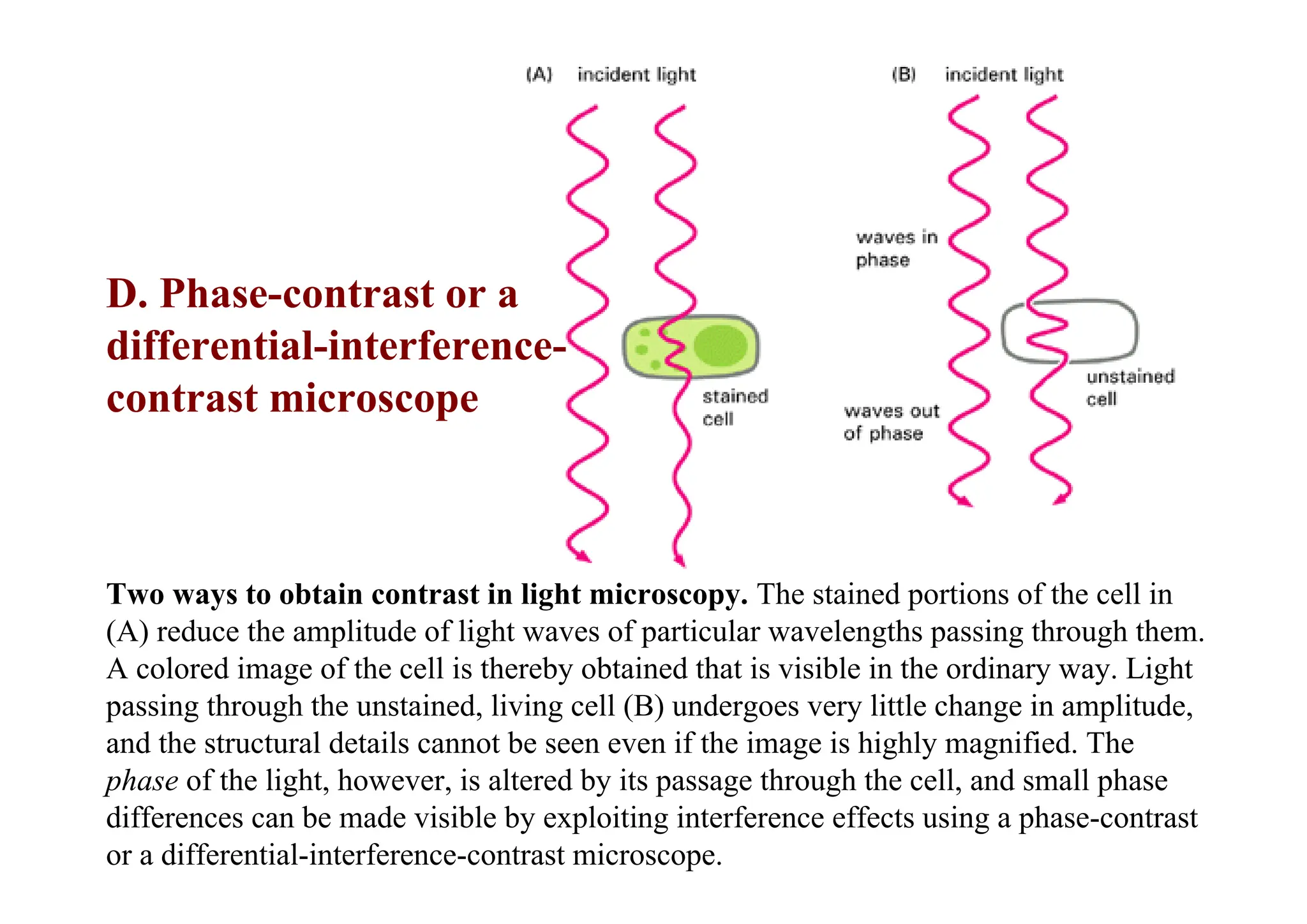
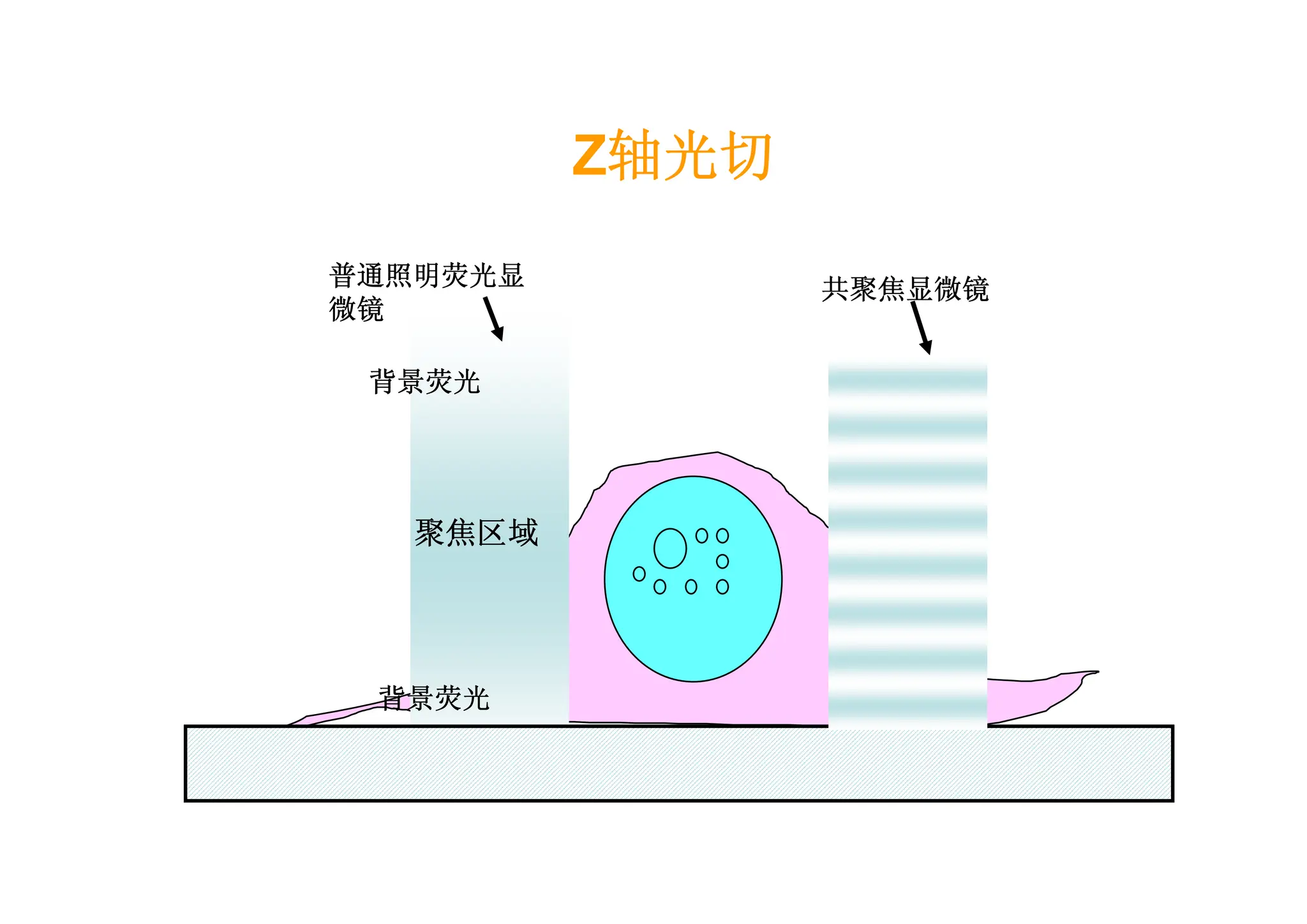
Two ways toobtain contrast in light microscopy. The stained portions of the cell in
(A) reduce the amplitude of light waves of particular wavelengths passing through them.
A colored image of the cell is thereby obtained that is visible in the ordinary way. Light
passing through the unstained, living cell (B) undergoes very little change in amplitude,
and the structural details cannot be seen even if the image is highly magnified. The
phase of the light, however, is altered by its passage through the cell, and small phase
differences can be made visible by exploiting interference effects using a phase-contrast
or a differential-interference-contrast microscope.
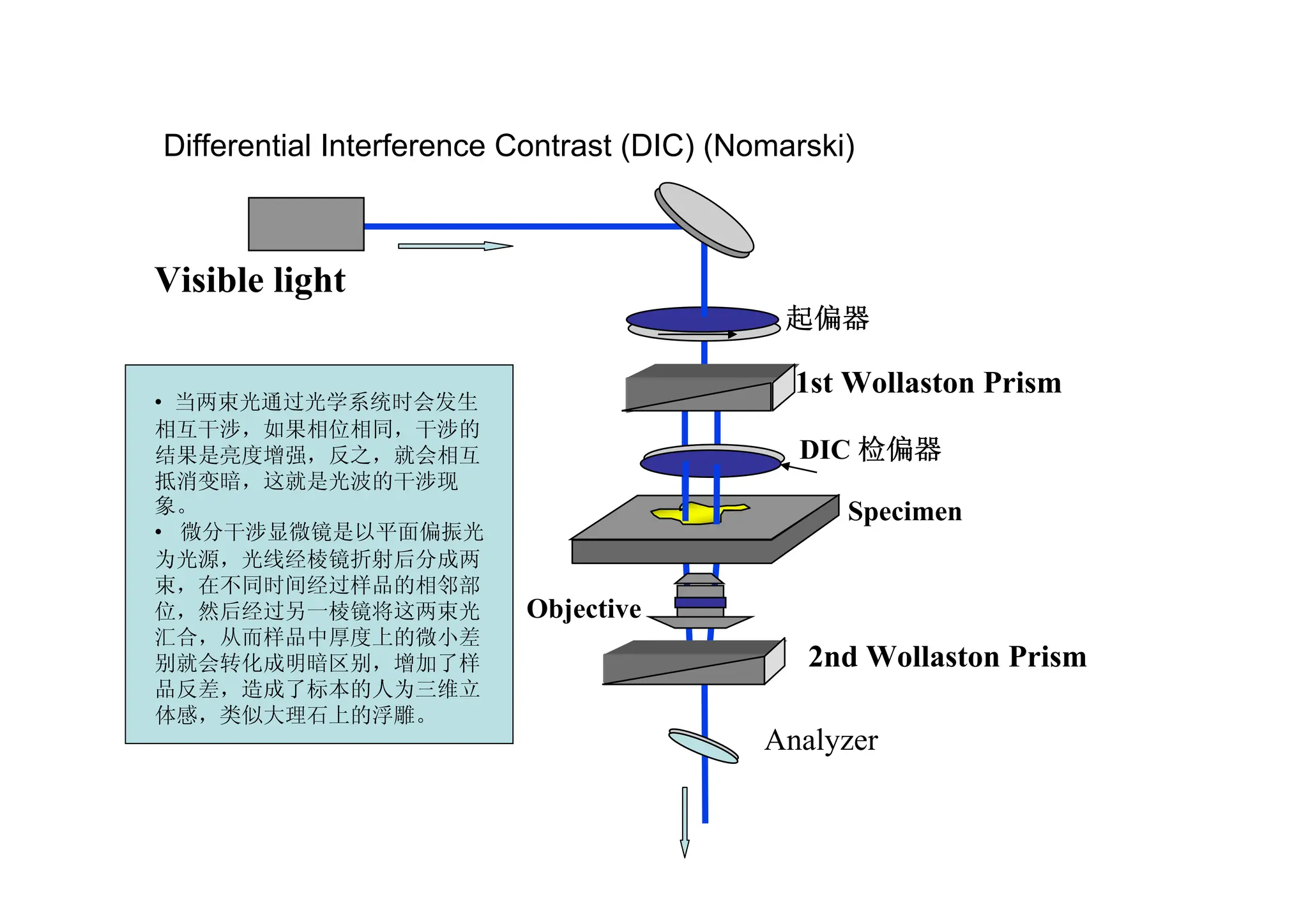
D. Phase-contrast or a
differential-interference-
contrast microscope
21.
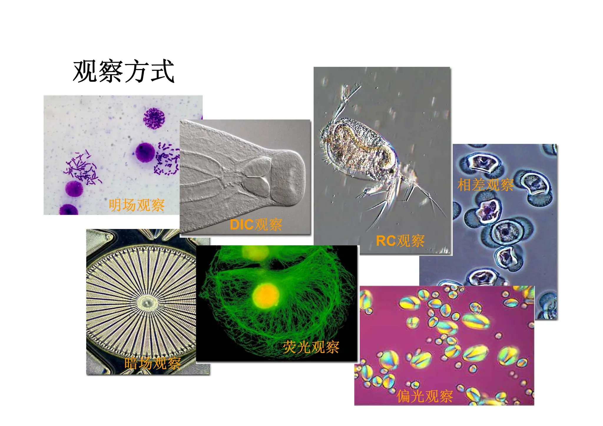
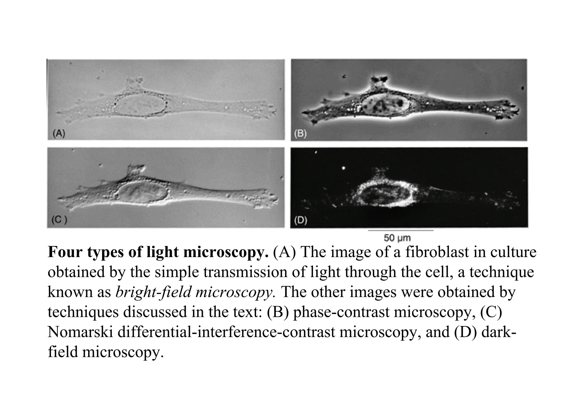
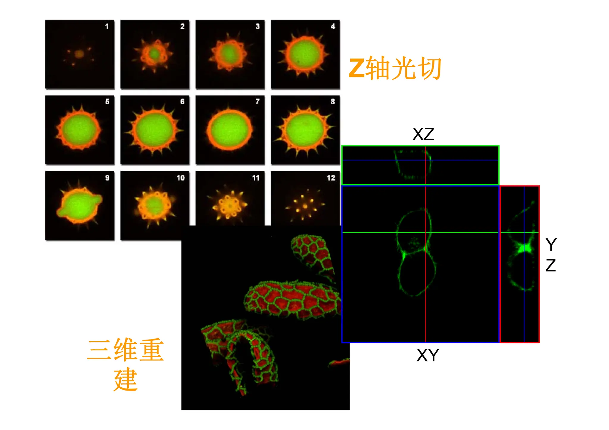
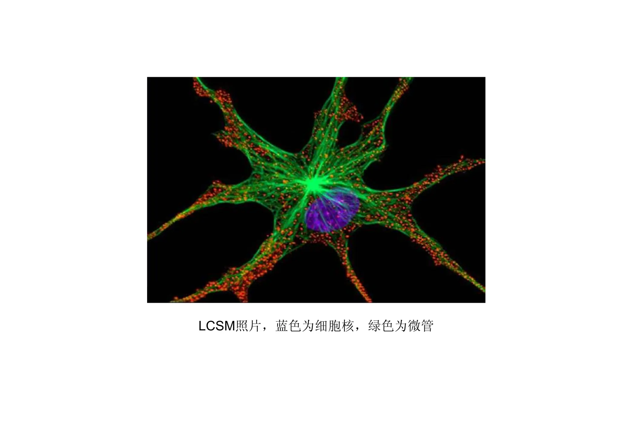
Four types oflight microscopy. (A) The image of a fibroblast in culture
obtained by the simple transmission of light through the cell, a technique
known as bright-field microscopy. The other images were obtained by
techniques discussed in the text: (B) phase-contrast microscopy, (C)
Nomarski differential-interference-contrast microscopy, and (D) dark-
field microscopy.
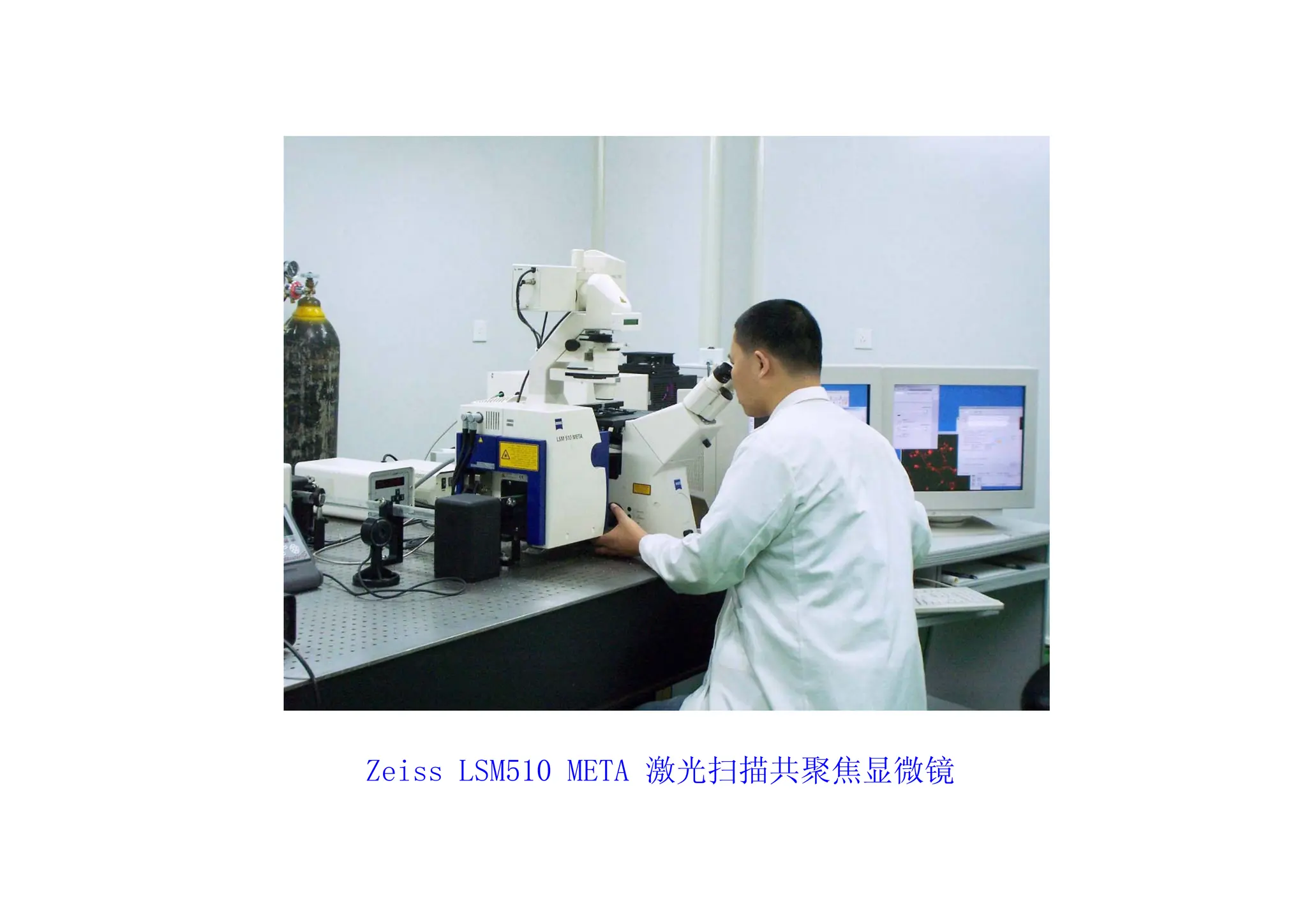
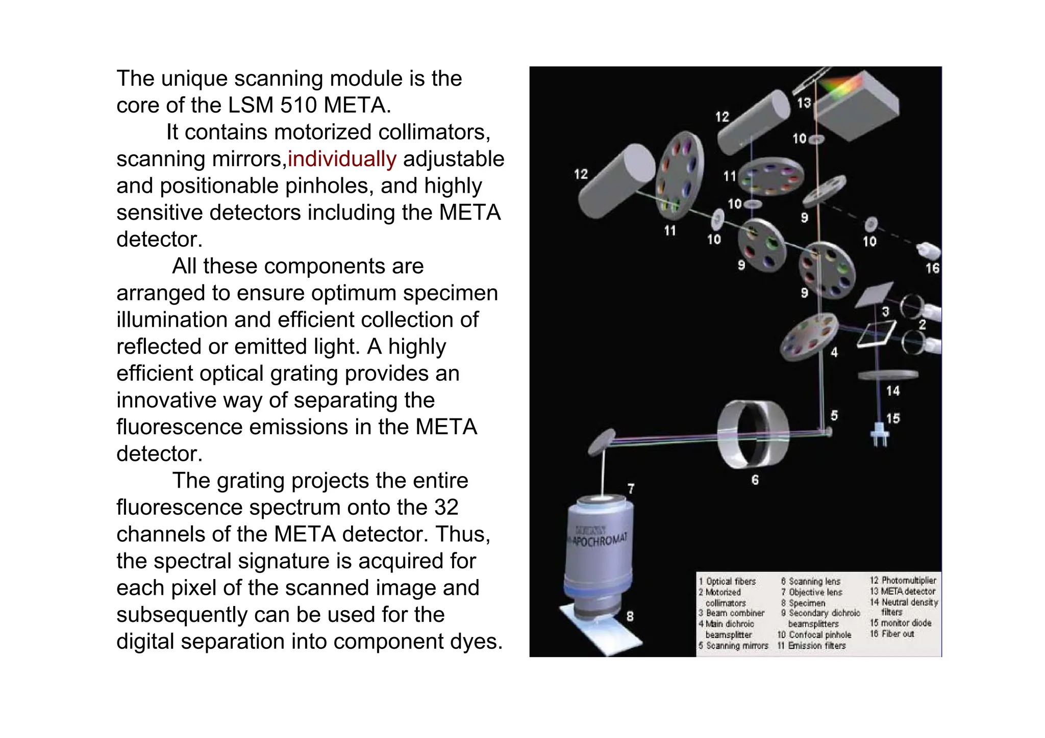
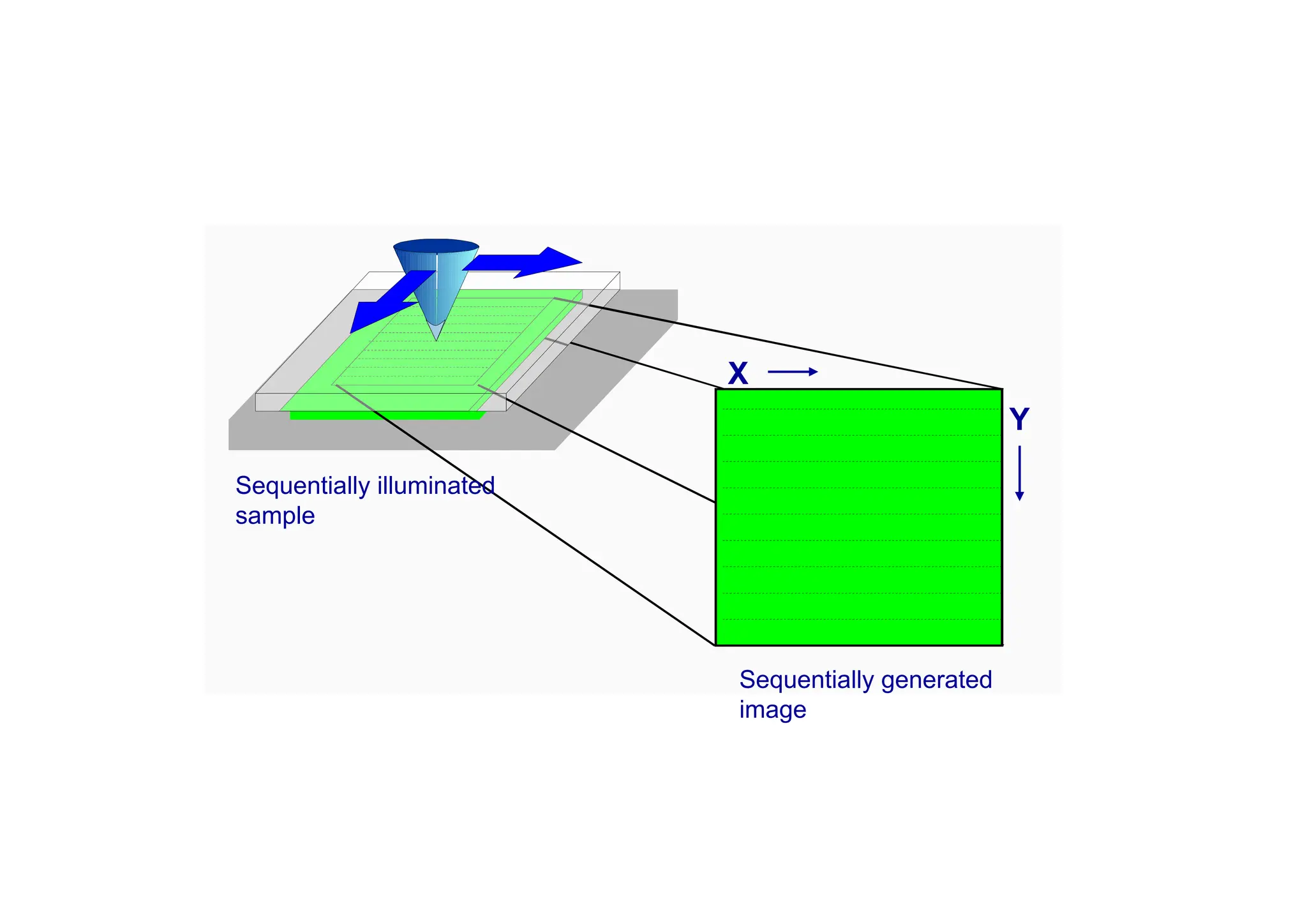
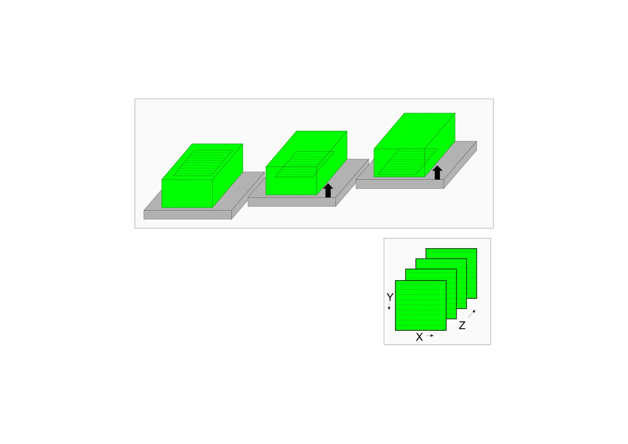
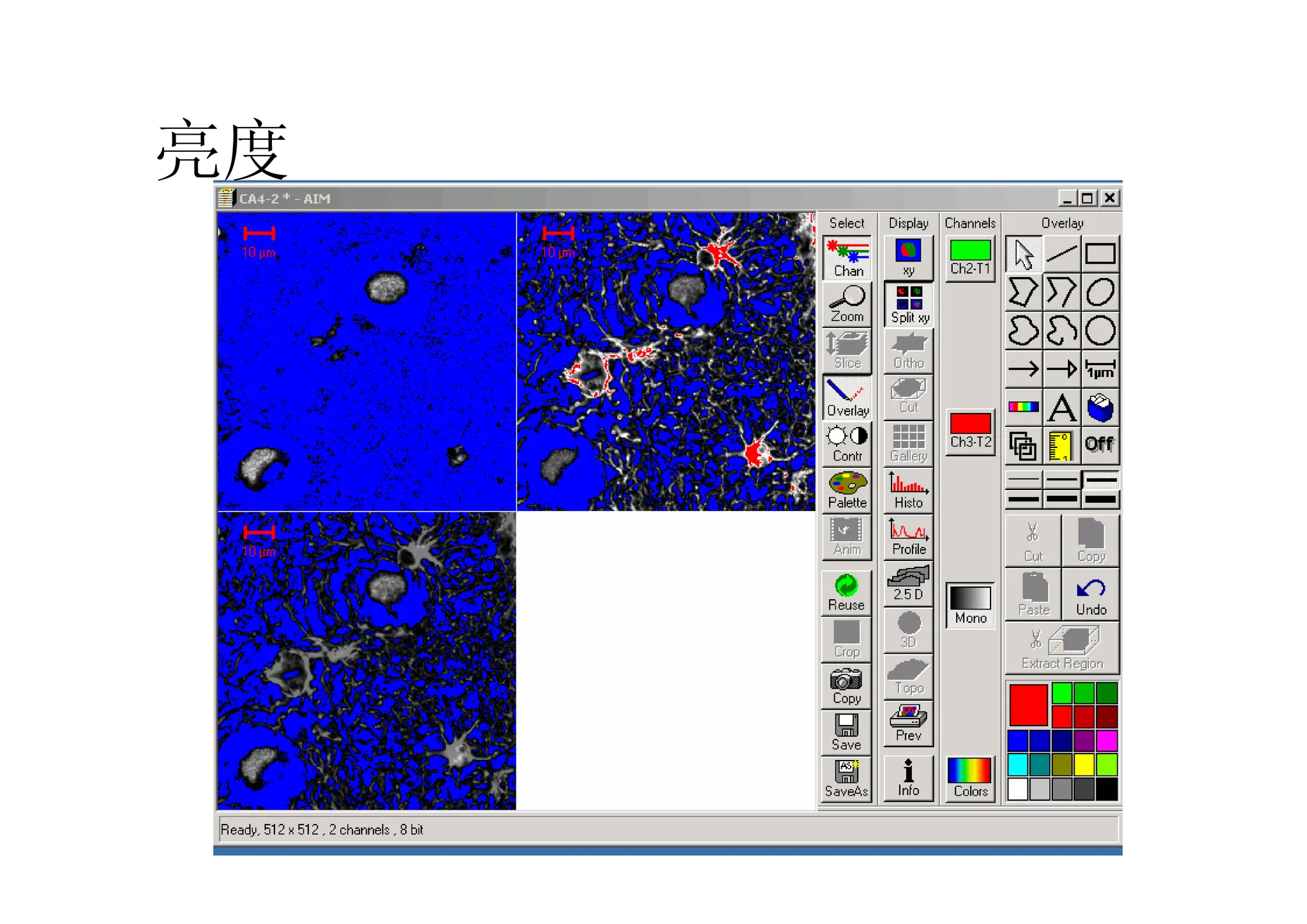
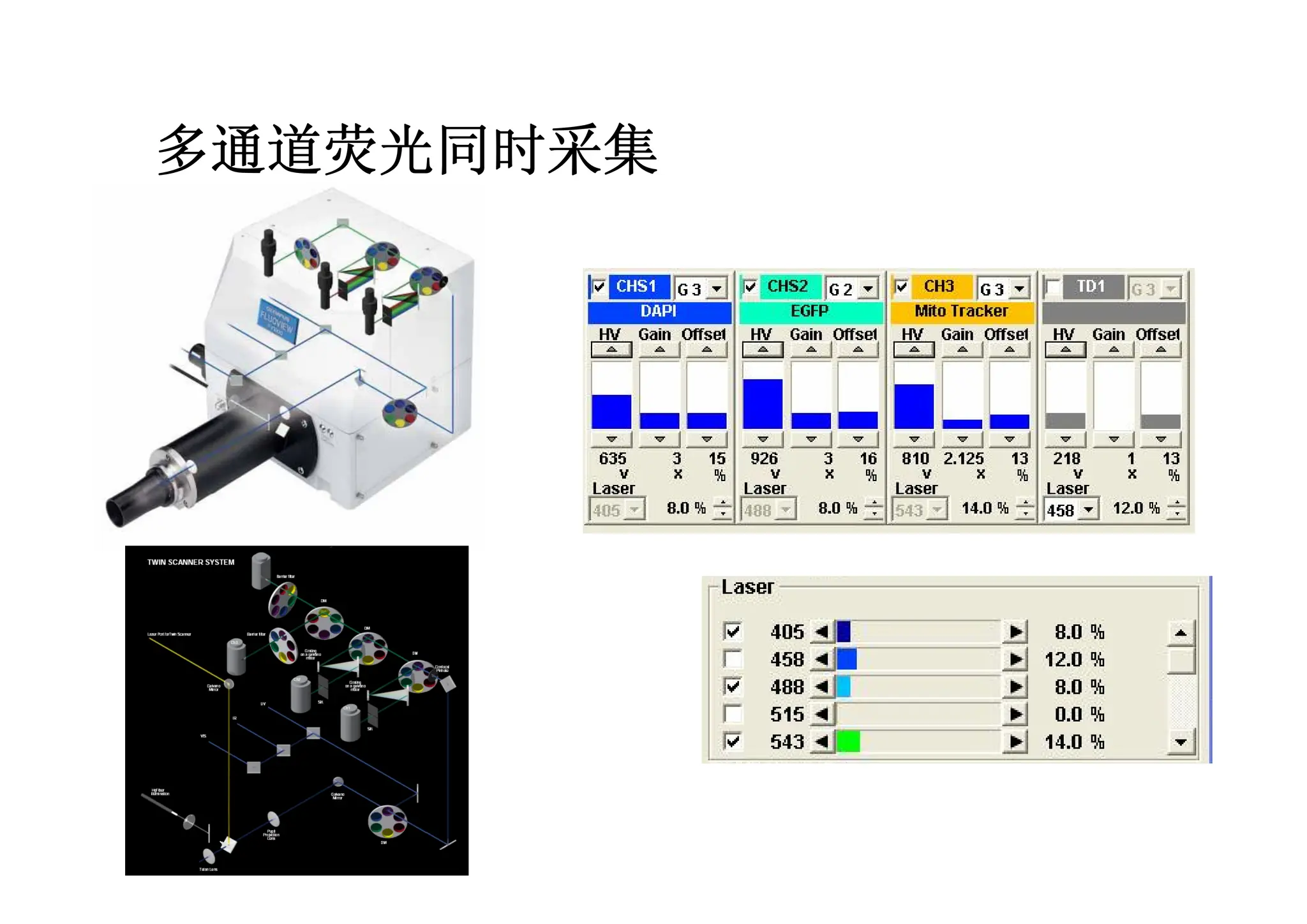
The unique scanningmodule is the
core of the LSM 510 META.
It contains motorized collimators,
scanning mirrors,individually adjustable
and positionable pinholes, and highly
sensitive detectors including the META
detector.
All these components are
arranged to ensure optimum specimen
illumination and efficient collection of
reflected or emitted light. A highly
efficient optical grating provides an
innovative way of separating the
fluorescence emissions in the META
detector.
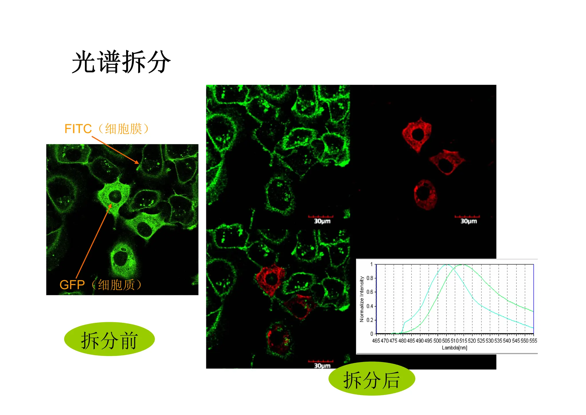
The grating projects the entire
fluorescence spectrum onto the 32
channels of the META detector. Thus,
the spectral signature is acquired for
each pixel of the scanned image and
subsequently can be used for the
digital separation into component dyes.