Test de daltonisme: Fitxes ishihara
•Download as PPT, PDF•
1 like•5,014 views
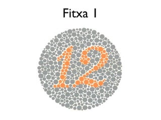
Tenim aquí un test per saber si podem patir daltonisme, si no veiem els nombres que apareixen dintre de les figures
Report
Share
Report
Share

Recommended
armd.pptx
Age Related Macular Degeneration (AMD) is a degenerative disorder affecting the macula characterized by drusen and RPE changes in the absence of other disorders. It is classified as dry or wet. Dry AMD includes early, intermediate, and dry advanced AMD. Wet AMD is neovascular and can be classic or occult. The risk increases with age, especially after age 50. Treatment includes antioxidants, lifestyle changes, and for wet AMD, anti-VEGF agents are the first line treatment administered every 4-8 weeks.
Age related macular degeneration
This document discusses age-related macular degeneration (AMD) and summarizes the findings of the AREDS study. It describes the stages of AMD from early to late dry and wet forms. The AREDS study formulation was modified to address issues like zinc absorption and carotene safety. The revised formula contains lower zinc, vitamins C and E, and lutein/zeaxanthine instead of beta-carotene. A risk scale is provided to estimate 5 and 10-year risks of advanced AMD based on risk factor points.
Age-related Macular Degeneration
Let's learn about age-related macular degeneration: Introduction, clinical types, pathogenesis & treatment. Few fundoscopic images are also added for extra learning. Happy Learning!
OPTIC DISC PIT Pathogenesis and Management
Congenital pit is an atypical coloboma usually located on the temporal edge of the disc, associated with irregular defects in the juxtapapillary choroid and pigment epithelium. Macular fibers passing through this area often are affected and corresponding changes in the retinal ganglion cell layer and in the visual field occur.
Ocular toxoplasmosis.pptx
An 8-year-old girl presented with complaints of sitting closer to the TV for 3 weeks. Her ocular examination revealed multiple patchy chorioretinal lesions in both eyes as well as a cataract in the right eye. Bloodwork was normal. The clinical presentation and bilateral lesions suggest a possible congenital infection like toxoplasmosis. Further follow up is needed to monitor the progression of the disease.
Ocular hypertension
This document discusses ocular hypertension and glaucoma. It defines glaucoma as a group of diseases involving optic nerve damage. It notes that ocular hypertension affects 4-10% of people over 40, and lists risk factors for developing glaucoma such as central corneal thickness under 555 microns. The OHTS study found that treated patients had a 1% risk per year of developing glaucoma compared to 2% for untreated patients. For treatment decisions, risks and benefits are weighed based on factors like life expectancy and tolerance. Laser trabeculoplasty may help lower pressure for some. The case study describes an unreliable 55-year-old female with risk factors like family history and lower corneal thickness who is being
ARMD 2016
ARMD should be detected and prevention should be tried , and wet armd treated with anti vegf drugs .
Glaucoma good
Glaucoma is a progressive optic neuropathy and leading cause of blindness. It is characterized by loss of retinal ganglion cells resulting in visual field loss. While elevated intraocular pressure is a major risk factor, glaucoma can occur with normal pressure as well. Diagnosis is based on visual field testing, optic nerve examination, and intraocular pressure measurement. Treatment may involve eye drop medications or surgeries like trabeculectomy and laser trabeculoplasty to lower pressure and prevent further vision loss.
Primary Angle Closure Glaucoma
This document discusses different types of angle closure conditions including primary angle closure suspect (PACS), primary angle closure (PAC), and primary angle closure glaucoma (PACG). It provides details on diagnosis, risk factors, symptoms, examination findings, treatment options including laser iridotomy, and management of acute angle closure attacks.
OCT in glaucoma ppt;1
1. Optical coherence tomography (OCT) uses light interferometry to perform high-resolution, cross-sectional imaging of the retina. It provides quantitative measurements of retinal nerve fiber layer thickness.
2. OCT images are analyzed to detect structural changes in the optic nerve head and retinal nerve fiber layer that can indicate glaucoma, often before visual field defects appear. Parameters like retinal nerve fiber layer thickness, cup-to-disc ratio, and nerve fiber layer deviation maps are used to diagnose and monitor glaucoma progression.
3. Macular ganglion cell complex thickness, which includes the retinal nerve fiber layer, ganglion cell layer, and inner plexiform layer, can also detect early glaucomatous loss
Amsler chart
The Amsler chart is used to evaluate the central 20 degrees of visual field to screen for and monitor macular diseases. It consists of a 10cm square grid with 400 smaller 5mm squares that the patient focuses on from 16 inches away while reporting any distortions, blurriness, or missing areas. There are 7 chart types including the standard high contrast grid, ones with diagonal lines, red squares, random dots, and varying line patterns to detect different visual field defects that may indicate conditions like macular edema, tumors, metamorphopsia, scotomas, or glaucoma.
Branch Retinal Vein Occlusion
This document discusses branch retinal vein occlusion (BRVO), including its pathogenesis, risk factors, clinical features, investigations, and management approaches. Some key points:
- BRVO is the second most common cause of vision loss due to retinal vascular disease, after diabetic retinopathy. It occurs most often in patients in their 50s-60s and is caused by obstruction of a branch retinal vein.
- Risk factors include hypertension, diabetes, hyperlipidemia, glaucoma, smoking, and age-related atherosclerosis. Laser treatment can help manage macular edema and neovascularization complications.
- Treatment aims to manage modifiable risk factors and sight-threatening complications like macular edema, non
Degeneración macular-Coriorretinopatia serosa central
Este documento resume dos enfermedades oculares: la degeneración macular relacionada con la edad (DMA) y la corioretinopatía serosa central. La DMA es una enfermedad progresiva y degenerativa de la macula que constituye la principal causa de pérdida de visión irreversible en personas mayores de 55 años. Puede presentarse como una forma atrófica no exudativa o como una forma exudativa neovascular. La corioretinopatía serosa central causa un desprendimiento seroso de la retina neurosensorial localizado, general
Pigment epithelial defect and intraretinal fluid
A simple and informative presentation on PED & IRF with pathophysiology, clinical examination, diagnostic imaging and one case study each for both PED & IRF
Intermeddiate & posterior uveitis dr.k.srikanth, 23.03.2016 revised
This document discusses different types of uveitis including intermediate uveitis and posterior uveitis. Intermediate uveitis presents with symptoms like floaters and deterioration of vision. Signs include anterior vitritis, snowball exudates near the ora, and peripheral periphlebitis. Posterior uveitis involves the retina and can cause symptoms such as floaters, diminished vision, and scotomas. Signs include vitritis, retinal/choroidal infiltration and exudation, and vessel sheathing. Treatment involves cycloplegia, corticosteroids, and immunosuppressants to relieve symptoms, prevent vision loss and complications.
Color blind test
This document contains a color blind test to see if individuals can differentiate between colors. It instructs those with red-green color blindness to trace lines connecting shapes of different colors, like purple and blue-green, to test whether they can distinguish between those hues. Depending on the type and severity of color blindness, the lines that can be traced may vary, such as only being able to see the purple line or seeing both lines but finding one easier to follow.
TASS vs Endophthalmitis
toxic anterior segment syndrome, TASS, endophthalmitis, purulent endophthalmitis, infection after cataract surgery, inflammation after cataract surgery, cataract extraction, phaco, phacoemulsification, eyepain, complication of cataract surgery,
Retinal detachment kalpana
This document discusses retinal detachment, specifically rhegmatogenous retinal detachment. It defines retinal detachment as the separation of the neural retina from the pigment epithelium of the retina. Rhegmatogenous retinal detachment results from a retinal break held open by vitreous traction, allowing liquefied vitreous to accumulate under the retina and separate it from the RPE. The document covers the epidemiology, risk factors, examination techniques including indirect ophthalmoscopy and ultrasound, and characteristics of rhegmatogenous retinal detachment.
Age related macular degeneration (
Age-related macular degeneration (AMD) is a common cause of vision loss in older adults. There are two main types: dry AMD, characterized by drusen and retinal pigment abnormalities, and wet AMD, characterized by abnormal blood vessel growth. Risk factors include increasing age, smoking, family history, and genetics. Diagnosis involves examination of the retina and macula using techniques like funduscopy, fluorescein angiography, and optical coherence tomography. Treatment options for wet AMD aim to stop blood vessel growth through therapies like anti-VEGF injections, photodynamic therapy, and laser photocoagulation. Lifestyle modifications and antioxidant supplements may help reduce risk of progression for dry AMD.
PRINCIPLES OF MANAGEMENT OF MACULAR HOLE
MANAGEMENT OF MACULAR HOLE, Ophthalmology presentation, eye care in the elderly , macular hole as a consequence of trauma, Vitreoretinal surgical cases, ,
Choroidal neovascular membranes (CNVM)
Choroidal neovascularization (CNV) involves the abnormal growth of new blood vessels from the choroid layer of the eye through Bruch's membrane. This can cause vision loss and is a common cause of wet macular degeneration. CNV occurs due to alterations in Bruch's membrane and high levels of vascular endothelial growth factor. It is classified based on its location relative to the retinal pigment epithelium and fovea. Symptoms include sudden vision loss and visual distortions. CNV is diagnosed through imaging like optical coherence tomography and fluorescein angiography and treated with injections of anti-VEGF drugs to inhibit blood vessel growth.
Diabetic retinopathy
Diabetic retinopathy etiology pathogenesis,classification grading diagnosis investigations management.
Common Causes of Uveitis Part1
www.ophthalclass.blogspot.com has the complete post.
In Part1 the topics discussed are the causes of anterior, intermediate, posterior and panuveitis. There is also a section on the associated features like history, demographics and examination findings that help to narrow down the differential diagnosis.
Posterior uveitis
This document discusses uveal tract diseases, specifically uveitis (inflammation of the uveal tract). It defines posterior uveitis as inflammation of the choroid. Common causes of posterior uveitis include infectious diseases like tuberculosis, fungi, viruses, and parasites, as well as non-infectious conditions like autoimmune diseases. Symptoms include vision defects, flashes of light, and positive or negative scotomas. Signs include vitreous opacities, active or healed patches of choroiditis. Choroiditis is classified based on location and number of lesions, and can lead to complications if not treated properly with corticosteroids, immunosuppressants, or specific treatments for the
Colorblindness
This document contains a table comparing the number of people who identified different keys as normal colors versus those identified by people with red-green color blindness and other types of color blindness. The table shows that people with red-green color blindness have more trouble distinguishing between red and green keys compared to those without color blindness.
Test Ishihara
This document describes the Ishihara color plates test, which contains 38 plates used to test for color blindness. Each plate is shown to individuals to identify numbers or patterns. The expected views of those with normal color vision are listed separately from the expected views of those with red-green color deficiencies. By comparing the individual's responses to the expected views for normal and deficient vision, the test can diagnose different types of color blindness.
More Related Content
What's hot
Glaucoma good
Glaucoma is a progressive optic neuropathy and leading cause of blindness. It is characterized by loss of retinal ganglion cells resulting in visual field loss. While elevated intraocular pressure is a major risk factor, glaucoma can occur with normal pressure as well. Diagnosis is based on visual field testing, optic nerve examination, and intraocular pressure measurement. Treatment may involve eye drop medications or surgeries like trabeculectomy and laser trabeculoplasty to lower pressure and prevent further vision loss.
Primary Angle Closure Glaucoma
This document discusses different types of angle closure conditions including primary angle closure suspect (PACS), primary angle closure (PAC), and primary angle closure glaucoma (PACG). It provides details on diagnosis, risk factors, symptoms, examination findings, treatment options including laser iridotomy, and management of acute angle closure attacks.
OCT in glaucoma ppt;1
1. Optical coherence tomography (OCT) uses light interferometry to perform high-resolution, cross-sectional imaging of the retina. It provides quantitative measurements of retinal nerve fiber layer thickness.
2. OCT images are analyzed to detect structural changes in the optic nerve head and retinal nerve fiber layer that can indicate glaucoma, often before visual field defects appear. Parameters like retinal nerve fiber layer thickness, cup-to-disc ratio, and nerve fiber layer deviation maps are used to diagnose and monitor glaucoma progression.
3. Macular ganglion cell complex thickness, which includes the retinal nerve fiber layer, ganglion cell layer, and inner plexiform layer, can also detect early glaucomatous loss
Amsler chart
The Amsler chart is used to evaluate the central 20 degrees of visual field to screen for and monitor macular diseases. It consists of a 10cm square grid with 400 smaller 5mm squares that the patient focuses on from 16 inches away while reporting any distortions, blurriness, or missing areas. There are 7 chart types including the standard high contrast grid, ones with diagonal lines, red squares, random dots, and varying line patterns to detect different visual field defects that may indicate conditions like macular edema, tumors, metamorphopsia, scotomas, or glaucoma.
Branch Retinal Vein Occlusion
This document discusses branch retinal vein occlusion (BRVO), including its pathogenesis, risk factors, clinical features, investigations, and management approaches. Some key points:
- BRVO is the second most common cause of vision loss due to retinal vascular disease, after diabetic retinopathy. It occurs most often in patients in their 50s-60s and is caused by obstruction of a branch retinal vein.
- Risk factors include hypertension, diabetes, hyperlipidemia, glaucoma, smoking, and age-related atherosclerosis. Laser treatment can help manage macular edema and neovascularization complications.
- Treatment aims to manage modifiable risk factors and sight-threatening complications like macular edema, non
Degeneración macular-Coriorretinopatia serosa central
Este documento resume dos enfermedades oculares: la degeneración macular relacionada con la edad (DMA) y la corioretinopatía serosa central. La DMA es una enfermedad progresiva y degenerativa de la macula que constituye la principal causa de pérdida de visión irreversible en personas mayores de 55 años. Puede presentarse como una forma atrófica no exudativa o como una forma exudativa neovascular. La corioretinopatía serosa central causa un desprendimiento seroso de la retina neurosensorial localizado, general
Pigment epithelial defect and intraretinal fluid
A simple and informative presentation on PED & IRF with pathophysiology, clinical examination, diagnostic imaging and one case study each for both PED & IRF
Intermeddiate & posterior uveitis dr.k.srikanth, 23.03.2016 revised
This document discusses different types of uveitis including intermediate uveitis and posterior uveitis. Intermediate uveitis presents with symptoms like floaters and deterioration of vision. Signs include anterior vitritis, snowball exudates near the ora, and peripheral periphlebitis. Posterior uveitis involves the retina and can cause symptoms such as floaters, diminished vision, and scotomas. Signs include vitritis, retinal/choroidal infiltration and exudation, and vessel sheathing. Treatment involves cycloplegia, corticosteroids, and immunosuppressants to relieve symptoms, prevent vision loss and complications.
Color blind test
This document contains a color blind test to see if individuals can differentiate between colors. It instructs those with red-green color blindness to trace lines connecting shapes of different colors, like purple and blue-green, to test whether they can distinguish between those hues. Depending on the type and severity of color blindness, the lines that can be traced may vary, such as only being able to see the purple line or seeing both lines but finding one easier to follow.
TASS vs Endophthalmitis
toxic anterior segment syndrome, TASS, endophthalmitis, purulent endophthalmitis, infection after cataract surgery, inflammation after cataract surgery, cataract extraction, phaco, phacoemulsification, eyepain, complication of cataract surgery,
Retinal detachment kalpana
This document discusses retinal detachment, specifically rhegmatogenous retinal detachment. It defines retinal detachment as the separation of the neural retina from the pigment epithelium of the retina. Rhegmatogenous retinal detachment results from a retinal break held open by vitreous traction, allowing liquefied vitreous to accumulate under the retina and separate it from the RPE. The document covers the epidemiology, risk factors, examination techniques including indirect ophthalmoscopy and ultrasound, and characteristics of rhegmatogenous retinal detachment.
Age related macular degeneration (
Age-related macular degeneration (AMD) is a common cause of vision loss in older adults. There are two main types: dry AMD, characterized by drusen and retinal pigment abnormalities, and wet AMD, characterized by abnormal blood vessel growth. Risk factors include increasing age, smoking, family history, and genetics. Diagnosis involves examination of the retina and macula using techniques like funduscopy, fluorescein angiography, and optical coherence tomography. Treatment options for wet AMD aim to stop blood vessel growth through therapies like anti-VEGF injections, photodynamic therapy, and laser photocoagulation. Lifestyle modifications and antioxidant supplements may help reduce risk of progression for dry AMD.
PRINCIPLES OF MANAGEMENT OF MACULAR HOLE
MANAGEMENT OF MACULAR HOLE, Ophthalmology presentation, eye care in the elderly , macular hole as a consequence of trauma, Vitreoretinal surgical cases, ,
Choroidal neovascular membranes (CNVM)
Choroidal neovascularization (CNV) involves the abnormal growth of new blood vessels from the choroid layer of the eye through Bruch's membrane. This can cause vision loss and is a common cause of wet macular degeneration. CNV occurs due to alterations in Bruch's membrane and high levels of vascular endothelial growth factor. It is classified based on its location relative to the retinal pigment epithelium and fovea. Symptoms include sudden vision loss and visual distortions. CNV is diagnosed through imaging like optical coherence tomography and fluorescein angiography and treated with injections of anti-VEGF drugs to inhibit blood vessel growth.
Diabetic retinopathy
Diabetic retinopathy etiology pathogenesis,classification grading diagnosis investigations management.
Common Causes of Uveitis Part1
www.ophthalclass.blogspot.com has the complete post.
In Part1 the topics discussed are the causes of anterior, intermediate, posterior and panuveitis. There is also a section on the associated features like history, demographics and examination findings that help to narrow down the differential diagnosis.
Posterior uveitis
This document discusses uveal tract diseases, specifically uveitis (inflammation of the uveal tract). It defines posterior uveitis as inflammation of the choroid. Common causes of posterior uveitis include infectious diseases like tuberculosis, fungi, viruses, and parasites, as well as non-infectious conditions like autoimmune diseases. Symptoms include vision defects, flashes of light, and positive or negative scotomas. Signs include vitreous opacities, active or healed patches of choroiditis. Choroiditis is classified based on location and number of lesions, and can lead to complications if not treated properly with corticosteroids, immunosuppressants, or specific treatments for the
What's hot (20)
Degeneración macular-Coriorretinopatia serosa central
Degeneración macular-Coriorretinopatia serosa central
Intermeddiate & posterior uveitis dr.k.srikanth, 23.03.2016 revised
Intermeddiate & posterior uveitis dr.k.srikanth, 23.03.2016 revised
Viewers also liked
Colorblindness
This document contains a table comparing the number of people who identified different keys as normal colors versus those identified by people with red-green color blindness and other types of color blindness. The table shows that people with red-green color blindness have more trouble distinguishing between red and green keys compared to those without color blindness.
Test Ishihara
This document describes the Ishihara color plates test, which contains 38 plates used to test for color blindness. Each plate is shown to individuals to identify numbers or patterns. The expected views of those with normal color vision are listed separately from the expected views of those with red-green color deficiencies. By comparing the individual's responses to the expected views for normal and deficient vision, the test can diagnose different types of color blindness.
Ishihara.14.plate.instructions
This document provides instructions for using the Ishihara Color Vision Test, which consists of a series of plates designed to test for color deficiencies. It summarizes:
- The test uses plates that people with normal color vision see numbers or patterns in, while those with red-green deficiencies may see something different or nothing at all.
- The results are analyzed to determine if the subject's color vision is normal, deficient, or if they have total color weakness or blindness. Most common are red-green deficiencies which are divided into protan and deutan types.
- Plates 1-11 are used initially to separate those with normal versus deficient color vision. The document also provides details on what different subjects should
Dvorine Color Test offered by Operture.com
Dvorine pseudo-isochromatic plates is an approved test for color deficiency. Only available at http://www.operture.com/shopping/product/2253-dvorine-pseudoisochromatic-plates-color-vision-test?search=dvorine
Daltonismo francisco
O documento descreve o daltonismo, uma perturbação genética da visão colorida caracterizada pela falta de reconhecimento de uma ou mais cores. Foi descoberto em 1794 por John Dalton, que sofria da anomalia. É herdado através do cromossomo X, portanto afeta mais homens do que mulheres. O teste de Ishihara é usado para detectar o daltonismo comparando a capacidade de identificar números em imagens coloridas.
Daltonismo e hemofilia
O documento discute a herança genética da hemofilia, uma doença ligada ao cromossomo X. Explica que Fernando é hemofílico apesar de seus pais normais Jorge e Clara, pois o bisavô de Jorge era hemofílico, indicando que Clara está errada em acreditar que a condição de Fernando veio de Jorge.
DALTONISMO
El daltonismo es un defecto genético que dificulta la distinción de colores. Se hereda por un gen recesivo en el cromosoma X. Existen varios tipos de daltonismo dependiendo del cono afectado, incluyendo la ceguera al color rojo, verde o azul, o una visión anómala de los colores. El daltonismo se diagnostica mediante pruebas con cartas Ishihara.
Genética - Daltonismo | Biologia 12º Ano
O documento discute a determinação do sexo humano e hereditariedade ligada ao sexo. O sexo é determinado pelos cromossomos X e Y, com mulheres tendo dois X e homens um X e um Y. A hereditariedade ligada ao sexo envolve genes no cromossomo X, com traços passados de mães para filhos de forma diferente de pais para filhas. O daltonismo é dado como exemplo de hereditariedade ligada ao sexo.
Colour Blindness Ishihara Charts
Color blindness, also known as color vision deficiency, is the inability to perceive differences between some colors that others can distinguish. It is often genetic but can also be caused by eye, nerve, or brain damage or chemical exposure. The most common types are red-green deficiencies but it is also possible to be deficient in blue perception or see only in black and white. Color blindness is diagnosed using tests that show numbers or patterns embedded in color plates that those with normal color vision can see but those who are color deficient may not be able to distinguish.
Tipos de daltonismo
El daltonismo es una anomalía permanente donde el ojo tiene dificultades para ver los colores de forma habitual debido a problemas con los pigmentos en las neuronas del ojo conocidas como conos. Los dos tipos más comunes son la dificultad para diferenciar entre rojo y verde, o entre azul y amarillo, dependiendo de qué pigmento falta. El tipo más serio es la acromatopsia donde todo se ve en tonos grises y no se pueden observar colores.
DALTONISMO
La exposición contiene introducción, tipos, genética, y diagnóstico. Está muy completa sobre el tema.
Test de Ishihara
Este documento presenta las 24 imágenes del test de Ishihara para la detección del daltonismo. Cada imagen muestra los números o figuras que pueden ser vistas por personas con visión normal, protanomalía, deuteranomalía o tritanomalía. El propósito es identificar posibles deficiencias en la percepción del color rojo, verde o azul a través de la incapacidad de distinguir los patrones de color en las placas Ishihara.
daltonismo
power point sobre el daltonismo sus tipos y caracteristicas sobre esta enfermedad hereditaria
la extensión esta en .potx cambienla a ppt :)
Daltonismo
O documento discute o daltonismo, uma perturbação genética na percepção de cores que afeta principalmente a diferenciação entre vermelho e verde. O distúrbio ocorre com mais frequência em homens e pode ser herdado. Existem testes para diagnosticar o grau em que a percepção de cores é afetada em cada pessoa.
Daltonismo
O Daltonismo é uma alteração na percepção de cores causada por deficiência nas células cones da retina responsáveis pela visão do vermelho e verde. É diagnosticado através do teste de Ishihara ou do Anomaloscópio de Nagel, e não tem cura definitiva, embora lentes com filtros seletivos possam melhorar o contraste. Afecta mais homens devido ser ligado ao cromossoma X.
Soal Tes Potensi Akademik (TPA)
Silahkan di download untuk latihan soal-soal TPA.
Semoga Bermanfaat.
Daltonismo
El daltonismo es una enfermedad hereditaria ligada al cromosoma X que causa dificultad para distinguir colores. Afecta aproximadamente al 8% de los hombres y 0.5% de las mujeres. Existen varios tipos de daltonismo dependiendo del pigmento defectuoso en la retina, incluyendo daltonismo acromático, monocromático y dicromático. No existe tratamiento para curar la enfermedad, aunque lentes especiales pueden ayudar.
Color Vision Testing
The document discusses color vision testing techniques and color deficiencies. It describes various color vision tests including pseudoisochromatic plates, lantern tests, arrangement tests, and anomaloscopes. It explains what each test screens for and its purpose. It also discusses the different types of color deficiencies including red, green, blue deficiencies and how color may appear to those with deficiencies.