1. Time Course for the Expression of the Ets-1
Transcriptional Regulator in Rats Following
Traumatic Brain Injury
Kayla Ruby Arroyo1, Gregory Ford2
1Spelman College Biology Department
2Biology Department, Morehouse College
<
Introduction
Methods
Surgical Procedure
Traumatic brain injury (TBI) is a significant health issue
predicted to become the third leading cause of disease burden
globally by 2030. It is estimated that ~3 million persons in the
United States are living with long-term or lifelong effects of
traumatic brain injury (TBI). Approximately 1.7 million new TBIs
are sustained each year resulting in 53,000 deaths.
TBI results from an insult to the brain caused by an external
force (i.e. car accident, fall, motor vehicle accident). The
neurological injury associated with TBI occurs in two stages,
commonly termed primary and secondary injury. Primary injury
is the initial injury to brain tissue, caused by mechanical
disruption . This leads to a secondary injury mediated by
inflammation. Inflammation is the body’s normal defense
against foreign invasion or tissue damage. While this process is
designed to be beneficial in keeping the body healthy and re-
establishing homeostasis, during TBI this process becomes
uncontrolled leading to cell death. The inflammatory cascade is
commonly initiated by the alteration of transcription factors,
influencing groups of genes.
In a previous study, the lab found a number of transcription
factors that increased in RNA expression following TBI. The
goal of this study to examine the protein expression of two of
these transcription factors, Oct-1 and Ets-1, in the rat brain
following TBI. This study will give incite into the transcriptional
regulation of inflammation following TBI.
Adult rats received a unilateral
controlled cortical impact
(CCI) and were sacrificed 3,
12 and 24h post-injury. The
ipsilateral hemi-brain tissue at
the site of the injury, the
corresponding contralateral
hemi-brain tissue, and naïve
(control) brain tissue were
used for histological analysis.
Controlled Cortical impact Device
D. F.E.
Conclusion
Acknowledgments
This research was made possible with the sponsorship of Spelman
College, MBRS-RISE and the NSF. Special thanks to Dr. Bryon Ford
and the Fordlab at Morehouse School of Medicine where this work
was performed.
After the TBI on the injured side, the time points of 3hr, 12hr,
and 24hr increased Ets-1 expression level. Therefore, the
results of this study that show an increase expression of Ets-1
could lead to the identification of Ets-1 and other transcription
factors as transcriptional regulators of inflammation following
trauma.
• Test for additional time points (6hr, 72hr, and 1week)
• Test other transcriptional factors including Oct-1 and Stat5,
which were also shown to increase in RNA expression in
previous studies.
• To quantify the increase expression level of Ets-1 at the
3hr, 12hr, and 24hr time points.
References
1. Zaloshnja E, Miller T, Langlois JA, Selassie AW: Prevalence of long-term
disability from traumatic brain injury in the civilian population of the United
States, 2005. J Head Trauma Rehabil 2008, 23(6):394–400.
2. Faul M, Xu L, Wald MM, Coronado VG: Traumatic brain injury in the United
States: emergency department visits, hospitalizations and deaths 2002–2006.
Atlanta (GA): Centers for Disease Control and Prevention, National Center for
injury Prevention and Control; 2010.
3. Coronado VG, Xu L, Basavaraju SV, McGuire LC, Wald MM, Faul MD,
Guzman BR, Hemphill JD: Surveillance for traumatic brain injury-related
deaths–United States, 1997–2007. MMWR Surveill Summ 2011, 60(5):1–32.
4. Selassie AW, Zaloshnja E, Langlois JA, Miller T, Jones P, Steiner C:
Incidence of long-term disability following traumatic brain injury
hospitalization,United States, 2003. J Head Trauma Rehabil 2008, 23(2):123–
131.
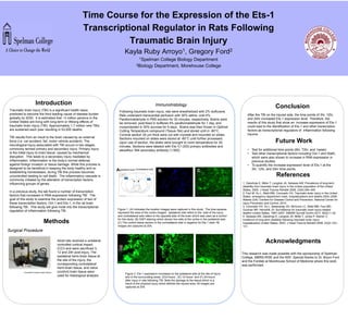
Figure 2. Ets-1 expression increased on the ipsilateral side at the site of injury
and in the surrounding areas (D)3-hours , (E) 12-hours and (F) 24-hours
after injury in rats following TBI. Note the damage to the tissue which is a
result of the physical injury which defines the injured area. All images are
captured at 20X.
Immunohistology
Following traumatic brain injury, rats were anesthetized with 2% isoflurane.
Rats underwent transcardial perfusion with 30% saline, cold 4%
Paraformaldehyde in PBS solution for 30 minutes, respectively. Brains were
be removed; post-fixed in buffered 4% paraformaldehyde for 1 day, and
cryoprotected in 30% sucrose for 5 days. Brains was then frozen in Optimal
Cutting Temperature compound (Tissue-Tek) and stored until in -80°C.
Coronal section 20 µm thick were cut with cryostat and mounted on slides.
Sections mounted on slides were stored at -80°C until further processed.
Upon use of section, the slides were brought to room temperature for 30
minutes. Sections were labeled with Ets-1(1:200) primary antibodies and
alexaflour 594 secondary antibody (1:500).
Ipsilateral side
Figure 1. (A) Indicates the location images were captured in this study. The blue squares
represent the area of the cortex imaged. Ipsilateral side refers to the side of the injury
and contralateral side refers to the opposite side of the brain which was used as a control
for this study. (B) DAPI staining which shows live cells at the cortex in the ipsilateral side.
(C) The control tissue section in the contralateral side in negative for Ets-1 stain. All
images are captured at 20X.
A. B. C.Contralateral side
Future Work