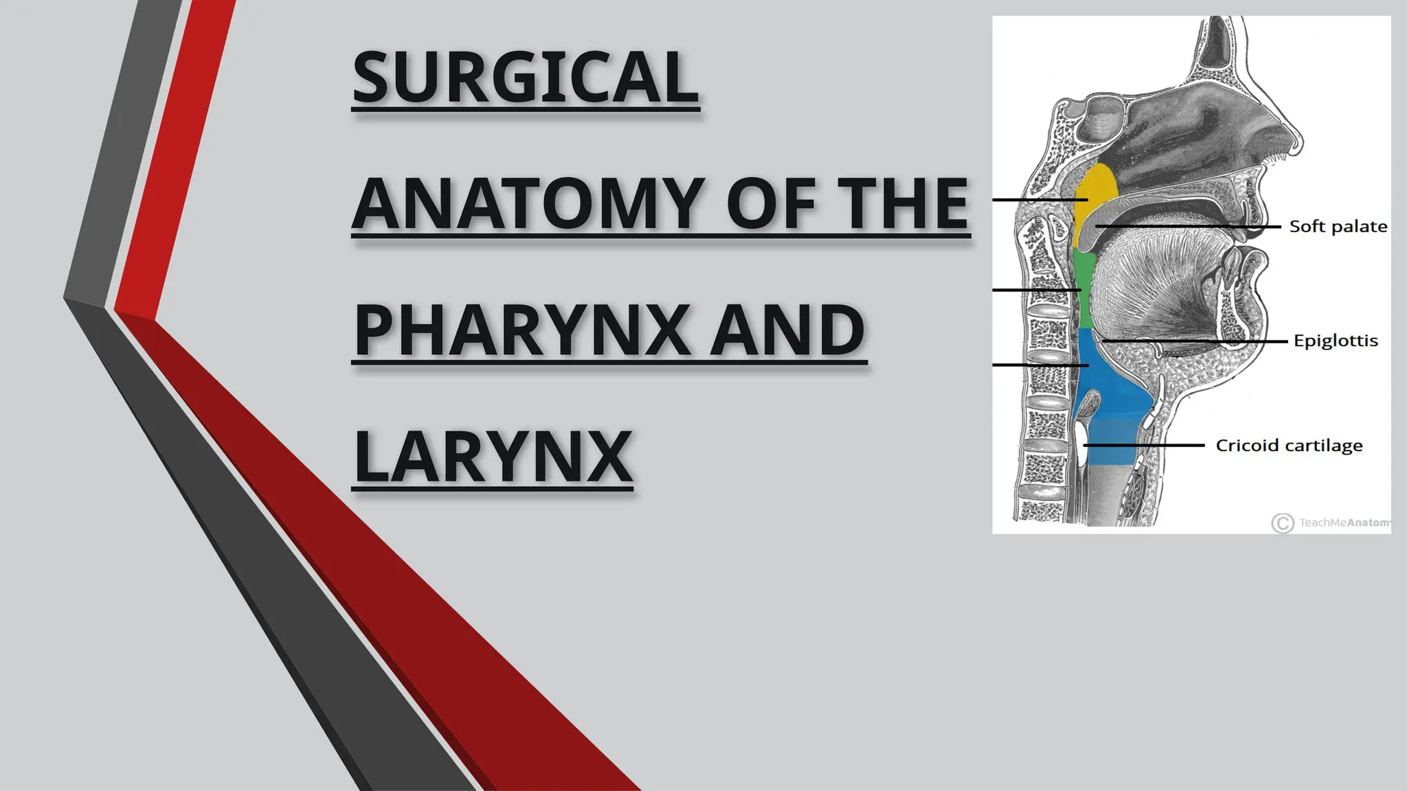
SURGICAL
ANATOMY OF THE
PHARYNX AND
LARYNX
CONTENTS
 Introduction
 Embryology of pharynx and larynx
 Anatomy of pharynx
 Anatomy of larynx
 Physiology of pharynx and larynx
 Examination
 Applied aspects
 Developmental anomalies
 Pharyngocutaneous fistula
 Laryngeal trauma
 Space infections
 Velopharyngeal insufficiency
 Obstructive sleep apnea
 Orthognathic surgery and pharyngeal space.
 Eagles syndrome.
 Zenker’s diverticulum.
Airway assessment and
management
Tracheostomy
Cricothyrotomy
Approaches to the
oropharynx
Conclusion
References
INTRODUCTION
PHARYNX
EMBRYOLOGY OF PHARYNX
 The primitive pharynx forms in the late
embryonic period as a dilation of the cranial
end of the foregut, lying between the
developing heart ventrally and the
chondrocranium rostrodorsally.
 The early pharynx is large relative to the
rest of the gut, is flattened ventrodorsally,
and gives rise to diverse structures from its
floor and side walls.
 The lateral aspects of the primitive pharynx
project a series of pouches between the
pharyngeal arches.
 The endodermal lining of the primitive
pharynx develops gradually from a
polyhedral cuboidal embryonic epithelium
into a respiratory mucous membrane.
 The cricothyroid and the constrictors
of the pharynx, palatopharyngeus
develop from occipital somites 2& 4.
 The third pharyngeal arch muscle ,
the stylopharyngeus, is derived from
seventh somitomere.
 The artery of the pharynx are derived
from the third arch artery.
Anatomy of pharynx
The wall of the pharynx
1. Mucosa & submucosa
2. Pharyngobasilar fascia
3. Longitudinal muscles
4. Circular muscles –
constrictors
5. Pharyngeal plexus of veins
& nerves
6. Buccopharyngeal fascia
Nasal part: the mucosa is
ciliated and resembles the
mucosa of the nose. It also
contains lymphoid nodules
that constitute the pharyngeal
tonsil (adenoids).
In the remainder of the
pharynx, the epithelium is
stratified squamous and the
mucosa is tightly attached to
the pharyngobasilar fascia
Mucous layer
MUSCLES OF PHARYNX
It consists of three
overlapping
constrictors:
Three
longitudinally
directed
muscles:
Origin: medial pterygoid plate,
pterygomandibular raphe, alveolar
process
Four parts-
Some fibers are attached to the
palatine aponeurosis and arise with
palatopharyngeal fibers. These fibers
as important in the formation of the
ridge of Passavant and they are
called the palatopharyngeal
sphincter.
The ridge of Passavant develops from
the passive folding of the pharyngeal
wall brought about by the elevation
of the pharynx through the
SUPERIOR CONSTRICTOR
MIDDLE CONSTRICTOR
Fan shaped muscle
Origin:
upper Fan-shaped border of the
greater cornu of the hyoid bone, from
the lesser cornu, and from the
stylohyoid ligament
Insertion:
Posterior median fibrous raphe
blending in the middle line with the
muscle of the opposite side.
INFERIOR CONSTRICTOR
Origin:
Thyropharyngeus arises from oblique line
of thyroid lamina, and by a small slip from
inferior cornu
Cricopharyngeus arises from the side of
the cricoid cartilage between attachment
of cricothyroid and articular facet for
inferior thyroid cornu
Insertion:
The upper part attaches to the median
raphe while the lower part forms a
circular band that lacks a median raphe
LONGITUDINAL MUSCLES
 Palatopharyngeus descends
from the sides of the palate and
runs longitudinally on the inner
aspect of constrictors.
 Salpingopharyngeus descends
from the auditory tube to merge
with the palatopharyngeus.
 Stylopharyngeus arises from
styloid process. Passes through
gap b/w sup. and middle
constrictor to run downwards on
the inner surface of middle and
inf. Constrictors
GAP BETWEEN PHARYGEAL MUSCLES &
STRUCTURES RELATED TO THEM
 SINUS OF MORGAGNI.
Structures- auditory tube, levator veli
palatine, ascending palatine artery.
 THE SUPERIOR AND MIDDLE
CONSTRICTORS:
Structures- Stylopharyngeus muscle and
glossopharyngeal N.
 THE MIDDLE AND INFERIOR
CONSTRICTOR muscle- internal
laryngeal N and superior laryngeal
vessels.
PARTS OF PHARYNX
 NASOPHARYNX: Lying behind
nasal fossae and above soft palate.
 OROPHARYNX: Lying behind
anterior pillars of fauces.
 LARYNGOPHARYNX: Lying behind
larynx.
 Lies behind the nasal cavities, above the soft palate.
`` Roof :
 It is formed by body of the sphenoid and the basilar part of the
occipital bone.
 A collection of lymphoid tissue, called the pharyngeal tonsil is present
in the submucosa of this region.
 Anterior wall :shows posterior nasal apertures
 Posterior wall :the anterior arch of the atlas.
 Lateral wall:
 On each side has the pharyngeal opening of the auditory tube.
 The posterior margin of the tube forms an elevation called the tubal
elevation.
 The salpingopharyngeus muscle produces a vertical fold of mucous
membrane called the salpingopharyngeal fold
Floor :
 soft palate
 pharyngeal isthmus.
NASOPHARYNX
PHARYNGEAL RECESS
 It is a small depression
in the lateral wall
behind the tubal
elevation.
 A collection of lymphoid
tissue in the submucosa
behind the opening of
the auditory tube is
called the Tubal tonsil.
PHARYNGEAL TONSIL
 It is a collection of lymphoid tissue
present in the wall of the pharyngeal
recess and the roof of nasal pharynx.
 Seen in children and sometimes in
adults.
 Enlargement of the pharyngeal
tonsil may interfere with the
nasal breathing & speech.
 It may obstruct the auditory
tube and lead to deafness
because of subsequent
absorption of air from the
tympanic cavity.
OROPHARYNX
In front:
communicates with the oral cavity through
orophayngeal isthmus.
Below:
it opens into the laryngophaynx at the level
of the upper border of epiglottis.
lateral wall :
palatopharyngeal arch , palatoglossal arch
and palatine tonsil
Posteriorly:
level with second, and upper part of the
third, cervical vertebrae.
WALDEYER’S RING
 pharyngeal tonsil (or "adenoids"), are located on
the roof of the nasopharynx, under the sphenoid
bone.
 2 tubal tonsils on each side, where each
auditory tube opens into the nasopharynx
 2 palatine tonsils (commonly called "the tonsils"),
are located in the oropharynx
 1 group of lingual tonsil are located on the back
part of the tongue
Peritonsillar abcess (quinsy), is a
complication of tonsillitis and consists of a
collection of pus beside the tonsil (peritonsillar
space).
Piriform fossa:
The fossa present on each side of the
inlet of larynx
It is bounded medially by the aryepiglottic
fold, laterally by the thyroid cartilage and
the thyrohyoid membrane.
LARYNGOPHARYNX
Hypopharynx.
€ It has four walls:
 Anterior wall
 Posterior wall
 Two Lateral walls.
€ Anterior wall:
 Laryngeal inlet.
 Posterior surface of the larynx.
€ Posterior wall: C3, C4, C5, and C6
vertebrae.
€ Lateral wall: thyroid cartilage and
thyrohyoid membrane. On each side of
laryngeal inlet, lies the piriform fossa .
PIRIFORM RECESS
o It is a deep recess broad above
and is narrow down below in
anterior part of lateral wall of
laryngopharynx, on each side
of laryngeal inlet.
o These recesses are generated
because of bulging of larynx
into laryngopharynx.
SIGNIFICANCE
The malignant tumor of the laryngopharynx may grow
in the space supplied by the piriform fossa without
producing symptoms until the patient presents with
metastatic lymphadenopathy.
The ingested foreign bodies (fish bones, safety pins)
are occasionally lodged into the piriform fossa.
Piriform fossae are dangerous sites for perforation by an
endoscope.
KILLIAN’S DEHISCENCE
It is named after the German ENT
surgeon Gustav Killian
A triangular area in the wall of the
pharynx between the thyropharyngeus
and cricopharyngeus of the
inferior constrictor of the pharynx .
This weak part lies below the level of the
vocal folds or upper border of the
cricoid lamina & is limited inferiorly by a
thick cricopharyngeal sphincter.
BLOOD SUPPLY
From the External Carotid Artery
& its branches
1- Tonsillar artery (from Facial
Artery)
2-Ascending palatine artery (from
Facial Artery)
3-Ascending pharyngeal Artery
(from external carotid)
4-Descending palatine artery
( from Maxillary artery).
5-Dorsalis lingulae artery (from
Lingual artery)
VENOUS AND LYMPHATIC DRAINAGE
The veins of the pharynx form a plexus on the posterior wall , this plexus
communicates above with the pterygoid plexus of veins and below with the
superior thyroid and lingual veins or directly drains into internal jugular.
Lymphatic drainage
• Roof and most of the posterior wall of the pharynx -the lateral
pharyngeal node
• Tonsillar region -into the deep cervical nodes;
• Laryngeal part of the pharynx group together in the piriform recess,
pierce the thyrohyoid membrane, and unite with other lymphatics to
go to the deep cervical nodes.
• The drainage is via lymphatics which pierce the superior constrictor
muscle and pass to the nodes along the internal jugular vein,
especially the tonsillar or jugulodigastric node at the angle of the
jaw.
NERVE SUPPLY
SENSORY
Nasopharynx,- by pharyngeal branch of
the pterygopalatine ganglion taking fibres
from maxillary division of trigeminal
nerve.
Oropharynx- glossopharyngeal nerve.
Laryngopharynx- Internal laryngeal nerve.
MOTOR
THE motor supply:
Derived from the cranial accessory N,
which via the branches of the vagus
to the pharyngeal plexus supply to
all the muscles of the pharynx.
Except,
Stylopharyngeus-glossopharyngeal
N.
the lower part of the inferior
constrictor- supplied by the recurrent
laryngeal branch of the vagus.
LARYNX
INTRODUCTION
 Lies in anterior midline of neck.
 From root of tongue to trachea.
 From laryngeal inlet upto lower
border of cricoid cartilage.
 Opposite to C3 to C6 vertebrae In
men.
 Slightly higher in female & children
 Until puberty there is little difference
b/w male & female larynx.
 After pubetry: Male larynx
undergoes considerable increase in
size; thyroid cartilage becomes
prominent called Adam’s apple
EMBRYOLOGY OF LARYNX
Cartilages are developed from 4th
and
6th
pharyngeal arch.
Hypoglossal cord derived from
occipital somites 1 to 4 contributes to
laryngeal muscle
ANATOMY OF LARYNX
9 Cartilages:
Connected by
Joints
Ligaments
Membranes
Moved by
8 muscles
Cavity- Mucous membrane
CARTILAGES OF LARYNX
HISTOLOGY OF LARYNGEAL CARTILAGES
Thyroid
Cricoid
Base of
arytenoids
Epiglottis
Cortniculate
Cuneiform
Process of
arytenoid
Hyaline cartilage
May ossify after 25yrs of age
Elasticus
Do not ossify
THYROID CARTILAGE
 Largest cartilage
 Shield shaped,open
posteriorly, angulated
anteriorly
 Protect larynx
 superior and inferior horn
 Inferior horn articulates via
synovial joint with cricoid
cartilage
 Laryngeal prominence i.e.
Adams apple – more
prominent in males
CRICOID CARTILAGE
Forms a complete ring around airway
Lies at the level of C 6 vertebra
Hyaline cartilage
Laminae gives attachment to fibers of
oesophagus and posterior
cricoarytenoid muscle
Cricothyroid and inferior constrictor
muscle take origin from arch.
The upper border of cricoid cartilage is
connected to thyroid cartilage by
cricothyroid membrane
EPIGLOTTIS
●Leaf shaped in adults and omega shaped in
children
●Stands vertically behind hyoid bone and
root of tongue
●Connected to body of hyoid and posterior
side of thyroid cartilage
●The anterior surface of epiglottis is
connected to tongue by a median
glossoepiglottic fold.
●The valleys on either side of glossoepiglottic
fold are termed as vallecula.
ARYTENOID CARTILAGE
Pyramid shaped
posterosuperior border of
the cricoid cartilage.
Has vocal process &
muscular process.
They influence the position
and tension of the
vocal folds.
More prone to damage
during laryngoscopy
CORNICULATE & CUNIEFORM
Corniculate cartilage –
Called as cartilage of
Santorini.
rests on apex of the
arytenoids
Cuneiform
cartilage(Wrisberg)
i. Lie in
aryepiglottic fold
Both these cartilages
strengthen arytenoids
JOINTS
CRICOTHYROID JOINT;
Rotatory movement around transverse
axis
CRICOARYTENOID JOINT:
Rotatory movement around vertical axis
MEMBRANES & LIGAMENTS OF LARYNX
EXTRINSIC:
 Thyrohyoid
membrane
 Hyoepiglottic
ligament
 Cricotracheal
ligament
INTRINSIC: FIBROELASTIC
MEMBRANE OF LARYNX
1. QUADRATE MEMBRANE
a)Aryepiglottic fold
b)Vestibular ligament
2. CRICOTHYROID LIGAMENT
a)Conus elasticus.
THYROHYOID MEMBRANE
Fibrous membrane that connects the
upper border of the thyroid cartilage
the lower border of the hyoid bone.
Its lateral portion is pierced by
The internal laryngeal nerve
Superior laryngeal artery
Serves as an anatomical
landmark for SLN block
EXTRINSIC
HYOEPIGLOTTIC
CRICOTRACHEAL
INTRINSIC
Part of fibro elastic membrane.
Lies just outside of mucous membrane.
FIBRO ELASTIC MEMBRANE divided into upper
& lower part by laryngeal ventricle
Quadrate membrane
Conus elasticus.
LARYNGEAL INLET
CAVITY OF LARYNX
Three distinct parts:
Supra glottis part/Vestibule
Glottic /Sinus/Ventricle
Infraglottic
VESTIBULE
Extends from the aditus to the vestibular
folds.
It is bounded by
 Epiglottis
 Aryepiglottic folds
 Arytenoid cartilages
 Arytenoid muscle.
Vocal fold or the true vocal cord
Lies inferior to the vestibular fold,
extends between thyroid cartilage and
the vocal process of the arytenoid
cartilage
Contains the
Vocal ligament
Vocalis portion of thyro artyenoid muscle
Functions to
Control flow of air through Rima glottidis
Produce sounds of speech
VENTRICULAR FOLD
The ventricle extends from the level of the vestibular
folds to the level of the vocal folds
The vocal folds are white and contain the vocal
ligaments.
The vocal folds and the slit between them constitute
the glottis.
The glottis is normally the narrowest portion of the
larynx, but its shape and size vary greatly with
movements of the arytenoids cartilages and the
vocal folds
SUBGLOTTIC/INFRAGLOTTIC
Lies between the vocal fold and the
lower border of the cricoid cartilage.
Is continuous inferiorly to the
cricothyroid membrane.
Note that an emergency airway made
through the cricothyroid membrane will
enter the airway below the vocal fold
where obstruction of the airway most
commonly occurs
RIMA GLOTTIDIS
Is the interval between the paired vocal folds
Is the narrowest part of the laryngeal cavity in
adults.
Opens and closes to regulate the passage of air
Note that the obstruction to the laryngeal airway at
the rima glottidis may result from
Aspirated food
Edema in the mucosa – resulting from an allergic
response
EXTRINSIC MUSCLES
The elevators and the depressors of
larynx
Elevators of the Larynx :
Stylohyoid
Mylohyoid
Digastric
Stylopharyngeus
Palatopharyngeus
Depressors of the Larynx:
Omohyoid
Sternohyoid
Sternothyroid
INTRINSIC MUSCLES
MUSCLES ACTING ON VOCAL CORDS
Length & tension
Cricothyroid
Thyroarytenoid
Abductor posterior cricoarytenoid
Adductor
Lateral cricoarytenoid
Thyroarytenoid
Transverse arytenoid
MUSCLES ACTING ON EPIGLOTTIS
Aryepiglotticus
Thyroepiglotticus
MUSCLES ACTING ON LARYNX
MOVEMENT MUSCLE
1.Elevation of larynx Thyrohyoid, mylohyoid
2.Depression Sternothyroid, sternohyoid
3.Opening of inlet of larynx Thyroepiglotticus
4. Closing inlet of larynx Aryepiglotticus
5.Abductor of vocal cord Post. cricoarytenoid
6. Adductor of vocal cord Lateral cricoarytenoid, transverse ,oblique
arytenoid
7.Tensor of vocal cord Cricothyroid
8. Relaxor of vocal cord Thyroarytenoid
NERVE SUPPLY
Sensory
Above the level of vocal fold- Internal laryngeal nerve
Below level of vocal fold- Recurrent laryngeal nerve
Motor
All intrinsic muscle- Recurrent laryngeal nerve
Cricothyroid- External laryngeal nerve
ARTERIAL & VENOUS SUPPLY
PHYSIOLOGY OF PHARYNX &
LARYNX
SPEECH
SWALLOWING
COUGHING
EXAMINATION
EXAMINATION OF NASOPHARYNX
Visualization :
Flexible fibreoptic telescope- assess the eustachian tube orifices, adenoids
and soft palate.
Radiographs :
Lateral neck radiographs
Contrast radiography
Videofluoroscopy – examine the palate and nasopharynx during the
evaluation of the velopharyngeal insufficiency.
CT, MRI – detect masses.
EXAMINATION OF HYPOPHARYNX
INDIRECT METHOD – LARYNGEAL MIRROR
DIRECT METHOD – LARYNGOSCOPE
CLINICAL EXAMINATION:
External palpation
Indirect laryngoscopy
COMPLETE EXAMINATION:
Direct laryngoscopy
Radiograph
Fiberoptic laryngoscopy
DIRECT METHOD
Direct method is - use of laryngoscope -visualize the glottis.
It consists of two parts: handle and the blade, which is available in two
basic styles- straight and curved.
The tip of the straight blade is placed under the tracheal surface of the
epiglottis during intubation.
The tip of the curved blade is placed in the vallecula, and elevation of the
surrounding tissues causes traction on the epiglottis, lifting it and thus
exposing the laryngeal aperture.
The handle is always held in the left hand the right hand should
contain a rigid suction device.
The patient’s mouth is cleared of any dentures and bridges if
present.
The blade is placed in the right side of the mouth. and should be
carefully advanced, displacing the tongue to the left.
Curved blades should be advanced gradually, while gentle lift is
applied along the axis of the handle, until the tip of the
epiglottis is seen.
Once this structure is seen, the tip of the blade is advanced into
the vallecula, revealing the vocal cords.
DEVELOPMENTAL ANAMOLIES
Laryngomalacia :
 Most common congenital laryngeal anomaly.
 May produce life- threatening obstructive
apnea, cor-pulmonale, and failure to thrive.
 It arises from a continued immaturity of the
larynx.
 Lesion resulting in collapse of the supraglottic
structures during inspiration, leading to airway
obstruction.
Supraglottic webs:
 Diaphragmatic growths of differing width that
partially occlude supraglottic lumen.
symptoms include voice changes and
dyspnea.
Laryngocele:
Air filled sac produced in larynx due to
dilatation of laryngeal saccule .
Occur congenitally or due to raised
intrathoracic pressure.
Occurs in persons engaged in playing
windpipe instruments or weight
lifting
Zenker’s diverticulum
LARYNGEAL TRAUMA
● Laryngotracheal injuries, in general necessitate immediate attention not only as a
lifesaving measure but also to prevent delayed complications.
Classification:
● Depending on site
SUPRAGLOTTIC
GLOTTIC
SUBGLOTTIC
TRACHEAL
● According to Richardson: Depending on tissue injured
CARTILAGE
MUCOSA
LIGAMENTS
JOINTS
● According to Olson: Depending on severity
MILD
MODRERATE
SEVERE
Based on Etiology
External trauma
Open & Closed
Intubation
Tracheostomy and Cricothyrotomy
Burns and scalds
Radiation
Iatrogenic: endoscopy, laser, etc.
Miscellaneous
Nasogastric tube
Foreign bodies
Closed Injuries :
Blunt trauma to the larynx is
usually caused by
motor vehicle accidents, but it
may be sustained
in sports (e.g., karate, basketball,
or ice hockey)
or it may result from attempts at
strangulation or
hanging.
SYMPTOMS
voice change
pain
dyspnea
dysphagia
cough
hemoptysis
SIGNS
mild external bruising or
laceration
flattening of neck with loss of
thyroid prominance
INTUBATION INJURIES
•Acute
• Edema
• Laceration
• Hematoma
• Vocal cord avulsion
• Arytenoids cartilage dislocation
•Delayed
• Supraglottic Stenosis
• Glottic Edema
• Cricoarytenoid joint dysfunction
• Granuloma
• Vocal cord paralysis
• Interaryteniod fibrosis
• Subglottic Stenosis
Space infections
PARAPHARYNGEAL SPACE
Include lateral pharyngeal and retropharyngeal
spaces.
These are the major pathways for the spread of
head and neck infections.
These spaces form a “ring” around the pharynx;
and together form a pathway for the spread of
orofacial infections in the neck and mediastinum.
The parapharyngeal spaces communicate directly
with both submandibular space anteroinferior;
and retromandibular space posteriorly.
LATERAL PHARYNGEAL SPACE
Potential cone shaped space with its base at the
base of skull and its apex at the greater horn
of the hyoid bone
Space is divided into two by the styloid process,
as anterior and posterior compartments.
Infection of this space is extremely serious owing
to the intimate relationship with the carotid
sheath.
May result from – pharyngitis, parotitis, ootitis,
mastotitis, and dental infection.
Anterior compartment infected- pain, fever,
chills, medial bulging of lat. Phar. wall – deviation
of the uvula, dysphagia, trismus etc.
Post comp- absence of trismus and visible
swelling, but respiratory obstruction – major
sign.
Treatment- antibiotics, surgical drainage and
Tracheostomy.
Retropharyngeal space
This space lies between visceral division of middle layer of deep cervical fascia
around pharyngeal constrictors and alar division of deep layer of deep cervical
fascia posteriorly.
It extends from skull base to tracheal bifurcation around T2 where visceral and
alar divisions fuse.
It primarily contains retro pharyngeal lymphatics.
Painful deglutition, and if swelling is marked in lower portion of pharynx,
obstructive symptoms such as snoring, choking or even dyspnoea may occur.
Edema may eventually affect base of tongue, epiglottis and aryepiglottic fold.
In 3 to 5 days duration, mass becomes fluctuant and ruptures by pointing usually
through anterior tonsillar pillar.
Pharyngocutaneous lesion
It is an abnormal communication between the pharyngeal mucosa and skin.
Etiologic factors:
 Head and neck cancer patients
 Severe arteriosclerosis and diabetes
 Residual microscopic or gross tumor at the surgical site or at the resection
margins leads to disturbances in wound healing, early dehiscence, and the
appearance of fistulization.
 Technical errors in reconstruction of the surgical site also cause these fistulas,
such as tension on the mucosal closure line, inability to invert the mucosal edges
kinking of the suture line, and reduced vascular supply of the suture line.
 A radiation dose of more than 5000 rads administered within 6 months prior to
surgery has the potential for surgical complications involving the pharyngeal
closure and the survival of skin flaps.
VELOPHARYNGEAL INSUFFICIENCY
Velopharyngeal insufficiency (VPI) - is a disorder resulting in the improper closing of the
velopharyngeal sphincter (soft palate muscle in the mouth) during speech, allowing air to
escape through the nose instead of the mouth.
Velopharyngeal mechanism:
The Velopharyngeal mechanism is a complex sphincter encompassing the soft palate and
posterior pharyngeal wall as well as the lateral pharyngeal walls and accessory
structures.
The constrictor muscles of the lateral pharyngeal walls create lateral closure of the sphincter.
The adenoid pad, tonsils, and Passavant’s ridge contribute to the posterior wall.
Assessment of the velopharyngeal mechanism
Speech pathology
Clinical examination
Videofluoroscopy
Nasopharyngoscopy
TREATMENT
NON SURGICAL - Speech therapy,
Prosthetic manipulation
SURGICAL - Posterior wall Augmentation,
Sphincter pharyngoplasty
Superiorly based pharyngeal flap
surgeries ,Furlow palatoplasty ( z – plasty )
SPHINCTER PHARYNGOPLASTY
The goal of this treatment is the creation of a dynamic sphincter from the
musculature of the posterior tonsillar pillar.
Disadvantages:
The potential for taking functional, active musculature and creating
nonfunctioning bands of scar tissue.
 Incision -posterior surface of the posterior tonsillar pillar - right and
left side..
 A high transverse incision - posterior pharyngeal wall, connecting the
two previous incisions.
 An incision - anterior edge of the posterior tonsillar pillar, just
posterior to the tonsil on both sides.
The palatopharyngeus muscle bulk is dissected.
The tips of the two flaps are sutured to each other.
The two flaps are sutured into the transverse incision in the posterior
pharyngeal wall.
These flaps will be above the resting levels of the posterior soft palate
and uvula.
Obstructive sleep apnea
Definition : OSAS is characterized by repetitive episodes of upper airway
obstruction that occur during sleep usually in association with a reduction in
blood oxygen saturation.
According to Guilleminault
It is defined as, 30 or more apneic episodes that last more than 10seconds each
that occur within an 8-hr sleep period.
Prevalence
The prevalence of OSAS in the middle-aged population (30 to 60 years) is 4% in
men and 2% in women. However, prevalence rises dramatically with age
CLINICAL MANIFESTATIONS
• Obstruction of the upper airway, fragmented sleep, and the respiratory and
cardiovascular consequences of disordered breathing.
• Excessive daytime somnolence is a key feature of OSAS resulting from
disrupted sleep.
• Snoring, ranging in severity from mild to extremely loud, is invariably present.
Diagnosis is based on
 History
 clinical findings on examination
 Polysomnography – gold standard investigation for the diagnosis of OSAS.
 Radiology, cephalometry.
 Fluoroscopy, endoscopy.
 CT, MRI.
OSAS CAN BE MANAGED
NONSURGICALLY OR SURGICALLY.
SURGICAL MANAGEMENT
Tracheostomy was introduced for this
purpose in the 1970s.
Fujita and others first described the
use of the uvulopalatopharyngoplasty
(UPPP) for the treatment of OSAS in 1981.
This procedure involves shortening the soft
palate, amputating the uvula, and
removing redundant lateral and posterior
pharyngeal wall mucosa from the oral
pharynx.
Use of orthognathic surgery to treat OSAS
began toward the end of the 1970s, when
mandibular advancement was reported to
have reversed the symptoms of OSAS.
Non-surgical management:
The most successful non-surgical treatment is
continuous positive airway pressure (CPAP)
through the nose. Nasal cpap is administered
when patient is asleep by tight fitting mask
that is connected to compressor..
Oral appliances- removable anterior
repositioning splints, tongue retaining device,
klearway titrable appliance
ORTHOGNATHIC SURGERY
PHARYNGEAL AIRWAY DIMENSIONS &
OSAS
• Mandibular setback surgery produces a shift in oropharyngeal characteristics to a
morphology commonly associated with sleep apnea. In the longer term, this may
confirm a predisposition to sleep-disordered breathing, especially when these patients
reach middle age and are more likely to exhibit other risk factors for sleep apnea.
(Turnbull et al Journal of Orthodontics 2000)
• Reported two case-patients patients who underwent posterior repositioning of the
mandible for treatment of prognathism subsequently developed OSAS and concluded
that pts having undergone bilateral mand. prognathism can develop OSAS since it
causes airway narrow with change in tongue and hyoid position.. (Riley et al JOMS
1987)
• Study conducted on 23 female adults showed that set back surgery could possibly
predispose to the development of OSAS but in patients who have other risk factors
such as obesity, short neck, macroglossia, large uvula, and excessive soft tissue around
the nasopharyngeal region( FENGSHAN CHEN et al American Association of OMFS)
AIRWAY ASSESSMENT
Eagle’s syndrome
A nagging or aching sensation in the throat somewhat
similar to chronic pharyngitis.
The Pain may radiate to the middle ear or mastoid region.
Frequently a sensation of a foreign body having lodged in
the pharynx is noticed. There may be difficulty in swallowing .
Eagle’s syndrome is the term given to the symptomatic
elongation of the styloid process or mineralization of the
stylohyoid or stylomandibular ligament.
The normal length of the styloid process varies greatly, although
in the majority of patients it is 20 to 30 mm.
TREATMENT
• The elongated styloid process syndrome can be managed either
conservatively or surgically.
• Conservative treatments include analgesics, antidepressant
medications, anticonvulsants, transpharyngeal injection of steroids
and lidocaine, diazepam, nonsteroidal anti-inflammatory drugs, and
the application of topical heat.
• The most effective treatment is the surgical shortening of the styloid
process either via an intraoral or external approach as it produces
better long-term results.
CARCINOMA OF LARYNX
AETIOLOGY
Smoking
Tobacco
Alcohol
Previous radiation
Occupational exposure
Hereditary
AJCC CLASSIFICATION
Supraglottic
Glottic
Subglottic
TREATMENT
Radiotherapy
Surgery – conservative / total
laryngotomy
Combined therapy
SYMPTOMS
Throat pain,Dysphagia,Referred ear
pain
Mass in the neck,Stridor, Hoarseness of
voice
CARCINOM
A
Airway assessment and
management
CRITICAL SITUATIONS IN WHICH AIRWAY
OBSTRUCTION OCCUR
1. Severe maxillofacial trauma
2. Chest injuries
3. Rapid loss of consciousness
4. Post extubation following orthognathic surgery- with IMF
5. Cervicofacial infection
6. Sudden obstruction during the surgical procedure
7. Induction of GA in patients with laryngeal tumors
Techniques available for airway
management
Airway Assessment
Specific tests :
Mallampatti test : The Mallampati classification correlates tongue size to
pharyngeal size.
This test is performed with the patient in the sitting position, head in a neutral
position, the mouth wide open and the tongue protruding to its maximum.
Classification is assigned according to the extent the base of tongue is able to mask
the visibility of pharyngeal structures into three classes:
Class I : Visualization of the soft palate, fauces; uvula, anterior and the posterior
pillars.
Class II : Visualization of the soft palate, fauces and uvula.
Class III : Visualization of soft palate and base of uvula.
In Samsoon and Young’s modification (1987)3 of the Mallampati classification, a
IV class was added.
Class IV: Only hard palate is visible. Soft palate is not visible at all.
Thyromental (T-M) distance (Patils test):It is defined as the distance
from the mentum to the thyroid notch while the patient’s anto-
occipital neck is fully extended.
This measurement helps in determining how readily the laryngeal
axis will fall in line with the pharyngeal axis when the atlanto-
occipital joint is extended.
Alignment of these two axes is difficult if the T-M distance is < 3
finger breadths or < 6 cm in adults;
6-6.5 cm is less difficult, while > 6.5 cm is normal.
Sterno-mental distance :
Savva (1948) estimated the distance from the suprasternal notch to
the mentum and investigated its possible correlation with
Mallampati class, jaw protrusion, inter-incisor gap, and
thyromental distance. It was measured with the head fully
extended on the neck with the mouth closed. A value of less than
12 cm is found to predict difficult intubation.
Mandibulo -hyoid distance: Measurement of length from chin (mental) to
hyoid should be at least 4 cm or three finger breadths. It was found
that laryngoscopy became more difficult as the vertical distance
between the mandible and hyoid bone increased.
LEMON AIRWAY ASSESSMENT METHOD
L- Look externally (facial trauma, large incisors, large
tongue)
E- Evaluate the 3-3-2 rule (incisor distance-3 finger
breadths, hyoid-mental distance-3 finger breadths,
thyroid-to-floor of mouth distance-2 finger breadths)
M- Mallampati (Mallampati score > 3).
O- Obstruction (presence of any condition like
epiglottis, peritonsillar abscess, trauma).
N- Neck mobility (limited neck mobility)
Patients in the difficult intubation group have higher LEMON
scores.
TRACHEOSTOMY
CONTRAINDICATIONS:
• In cases where the patient
airway can be safely secured
by other means
• In presence of expanding
hematoma.
INDICATIONS:
Tracheostomy needed to relieve respiratory
obstruction.
Needed to prevent aspiration of fluids, pus or
blood from the trachea.
Indicated in certain diseases which lead to
retention of secretion in lower respiratory
tract.
Indicated in certain conditions leading to
respiratory insufficiency.
Muscular spasm and recurrent laryngeal nerve
spasm as in tetanus .
COMPLICATIONS
●During surgery
●Postoperative complications:
●Surgical emphysema of neck & chest
●Displacement of tube
●High tracheostomy may damage
cricoid cartilage
●Damage to tracheal rings
●Pulmonary infection
●Fatal hemorrhage may occur due to
erosion of great vessels by tube end.
CARE FOR
TRACHEOSTOMY:
• Proper positioning of
tube & proper placement
• Removal of secretions.
• Cleaning of
tracheostomy tube.
• Wound is properly
dressed to avoid
infection
• If cuffed tube is used ,
should be periodically
deflated
Procedure
• Incisions : both horizontal and vertical
incisions are advocated.
• Vertical incision:
• Advocated in emergency conditions,
to maintain midline dissection and to
reduce the potential for anatomic
damage when the direction of the
incision is changed.
• It is made from inferior to the cricoid
cartilage to the suprasternal notch
and is carried through the
subcutaneous tissue and platysma
muscle before the dissection.
• Horizontal incisions:
• This is done for improved cosmetic results.
• A 4 to 5-cm incision is made approximately
2cm below the cricoid cartilage. the
incision is carried through the
subcutaneous tissue and platysma muscle
until the superficial layer of the deep
cervical fascia is identified.
• Two principles must adhere for entrance into
the trachea:
• Cricoid cartilage and the first tracheal ring
must not be cut or injured.
• The incision into the trachea must not
extend below the fourth tracheal ring.
• A Tracheostomy hook is placed between the first
and the second tracheal rings to elevate the trachea
into the surgical area.
• An appropriate tracheostomy tube is selected to
occupy 2/3 to ¾ of the tracheal lumen diameter and
is inserted through the 2nd
and 3rd
tracheal rings.
• Complications of tracheostomy:
• Hemorrhage
• Aspiration
• Vocal cord paralysis
• Infection.
CRICOTHYROTOMY
Indications :
a. Obstruction of the airway from massive facial trauma is the most
common indication.
b. Oropharyngeal obstruction; edema secondary to infection;
foreign body, and mass lesions.
c. Conditions in which tracheal intubation is contraindicated.
Contraindications :
d. Age – in children << than 11yrs.
e. Crush injury to the larynx.
f. Preexisting laryngeal or tracheal pathology.
Approaches to pharynx
Approaches through the oral cavity:
The transoral approach
Median labio-mandibulo glossotomy,
Mandibulectomy
Approaches to the oropharynx through the neck/ transcervical:
Anterior pharyngotomy
High lateral pharyngotomy
Combination of Pharyngotomy with partial or total
Laryngotomy :
Suprahyoid supraglottic laryngotomy
TRANSORAL APPROACH
It is appropriate for lesions of the faucial
arches, tonsils, and upper posterior
pharyngeal wall.
It is recommended that only small lesions
(1.5 cm or less) of the posterior wall be
approached transorally, as deep
margin dissection can be difficult with
larger lesions.
MEDIAN LABIO-MANDIBULO GLOSSOTOMY
Trotter’s procedure
It is useful for lesions of the base of tongue,
upper posterior pharyngeal wall, soft palate,
and nasopharynx.
It is performed by exposing the mandible via a
lip-splitting incision.
The entire tongue can be split to access more
posterior lesions.
This approach preserves all sensation and has
minimal postoperative morbidity.
It is best for midline lesions as it allows little
lateral exposure
LIP SPLIT MANDIBULOTOMY / MANDIBULAR
SWING
● This procedure provides excellent
exposure for lesions of the entire
tongue, soft palate, posterior
pharyngeal wall, and tonsillar
fossae.
● After the mandible has been split
the lingual floor of mouth mucosa
on the side of the lesion is incised.
● The mylohyoid and digastric
muscles are divided, leaving a cuff
of tissue on either side for
reapproximation.
ANTERIOR PHARYNGOTOMY
 The suprahyoid approach to the
oropharynx is best used for exposure of
lesions of the tongue-base, faucial
arches, suprahyoid epiglottis, and low
posterior pharyngeal wall lesions that
cannot be excised transorally.
 Flap is raised and the hyoid bone
identified.
 The hyoid is grasped and retracted
inferiorly.
 The intrinsic muscles of the tongue are dissected from the hyoid and the
hyoepiglottic ligament. This is the point of precise entry into the oropharynx.
 Once the initial pharyngotomy has been made, it can be widened so that the
lesion may be respected under direct vision.
CONCLUSION
The pharynx and larynx perform vital functions, which may be affected
pre or post operatively. Hence it is essential to have a sound knowledge
of the surgical anatomy of pharynx and larynx before carrying out any
surgical procedure in this region.
REFERENCES
 Anatomy for surgeons – Hollinshead
 Gray’s Anatomy - 39th
Edition
 Oral anatomy – Sicher
 Oral and Maxillofacial trauma – Vol.1 – Fonseca
 Oral and Maxillofacial surgery – Vol 6 – Fonseca
 Otorhinolaryngology – Head and Neck surgery – 15th
edition – Ballenger.
 Last’s anatomy: regional and applied
 Complications of head and neck surgery – Krespi and Osoff
 Otorhinolaryngology – Vol.3, 3rd
Edition.
 Paediatric orotlaringology – Vol.2
 Head and Neck Surgical pathology – Ben Z. Pilch
 Oral and Maxillofacial infections – 4th
Edition – Topazian
 Human embryology – Inderbir Singh.
 Maxillofacial injuries – Rowe and Williams Vol 1
 Comprehensive surgical atlas in otolaryngology and head and neck surgery
 Atlas of the Oral and Maxillofacial Surgery Clinics of North America – 2003
THANK YOU
SURGICAL
ANATOMY OF THE
PHARYNX AND
LARYNX
MODERATOR PRESENTER
DR. AMITH HADHIMANE DR. PIYUSH DALMIA

Surgical Anatomy Of Pharynx And Larynx..pptx

  • 1.
  • 2.
    CONTENTS  Introduction  Embryologyof pharynx and larynx  Anatomy of pharynx  Anatomy of larynx  Physiology of pharynx and larynx  Examination  Applied aspects  Developmental anomalies  Pharyngocutaneous fistula  Laryngeal trauma  Space infections  Velopharyngeal insufficiency  Obstructive sleep apnea  Orthognathic surgery and pharyngeal space.  Eagles syndrome.  Zenker’s diverticulum. Airway assessment and management Tracheostomy Cricothyrotomy Approaches to the oropharynx Conclusion References
  • 3.
  • 4.
  • 5.
    EMBRYOLOGY OF PHARYNX The primitive pharynx forms in the late embryonic period as a dilation of the cranial end of the foregut, lying between the developing heart ventrally and the chondrocranium rostrodorsally.  The early pharynx is large relative to the rest of the gut, is flattened ventrodorsally, and gives rise to diverse structures from its floor and side walls.  The lateral aspects of the primitive pharynx project a series of pouches between the pharyngeal arches.  The endodermal lining of the primitive pharynx develops gradually from a polyhedral cuboidal embryonic epithelium into a respiratory mucous membrane.
  • 6.
     The cricothyroidand the constrictors of the pharynx, palatopharyngeus develop from occipital somites 2& 4.  The third pharyngeal arch muscle , the stylopharyngeus, is derived from seventh somitomere.  The artery of the pharynx are derived from the third arch artery.
  • 7.
    Anatomy of pharynx Thewall of the pharynx 1. Mucosa & submucosa 2. Pharyngobasilar fascia 3. Longitudinal muscles 4. Circular muscles – constrictors 5. Pharyngeal plexus of veins & nerves 6. Buccopharyngeal fascia
  • 8.
    Nasal part: themucosa is ciliated and resembles the mucosa of the nose. It also contains lymphoid nodules that constitute the pharyngeal tonsil (adenoids). In the remainder of the pharynx, the epithelium is stratified squamous and the mucosa is tightly attached to the pharyngobasilar fascia Mucous layer
  • 9.
    MUSCLES OF PHARYNX Itconsists of three overlapping constrictors: Three longitudinally directed muscles:
  • 10.
    Origin: medial pterygoidplate, pterygomandibular raphe, alveolar process Four parts- Some fibers are attached to the palatine aponeurosis and arise with palatopharyngeal fibers. These fibers as important in the formation of the ridge of Passavant and they are called the palatopharyngeal sphincter. The ridge of Passavant develops from the passive folding of the pharyngeal wall brought about by the elevation of the pharynx through the SUPERIOR CONSTRICTOR
  • 11.
    MIDDLE CONSTRICTOR Fan shapedmuscle Origin: upper Fan-shaped border of the greater cornu of the hyoid bone, from the lesser cornu, and from the stylohyoid ligament Insertion: Posterior median fibrous raphe blending in the middle line with the muscle of the opposite side.
  • 12.
    INFERIOR CONSTRICTOR Origin: Thyropharyngeus arisesfrom oblique line of thyroid lamina, and by a small slip from inferior cornu Cricopharyngeus arises from the side of the cricoid cartilage between attachment of cricothyroid and articular facet for inferior thyroid cornu Insertion: The upper part attaches to the median raphe while the lower part forms a circular band that lacks a median raphe
  • 13.
    LONGITUDINAL MUSCLES  Palatopharyngeusdescends from the sides of the palate and runs longitudinally on the inner aspect of constrictors.  Salpingopharyngeus descends from the auditory tube to merge with the palatopharyngeus.  Stylopharyngeus arises from styloid process. Passes through gap b/w sup. and middle constrictor to run downwards on the inner surface of middle and inf. Constrictors
  • 14.
    GAP BETWEEN PHARYGEALMUSCLES & STRUCTURES RELATED TO THEM  SINUS OF MORGAGNI. Structures- auditory tube, levator veli palatine, ascending palatine artery.  THE SUPERIOR AND MIDDLE CONSTRICTORS: Structures- Stylopharyngeus muscle and glossopharyngeal N.  THE MIDDLE AND INFERIOR CONSTRICTOR muscle- internal laryngeal N and superior laryngeal vessels.
  • 15.
    PARTS OF PHARYNX NASOPHARYNX: Lying behind nasal fossae and above soft palate.  OROPHARYNX: Lying behind anterior pillars of fauces.  LARYNGOPHARYNX: Lying behind larynx.
  • 16.
     Lies behindthe nasal cavities, above the soft palate. `` Roof :  It is formed by body of the sphenoid and the basilar part of the occipital bone.  A collection of lymphoid tissue, called the pharyngeal tonsil is present in the submucosa of this region.  Anterior wall :shows posterior nasal apertures  Posterior wall :the anterior arch of the atlas.  Lateral wall:  On each side has the pharyngeal opening of the auditory tube.  The posterior margin of the tube forms an elevation called the tubal elevation.  The salpingopharyngeus muscle produces a vertical fold of mucous membrane called the salpingopharyngeal fold Floor :  soft palate  pharyngeal isthmus. NASOPHARYNX
  • 17.
    PHARYNGEAL RECESS  Itis a small depression in the lateral wall behind the tubal elevation.  A collection of lymphoid tissue in the submucosa behind the opening of the auditory tube is called the Tubal tonsil.
  • 18.
    PHARYNGEAL TONSIL  Itis a collection of lymphoid tissue present in the wall of the pharyngeal recess and the roof of nasal pharynx.  Seen in children and sometimes in adults.  Enlargement of the pharyngeal tonsil may interfere with the nasal breathing & speech.  It may obstruct the auditory tube and lead to deafness because of subsequent absorption of air from the tympanic cavity.
  • 19.
    OROPHARYNX In front: communicates withthe oral cavity through orophayngeal isthmus. Below: it opens into the laryngophaynx at the level of the upper border of epiglottis. lateral wall : palatopharyngeal arch , palatoglossal arch and palatine tonsil Posteriorly: level with second, and upper part of the third, cervical vertebrae.
  • 20.
    WALDEYER’S RING  pharyngealtonsil (or "adenoids"), are located on the roof of the nasopharynx, under the sphenoid bone.  2 tubal tonsils on each side, where each auditory tube opens into the nasopharynx  2 palatine tonsils (commonly called "the tonsils"), are located in the oropharynx  1 group of lingual tonsil are located on the back part of the tongue
  • 21.
    Peritonsillar abcess (quinsy),is a complication of tonsillitis and consists of a collection of pus beside the tonsil (peritonsillar space). Piriform fossa: The fossa present on each side of the inlet of larynx It is bounded medially by the aryepiglottic fold, laterally by the thyroid cartilage and the thyrohyoid membrane.
  • 22.
    LARYNGOPHARYNX Hypopharynx. € It hasfour walls:  Anterior wall  Posterior wall  Two Lateral walls. € Anterior wall:  Laryngeal inlet.  Posterior surface of the larynx. € Posterior wall: C3, C4, C5, and C6 vertebrae. € Lateral wall: thyroid cartilage and thyrohyoid membrane. On each side of laryngeal inlet, lies the piriform fossa .
  • 23.
    PIRIFORM RECESS o Itis a deep recess broad above and is narrow down below in anterior part of lateral wall of laryngopharynx, on each side of laryngeal inlet. o These recesses are generated because of bulging of larynx into laryngopharynx.
  • 24.
    SIGNIFICANCE The malignant tumorof the laryngopharynx may grow in the space supplied by the piriform fossa without producing symptoms until the patient presents with metastatic lymphadenopathy. The ingested foreign bodies (fish bones, safety pins) are occasionally lodged into the piriform fossa. Piriform fossae are dangerous sites for perforation by an endoscope.
  • 25.
    KILLIAN’S DEHISCENCE It isnamed after the German ENT surgeon Gustav Killian A triangular area in the wall of the pharynx between the thyropharyngeus and cricopharyngeus of the inferior constrictor of the pharynx . This weak part lies below the level of the vocal folds or upper border of the cricoid lamina & is limited inferiorly by a thick cricopharyngeal sphincter.
  • 26.
    BLOOD SUPPLY From theExternal Carotid Artery & its branches 1- Tonsillar artery (from Facial Artery) 2-Ascending palatine artery (from Facial Artery) 3-Ascending pharyngeal Artery (from external carotid) 4-Descending palatine artery ( from Maxillary artery). 5-Dorsalis lingulae artery (from Lingual artery)
  • 27.
    VENOUS AND LYMPHATICDRAINAGE The veins of the pharynx form a plexus on the posterior wall , this plexus communicates above with the pterygoid plexus of veins and below with the superior thyroid and lingual veins or directly drains into internal jugular. Lymphatic drainage • Roof and most of the posterior wall of the pharynx -the lateral pharyngeal node • Tonsillar region -into the deep cervical nodes; • Laryngeal part of the pharynx group together in the piriform recess, pierce the thyrohyoid membrane, and unite with other lymphatics to go to the deep cervical nodes. • The drainage is via lymphatics which pierce the superior constrictor muscle and pass to the nodes along the internal jugular vein, especially the tonsillar or jugulodigastric node at the angle of the jaw.
  • 28.
    NERVE SUPPLY SENSORY Nasopharynx,- bypharyngeal branch of the pterygopalatine ganglion taking fibres from maxillary division of trigeminal nerve. Oropharynx- glossopharyngeal nerve. Laryngopharynx- Internal laryngeal nerve. MOTOR THE motor supply: Derived from the cranial accessory N, which via the branches of the vagus to the pharyngeal plexus supply to all the muscles of the pharynx. Except, Stylopharyngeus-glossopharyngeal N. the lower part of the inferior constrictor- supplied by the recurrent laryngeal branch of the vagus.
  • 29.
  • 30.
    INTRODUCTION  Lies inanterior midline of neck.  From root of tongue to trachea.  From laryngeal inlet upto lower border of cricoid cartilage.  Opposite to C3 to C6 vertebrae In men.  Slightly higher in female & children  Until puberty there is little difference b/w male & female larynx.  After pubetry: Male larynx undergoes considerable increase in size; thyroid cartilage becomes prominent called Adam’s apple
  • 31.
    EMBRYOLOGY OF LARYNX Cartilagesare developed from 4th and 6th pharyngeal arch. Hypoglossal cord derived from occipital somites 1 to 4 contributes to laryngeal muscle
  • 32.
    ANATOMY OF LARYNX 9Cartilages: Connected by Joints Ligaments Membranes Moved by 8 muscles Cavity- Mucous membrane
  • 33.
  • 34.
    HISTOLOGY OF LARYNGEALCARTILAGES Thyroid Cricoid Base of arytenoids Epiglottis Cortniculate Cuneiform Process of arytenoid Hyaline cartilage May ossify after 25yrs of age Elasticus Do not ossify
  • 35.
    THYROID CARTILAGE  Largestcartilage  Shield shaped,open posteriorly, angulated anteriorly  Protect larynx  superior and inferior horn  Inferior horn articulates via synovial joint with cricoid cartilage  Laryngeal prominence i.e. Adams apple – more prominent in males
  • 36.
    CRICOID CARTILAGE Forms acomplete ring around airway Lies at the level of C 6 vertebra Hyaline cartilage Laminae gives attachment to fibers of oesophagus and posterior cricoarytenoid muscle Cricothyroid and inferior constrictor muscle take origin from arch. The upper border of cricoid cartilage is connected to thyroid cartilage by cricothyroid membrane
  • 37.
    EPIGLOTTIS ●Leaf shaped inadults and omega shaped in children ●Stands vertically behind hyoid bone and root of tongue ●Connected to body of hyoid and posterior side of thyroid cartilage ●The anterior surface of epiglottis is connected to tongue by a median glossoepiglottic fold. ●The valleys on either side of glossoepiglottic fold are termed as vallecula.
  • 38.
    ARYTENOID CARTILAGE Pyramid shaped posterosuperiorborder of the cricoid cartilage. Has vocal process & muscular process. They influence the position and tension of the vocal folds. More prone to damage during laryngoscopy
  • 39.
    CORNICULATE & CUNIEFORM Corniculatecartilage – Called as cartilage of Santorini. rests on apex of the arytenoids Cuneiform cartilage(Wrisberg) i. Lie in aryepiglottic fold Both these cartilages strengthen arytenoids
  • 40.
    JOINTS CRICOTHYROID JOINT; Rotatory movementaround transverse axis CRICOARYTENOID JOINT: Rotatory movement around vertical axis
  • 41.
    MEMBRANES & LIGAMENTSOF LARYNX EXTRINSIC:  Thyrohyoid membrane  Hyoepiglottic ligament  Cricotracheal ligament INTRINSIC: FIBROELASTIC MEMBRANE OF LARYNX 1. QUADRATE MEMBRANE a)Aryepiglottic fold b)Vestibular ligament 2. CRICOTHYROID LIGAMENT a)Conus elasticus.
  • 42.
    THYROHYOID MEMBRANE Fibrous membranethat connects the upper border of the thyroid cartilage the lower border of the hyoid bone. Its lateral portion is pierced by The internal laryngeal nerve Superior laryngeal artery Serves as an anatomical landmark for SLN block
  • 43.
  • 44.
    INTRINSIC Part of fibroelastic membrane. Lies just outside of mucous membrane. FIBRO ELASTIC MEMBRANE divided into upper & lower part by laryngeal ventricle Quadrate membrane Conus elasticus.
  • 45.
  • 46.
    CAVITY OF LARYNX Threedistinct parts: Supra glottis part/Vestibule Glottic /Sinus/Ventricle Infraglottic
  • 47.
    VESTIBULE Extends from theaditus to the vestibular folds. It is bounded by  Epiglottis  Aryepiglottic folds  Arytenoid cartilages  Arytenoid muscle.
  • 48.
    Vocal fold orthe true vocal cord Lies inferior to the vestibular fold, extends between thyroid cartilage and the vocal process of the arytenoid cartilage Contains the Vocal ligament Vocalis portion of thyro artyenoid muscle Functions to Control flow of air through Rima glottidis Produce sounds of speech
  • 49.
    VENTRICULAR FOLD The ventricleextends from the level of the vestibular folds to the level of the vocal folds The vocal folds are white and contain the vocal ligaments. The vocal folds and the slit between them constitute the glottis. The glottis is normally the narrowest portion of the larynx, but its shape and size vary greatly with movements of the arytenoids cartilages and the vocal folds
  • 50.
    SUBGLOTTIC/INFRAGLOTTIC Lies between thevocal fold and the lower border of the cricoid cartilage. Is continuous inferiorly to the cricothyroid membrane. Note that an emergency airway made through the cricothyroid membrane will enter the airway below the vocal fold where obstruction of the airway most commonly occurs
  • 51.
    RIMA GLOTTIDIS Is theinterval between the paired vocal folds Is the narrowest part of the laryngeal cavity in adults. Opens and closes to regulate the passage of air Note that the obstruction to the laryngeal airway at the rima glottidis may result from Aspirated food Edema in the mucosa – resulting from an allergic response
  • 52.
    EXTRINSIC MUSCLES The elevatorsand the depressors of larynx Elevators of the Larynx : Stylohyoid Mylohyoid Digastric Stylopharyngeus Palatopharyngeus Depressors of the Larynx: Omohyoid Sternohyoid Sternothyroid
  • 53.
    INTRINSIC MUSCLES MUSCLES ACTINGON VOCAL CORDS Length & tension Cricothyroid Thyroarytenoid Abductor posterior cricoarytenoid Adductor Lateral cricoarytenoid Thyroarytenoid Transverse arytenoid MUSCLES ACTING ON EPIGLOTTIS Aryepiglotticus Thyroepiglotticus
  • 54.
    MUSCLES ACTING ONLARYNX MOVEMENT MUSCLE 1.Elevation of larynx Thyrohyoid, mylohyoid 2.Depression Sternothyroid, sternohyoid 3.Opening of inlet of larynx Thyroepiglotticus 4. Closing inlet of larynx Aryepiglotticus 5.Abductor of vocal cord Post. cricoarytenoid 6. Adductor of vocal cord Lateral cricoarytenoid, transverse ,oblique arytenoid 7.Tensor of vocal cord Cricothyroid 8. Relaxor of vocal cord Thyroarytenoid
  • 55.
    NERVE SUPPLY Sensory Above thelevel of vocal fold- Internal laryngeal nerve Below level of vocal fold- Recurrent laryngeal nerve Motor All intrinsic muscle- Recurrent laryngeal nerve Cricothyroid- External laryngeal nerve
  • 56.
  • 57.
  • 58.
  • 59.
  • 60.
  • 61.
  • 62.
    EXAMINATION OF NASOPHARYNX Visualization: Flexible fibreoptic telescope- assess the eustachian tube orifices, adenoids and soft palate. Radiographs : Lateral neck radiographs Contrast radiography Videofluoroscopy – examine the palate and nasopharynx during the evaluation of the velopharyngeal insufficiency. CT, MRI – detect masses.
  • 63.
    EXAMINATION OF HYPOPHARYNX INDIRECTMETHOD – LARYNGEAL MIRROR DIRECT METHOD – LARYNGOSCOPE CLINICAL EXAMINATION: External palpation Indirect laryngoscopy COMPLETE EXAMINATION: Direct laryngoscopy Radiograph Fiberoptic laryngoscopy
  • 64.
    DIRECT METHOD Direct methodis - use of laryngoscope -visualize the glottis. It consists of two parts: handle and the blade, which is available in two basic styles- straight and curved. The tip of the straight blade is placed under the tracheal surface of the epiglottis during intubation. The tip of the curved blade is placed in the vallecula, and elevation of the surrounding tissues causes traction on the epiglottis, lifting it and thus exposing the laryngeal aperture.
  • 65.
    The handle isalways held in the left hand the right hand should contain a rigid suction device. The patient’s mouth is cleared of any dentures and bridges if present. The blade is placed in the right side of the mouth. and should be carefully advanced, displacing the tongue to the left. Curved blades should be advanced gradually, while gentle lift is applied along the axis of the handle, until the tip of the epiglottis is seen. Once this structure is seen, the tip of the blade is advanced into the vallecula, revealing the vocal cords.
  • 67.
    DEVELOPMENTAL ANAMOLIES Laryngomalacia : Most common congenital laryngeal anomaly.  May produce life- threatening obstructive apnea, cor-pulmonale, and failure to thrive.  It arises from a continued immaturity of the larynx.  Lesion resulting in collapse of the supraglottic structures during inspiration, leading to airway obstruction. Supraglottic webs:  Diaphragmatic growths of differing width that partially occlude supraglottic lumen. symptoms include voice changes and dyspnea.
  • 68.
    Laryngocele: Air filled sacproduced in larynx due to dilatation of laryngeal saccule . Occur congenitally or due to raised intrathoracic pressure. Occurs in persons engaged in playing windpipe instruments or weight lifting
  • 69.
  • 70.
    LARYNGEAL TRAUMA ● Laryngotrachealinjuries, in general necessitate immediate attention not only as a lifesaving measure but also to prevent delayed complications. Classification: ● Depending on site SUPRAGLOTTIC GLOTTIC SUBGLOTTIC TRACHEAL ● According to Richardson: Depending on tissue injured CARTILAGE MUCOSA LIGAMENTS JOINTS ● According to Olson: Depending on severity MILD MODRERATE SEVERE
  • 71.
    Based on Etiology Externaltrauma Open & Closed Intubation Tracheostomy and Cricothyrotomy Burns and scalds Radiation Iatrogenic: endoscopy, laser, etc. Miscellaneous Nasogastric tube Foreign bodies
  • 72.
    Closed Injuries : Blunttrauma to the larynx is usually caused by motor vehicle accidents, but it may be sustained in sports (e.g., karate, basketball, or ice hockey) or it may result from attempts at strangulation or hanging.
  • 73.
    SYMPTOMS voice change pain dyspnea dysphagia cough hemoptysis SIGNS mild externalbruising or laceration flattening of neck with loss of thyroid prominance
  • 74.
    INTUBATION INJURIES •Acute • Edema •Laceration • Hematoma • Vocal cord avulsion • Arytenoids cartilage dislocation •Delayed • Supraglottic Stenosis • Glottic Edema • Cricoarytenoid joint dysfunction • Granuloma • Vocal cord paralysis • Interaryteniod fibrosis • Subglottic Stenosis
  • 75.
  • 76.
    PARAPHARYNGEAL SPACE Include lateralpharyngeal and retropharyngeal spaces. These are the major pathways for the spread of head and neck infections. These spaces form a “ring” around the pharynx; and together form a pathway for the spread of orofacial infections in the neck and mediastinum. The parapharyngeal spaces communicate directly with both submandibular space anteroinferior; and retromandibular space posteriorly.
  • 77.
    LATERAL PHARYNGEAL SPACE Potentialcone shaped space with its base at the base of skull and its apex at the greater horn of the hyoid bone Space is divided into two by the styloid process, as anterior and posterior compartments. Infection of this space is extremely serious owing to the intimate relationship with the carotid sheath.
  • 78.
    May result from– pharyngitis, parotitis, ootitis, mastotitis, and dental infection. Anterior compartment infected- pain, fever, chills, medial bulging of lat. Phar. wall – deviation of the uvula, dysphagia, trismus etc. Post comp- absence of trismus and visible swelling, but respiratory obstruction – major sign. Treatment- antibiotics, surgical drainage and Tracheostomy.
  • 79.
    Retropharyngeal space This spacelies between visceral division of middle layer of deep cervical fascia around pharyngeal constrictors and alar division of deep layer of deep cervical fascia posteriorly. It extends from skull base to tracheal bifurcation around T2 where visceral and alar divisions fuse. It primarily contains retro pharyngeal lymphatics. Painful deglutition, and if swelling is marked in lower portion of pharynx, obstructive symptoms such as snoring, choking or even dyspnoea may occur. Edema may eventually affect base of tongue, epiglottis and aryepiglottic fold. In 3 to 5 days duration, mass becomes fluctuant and ruptures by pointing usually through anterior tonsillar pillar.
  • 80.
    Pharyngocutaneous lesion It isan abnormal communication between the pharyngeal mucosa and skin. Etiologic factors:  Head and neck cancer patients  Severe arteriosclerosis and diabetes  Residual microscopic or gross tumor at the surgical site or at the resection margins leads to disturbances in wound healing, early dehiscence, and the appearance of fistulization.  Technical errors in reconstruction of the surgical site also cause these fistulas, such as tension on the mucosal closure line, inability to invert the mucosal edges kinking of the suture line, and reduced vascular supply of the suture line.  A radiation dose of more than 5000 rads administered within 6 months prior to surgery has the potential for surgical complications involving the pharyngeal closure and the survival of skin flaps.
  • 81.
    VELOPHARYNGEAL INSUFFICIENCY Velopharyngeal insufficiency(VPI) - is a disorder resulting in the improper closing of the velopharyngeal sphincter (soft palate muscle in the mouth) during speech, allowing air to escape through the nose instead of the mouth. Velopharyngeal mechanism: The Velopharyngeal mechanism is a complex sphincter encompassing the soft palate and posterior pharyngeal wall as well as the lateral pharyngeal walls and accessory structures. The constrictor muscles of the lateral pharyngeal walls create lateral closure of the sphincter. The adenoid pad, tonsils, and Passavant’s ridge contribute to the posterior wall.
  • 82.
    Assessment of thevelopharyngeal mechanism Speech pathology Clinical examination Videofluoroscopy Nasopharyngoscopy TREATMENT NON SURGICAL - Speech therapy, Prosthetic manipulation SURGICAL - Posterior wall Augmentation, Sphincter pharyngoplasty Superiorly based pharyngeal flap surgeries ,Furlow palatoplasty ( z – plasty )
  • 84.
    SPHINCTER PHARYNGOPLASTY The goalof this treatment is the creation of a dynamic sphincter from the musculature of the posterior tonsillar pillar. Disadvantages: The potential for taking functional, active musculature and creating nonfunctioning bands of scar tissue.  Incision -posterior surface of the posterior tonsillar pillar - right and left side..  A high transverse incision - posterior pharyngeal wall, connecting the two previous incisions.  An incision - anterior edge of the posterior tonsillar pillar, just posterior to the tonsil on both sides.
  • 85.
    The palatopharyngeus musclebulk is dissected. The tips of the two flaps are sutured to each other. The two flaps are sutured into the transverse incision in the posterior pharyngeal wall. These flaps will be above the resting levels of the posterior soft palate and uvula.
  • 86.
    Obstructive sleep apnea Definition: OSAS is characterized by repetitive episodes of upper airway obstruction that occur during sleep usually in association with a reduction in blood oxygen saturation. According to Guilleminault It is defined as, 30 or more apneic episodes that last more than 10seconds each that occur within an 8-hr sleep period. Prevalence The prevalence of OSAS in the middle-aged population (30 to 60 years) is 4% in men and 2% in women. However, prevalence rises dramatically with age
  • 87.
    CLINICAL MANIFESTATIONS • Obstructionof the upper airway, fragmented sleep, and the respiratory and cardiovascular consequences of disordered breathing. • Excessive daytime somnolence is a key feature of OSAS resulting from disrupted sleep. • Snoring, ranging in severity from mild to extremely loud, is invariably present. Diagnosis is based on  History  clinical findings on examination  Polysomnography – gold standard investigation for the diagnosis of OSAS.  Radiology, cephalometry.  Fluoroscopy, endoscopy.  CT, MRI.
  • 88.
    OSAS CAN BEMANAGED NONSURGICALLY OR SURGICALLY. SURGICAL MANAGEMENT Tracheostomy was introduced for this purpose in the 1970s. Fujita and others first described the use of the uvulopalatopharyngoplasty (UPPP) for the treatment of OSAS in 1981. This procedure involves shortening the soft palate, amputating the uvula, and removing redundant lateral and posterior pharyngeal wall mucosa from the oral pharynx. Use of orthognathic surgery to treat OSAS began toward the end of the 1970s, when mandibular advancement was reported to have reversed the symptoms of OSAS. Non-surgical management: The most successful non-surgical treatment is continuous positive airway pressure (CPAP) through the nose. Nasal cpap is administered when patient is asleep by tight fitting mask that is connected to compressor.. Oral appliances- removable anterior repositioning splints, tongue retaining device, klearway titrable appliance
  • 89.
    ORTHOGNATHIC SURGERY PHARYNGEAL AIRWAYDIMENSIONS & OSAS • Mandibular setback surgery produces a shift in oropharyngeal characteristics to a morphology commonly associated with sleep apnea. In the longer term, this may confirm a predisposition to sleep-disordered breathing, especially when these patients reach middle age and are more likely to exhibit other risk factors for sleep apnea. (Turnbull et al Journal of Orthodontics 2000) • Reported two case-patients patients who underwent posterior repositioning of the mandible for treatment of prognathism subsequently developed OSAS and concluded that pts having undergone bilateral mand. prognathism can develop OSAS since it causes airway narrow with change in tongue and hyoid position.. (Riley et al JOMS 1987) • Study conducted on 23 female adults showed that set back surgery could possibly predispose to the development of OSAS but in patients who have other risk factors such as obesity, short neck, macroglossia, large uvula, and excessive soft tissue around the nasopharyngeal region( FENGSHAN CHEN et al American Association of OMFS)
  • 90.
  • 91.
    Eagle’s syndrome A naggingor aching sensation in the throat somewhat similar to chronic pharyngitis. The Pain may radiate to the middle ear or mastoid region. Frequently a sensation of a foreign body having lodged in the pharynx is noticed. There may be difficulty in swallowing . Eagle’s syndrome is the term given to the symptomatic elongation of the styloid process or mineralization of the stylohyoid or stylomandibular ligament. The normal length of the styloid process varies greatly, although in the majority of patients it is 20 to 30 mm.
  • 92.
    TREATMENT • The elongatedstyloid process syndrome can be managed either conservatively or surgically. • Conservative treatments include analgesics, antidepressant medications, anticonvulsants, transpharyngeal injection of steroids and lidocaine, diazepam, nonsteroidal anti-inflammatory drugs, and the application of topical heat. • The most effective treatment is the surgical shortening of the styloid process either via an intraoral or external approach as it produces better long-term results.
  • 93.
    CARCINOMA OF LARYNX AETIOLOGY Smoking Tobacco Alcohol Previousradiation Occupational exposure Hereditary AJCC CLASSIFICATION Supraglottic Glottic Subglottic TREATMENT Radiotherapy Surgery – conservative / total laryngotomy Combined therapy SYMPTOMS Throat pain,Dysphagia,Referred ear pain Mass in the neck,Stridor, Hoarseness of voice CARCINOM A
  • 94.
  • 95.
    CRITICAL SITUATIONS INWHICH AIRWAY OBSTRUCTION OCCUR 1. Severe maxillofacial trauma 2. Chest injuries 3. Rapid loss of consciousness 4. Post extubation following orthognathic surgery- with IMF 5. Cervicofacial infection 6. Sudden obstruction during the surgical procedure 7. Induction of GA in patients with laryngeal tumors
  • 96.
    Techniques available forairway management
  • 97.
    Airway Assessment Specific tests: Mallampatti test : The Mallampati classification correlates tongue size to pharyngeal size. This test is performed with the patient in the sitting position, head in a neutral position, the mouth wide open and the tongue protruding to its maximum. Classification is assigned according to the extent the base of tongue is able to mask the visibility of pharyngeal structures into three classes: Class I : Visualization of the soft palate, fauces; uvula, anterior and the posterior pillars. Class II : Visualization of the soft palate, fauces and uvula. Class III : Visualization of soft palate and base of uvula. In Samsoon and Young’s modification (1987)3 of the Mallampati classification, a IV class was added. Class IV: Only hard palate is visible. Soft palate is not visible at all.
  • 98.
    Thyromental (T-M) distance(Patils test):It is defined as the distance from the mentum to the thyroid notch while the patient’s anto- occipital neck is fully extended. This measurement helps in determining how readily the laryngeal axis will fall in line with the pharyngeal axis when the atlanto- occipital joint is extended. Alignment of these two axes is difficult if the T-M distance is < 3 finger breadths or < 6 cm in adults; 6-6.5 cm is less difficult, while > 6.5 cm is normal. Sterno-mental distance : Savva (1948) estimated the distance from the suprasternal notch to the mentum and investigated its possible correlation with Mallampati class, jaw protrusion, inter-incisor gap, and thyromental distance. It was measured with the head fully extended on the neck with the mouth closed. A value of less than 12 cm is found to predict difficult intubation. Mandibulo -hyoid distance: Measurement of length from chin (mental) to hyoid should be at least 4 cm or three finger breadths. It was found that laryngoscopy became more difficult as the vertical distance between the mandible and hyoid bone increased.
  • 99.
    LEMON AIRWAY ASSESSMENTMETHOD L- Look externally (facial trauma, large incisors, large tongue) E- Evaluate the 3-3-2 rule (incisor distance-3 finger breadths, hyoid-mental distance-3 finger breadths, thyroid-to-floor of mouth distance-2 finger breadths) M- Mallampati (Mallampati score > 3). O- Obstruction (presence of any condition like epiglottis, peritonsillar abscess, trauma). N- Neck mobility (limited neck mobility) Patients in the difficult intubation group have higher LEMON scores.
  • 100.
    TRACHEOSTOMY CONTRAINDICATIONS: • In caseswhere the patient airway can be safely secured by other means • In presence of expanding hematoma. INDICATIONS: Tracheostomy needed to relieve respiratory obstruction. Needed to prevent aspiration of fluids, pus or blood from the trachea. Indicated in certain diseases which lead to retention of secretion in lower respiratory tract. Indicated in certain conditions leading to respiratory insufficiency. Muscular spasm and recurrent laryngeal nerve spasm as in tetanus .
  • 101.
    COMPLICATIONS ●During surgery ●Postoperative complications: ●Surgicalemphysema of neck & chest ●Displacement of tube ●High tracheostomy may damage cricoid cartilage ●Damage to tracheal rings ●Pulmonary infection ●Fatal hemorrhage may occur due to erosion of great vessels by tube end. CARE FOR TRACHEOSTOMY: • Proper positioning of tube & proper placement • Removal of secretions. • Cleaning of tracheostomy tube. • Wound is properly dressed to avoid infection • If cuffed tube is used , should be periodically deflated
  • 102.
    Procedure • Incisions :both horizontal and vertical incisions are advocated. • Vertical incision: • Advocated in emergency conditions, to maintain midline dissection and to reduce the potential for anatomic damage when the direction of the incision is changed. • It is made from inferior to the cricoid cartilage to the suprasternal notch and is carried through the subcutaneous tissue and platysma muscle before the dissection.
  • 103.
    • Horizontal incisions: •This is done for improved cosmetic results. • A 4 to 5-cm incision is made approximately 2cm below the cricoid cartilage. the incision is carried through the subcutaneous tissue and platysma muscle until the superficial layer of the deep cervical fascia is identified. • Two principles must adhere for entrance into the trachea: • Cricoid cartilage and the first tracheal ring must not be cut or injured. • The incision into the trachea must not extend below the fourth tracheal ring.
  • 104.
    • A Tracheostomyhook is placed between the first and the second tracheal rings to elevate the trachea into the surgical area. • An appropriate tracheostomy tube is selected to occupy 2/3 to ¾ of the tracheal lumen diameter and is inserted through the 2nd and 3rd tracheal rings. • Complications of tracheostomy: • Hemorrhage • Aspiration • Vocal cord paralysis • Infection.
  • 105.
    CRICOTHYROTOMY Indications : a. Obstructionof the airway from massive facial trauma is the most common indication. b. Oropharyngeal obstruction; edema secondary to infection; foreign body, and mass lesions. c. Conditions in which tracheal intubation is contraindicated. Contraindications : d. Age – in children << than 11yrs. e. Crush injury to the larynx. f. Preexisting laryngeal or tracheal pathology.
  • 106.
    Approaches to pharynx Approachesthrough the oral cavity: The transoral approach Median labio-mandibulo glossotomy, Mandibulectomy Approaches to the oropharynx through the neck/ transcervical: Anterior pharyngotomy High lateral pharyngotomy Combination of Pharyngotomy with partial or total Laryngotomy : Suprahyoid supraglottic laryngotomy
  • 107.
    TRANSORAL APPROACH It isappropriate for lesions of the faucial arches, tonsils, and upper posterior pharyngeal wall. It is recommended that only small lesions (1.5 cm or less) of the posterior wall be approached transorally, as deep margin dissection can be difficult with larger lesions.
  • 108.
    MEDIAN LABIO-MANDIBULO GLOSSOTOMY Trotter’sprocedure It is useful for lesions of the base of tongue, upper posterior pharyngeal wall, soft palate, and nasopharynx. It is performed by exposing the mandible via a lip-splitting incision. The entire tongue can be split to access more posterior lesions. This approach preserves all sensation and has minimal postoperative morbidity. It is best for midline lesions as it allows little lateral exposure
  • 109.
    LIP SPLIT MANDIBULOTOMY/ MANDIBULAR SWING ● This procedure provides excellent exposure for lesions of the entire tongue, soft palate, posterior pharyngeal wall, and tonsillar fossae. ● After the mandible has been split the lingual floor of mouth mucosa on the side of the lesion is incised. ● The mylohyoid and digastric muscles are divided, leaving a cuff of tissue on either side for reapproximation.
  • 110.
    ANTERIOR PHARYNGOTOMY  Thesuprahyoid approach to the oropharynx is best used for exposure of lesions of the tongue-base, faucial arches, suprahyoid epiglottis, and low posterior pharyngeal wall lesions that cannot be excised transorally.  Flap is raised and the hyoid bone identified.  The hyoid is grasped and retracted inferiorly.
  • 111.
     The intrinsicmuscles of the tongue are dissected from the hyoid and the hyoepiglottic ligament. This is the point of precise entry into the oropharynx.  Once the initial pharyngotomy has been made, it can be widened so that the lesion may be respected under direct vision.
  • 112.
    CONCLUSION The pharynx andlarynx perform vital functions, which may be affected pre or post operatively. Hence it is essential to have a sound knowledge of the surgical anatomy of pharynx and larynx before carrying out any surgical procedure in this region.
  • 113.
    REFERENCES  Anatomy forsurgeons – Hollinshead  Gray’s Anatomy - 39th Edition  Oral anatomy – Sicher  Oral and Maxillofacial trauma – Vol.1 – Fonseca  Oral and Maxillofacial surgery – Vol 6 – Fonseca  Otorhinolaryngology – Head and Neck surgery – 15th edition – Ballenger.  Last’s anatomy: regional and applied  Complications of head and neck surgery – Krespi and Osoff  Otorhinolaryngology – Vol.3, 3rd Edition.  Paediatric orotlaringology – Vol.2  Head and Neck Surgical pathology – Ben Z. Pilch  Oral and Maxillofacial infections – 4th Edition – Topazian  Human embryology – Inderbir Singh.  Maxillofacial injuries – Rowe and Williams Vol 1  Comprehensive surgical atlas in otolaryngology and head and neck surgery  Atlas of the Oral and Maxillofacial Surgery Clinics of North America – 2003
  • 114.
  • 115.
    SURGICAL ANATOMY OF THE PHARYNXAND LARYNX MODERATOR PRESENTER DR. AMITH HADHIMANE DR. PIYUSH DALMIA

Editor's Notes

  • #1 Good morning HOD sir, staff members, seniors and my colleague. My todays seminar topic is on
  • #3 The pharynx and the larynx are two hollow tubes which perform the important functions of swallowing, respiration, and phonation . The functions of these structures are affected by systemic diseases, craniofacial trauma, space infections, etc. Through knowledge of Surgical anatomy of these structures is imp to avoid the inadvertent effects of hemorrhage and airway obstruction.
  • #4 It is musculofascial tube. Shaped like an inverted cone. It is about 12-14 cms long. It hangs down from the pharyngeal tubercle at the skull base and fuses with the oesophagus below at the level of lower border of the cricoid cartilage. The pharynx acts as a single physiological structure in its role as the common aero digestive pathway and its mucosal lining is shared with that of the auditory tubes, nasal cavity, oral cavity and larynx. Functionally, in addition via the Eustachian tube, it also assists in equalization of pressure within middle ear.
  • #10  INSERTION: Median raphe, being also prolonged by means of an aponeurosis to the pharyngeal spine on the basilar part of the occipital bone
  • #16 It is the opening in the floor bounded anteriorly by the soft palate, laterally by the palatopharyngeal arches and posterior by the pharyngeal wall. Above the isthmus, the posterior part of the nasopharynx is wide, the lateral extension on each side constituting the pharyngeal recess( fossa of rosenmuller)
  • #20 It is a lymphoid tissue ring located in the pharynx Function as a barrier to infection especially in the first few years of life
  • #21 Tonsillectomy> In dissection, an incision is made in the mucosa of the anterior pillar immediately in front of the tonsil; the gland is then freed by blunt dissection until it remains attached only by its pedicle of vessels near its lower pole. This pedicle is then crushed and divided by means of a wire snare. The guillotine is applied so that the tonsil bulges through the ring in the instrument. The tonsil is then removed by closing the blade of the guillotine.
  • #25 Synonyms: Laimer triangle Laimer-Haeckermann area Laimer-Killian triangle
  • #31 DEVELOPMENT OF LARYNX OCCURS IN THE 4TH WEEK OF INTRA UTERINE LIFE, THE DEVELOPMENT OF THE LARYNX START IN THE FORM OF LARYNGOTRACHEAL GROOVE IN THE VENTRAL WALL OF THE PHARYNX, THE GROOVE GRADUALLY DEEPENS AND ITS EDGES FUSES TO FORM A SEPTUM, THIS SEPTUM SEPARATES THE LARYNGOTRACHEAL TUBE FROM PHARYNX AND ESOPHAGUS.
  • #33 SINCE THE LARYNX, AS A PORTION OF THE AIR PASSAGE WAY, IT MUST REMAIN OPEN EXCEPT WHEN IT IS REFLEXLY OR VOLUNTARILY CLOSED, IT IS SURROUNDED BY CARTILAGE THAT PREVENTS ITS COLLAPSE AND ALSO GIVE ATTACHMENT TO ITS MUSCLES. LARYNX CONTAINS NINE CARTILAGES – 3 R UNPAIRED AND 3 PAIRED UNPAIRED CARILAGES – THYROID, CRICOID, EPIGLOTTIS. 3 PAIRED – ARYTENOID, CORNICULATE, CUNEIFORM.
  • #34 Thyroid , cricoid and basal parts of arytenoid cartilages are made up of hyaline cartilage. They may ossify after age of 25 yrs Other cartilages are made up of elastic cartilage and do not ossify.
  • #35 THIS CARTILAGE IS V- SHAPED IN CROSS SECTION, IT CONSISTS OF RIGHT AND LEFT LAMINA, EACH LAMINA IS ROUGHLY QUADRILATERAL, THE LAMINA IS PLACED OBLIQUELY RELATIVE TO THE MIDLINE, THEIR POSTERIOR BORDERS ARE FAR APART, ANTERIOR BORDERS APPROACH EACH OTHER AT AN ANGLE ABOUT 90 DEGREE IN MALE AND 120 DEGREE IN FEMALE. LOWER PART OF THE ANTERIOR BORDER OF THE RIGHT AND LEFT LAMINA FUSE TO FORM MEDIAN PROJECTION CALLED LARYNGEAL PROMINENCE AND UPPER PART OF THE ANTERIOR BORDER DO NOT MEET, THEY ARE SEPARATED BY THE superior THYROID NOTCH, THE POSTERIOR BORDERS ARE FREE. THE SUPERIOR CORNUA IS CONNECTED TO THE GREATER CORNUA OF THE HYOID BONE BY THE LATERAL THYROHYOID LIGAMENT, THE INFERIOR CORNUA ARTICULATES WITH THE CRICOID CARTILAGE TO FORM CRICOTHYROID JOINT. INFERIOR BORDER OF THE THYROID CARTILAGE IS CONVEX IN FRONT AND CONCAVE BEHIND, IN THE MEDIAN PLANE IT IS CONNECTED TO THE CRICOID CARTILAGE BY THE CONUS ELASTICUS.
  • #36 THIS CARTILAGE IS SHAPED LYKE A RING, IT ENCIRCLES THE LARYNX BELOW THE THYROID CARTILAGE , IT IS THICKER AND STRONGER THAN THE THYROID CARTILAGE. The RING HAS A NARROW ANTERIOR PART CALLED ARCH AND BROAD POSTERIOR PART CALLED LAMINA, THE LAMINA PROJECTS UPWARDS BEHIND THE THYROID CARTILAGE, AND ARTICULATES SUPERIORLY WITH THE ARYTENOID CARTILAGE. ATTACMMENTS - ANTERIOR PART OF ARCH OF CRICOID GIVES ORIGIN TO TRIANGULAR CRICOTHYROID MUSCLE, ANTERIOLATERAL ASPECT OF THE ARCH GIVES ORIGIN TO LATERAL CRICOARYTENOID MUSCLE, LAMINA OF CRICOID CARTILAGE GIVES ORIGIN TO POSTERIOR CRICOARYTENOID MUSCLE.
  • #37 THIS IS A LEAF SHAPED CARTILAGE PLACED IN THE ANTERIOR WALL OF THE UPPER PART OF THE LARYNX, ITS UPPER END IS BROAD AND FREE, AND PROJECTS UPWARD BEHIND THE HYOID BONE AND THE TONGUE., THE LOWER END OR STALK IS POINTED AND IS ATTACHED TO THE UPPER PART OF THE ANGLE BETWEEN THE TWO LAMINA OF THE THYROID CARTILAGE. THYROEPIGLOTTIC MUSCLE IS ATTACHED BETWEEN THE THYROID CARTILAGE AND MARGINS OF EPIGLOTTIS, IT KEEPS THE INLET OF THE LARYNX PATENT FOR BREATHING. ARYEPIGLOTIC MUSCLE CLOSES INLET DURING SWALLOWING.
  • #38 THERE ARE TWO SMALL PYRAMID SHAPED CARTILAGES LYING ON THE UPPER PART OF THE LAMINA OF THE CRICOID CARTILAGE, THE APEX OF THE ARYTENOID CARTILAGE IS CURVED POSTEROMEDIALLY AND ARTICULATES WITH THE CORNICULATE CARTILAGE. ITS BASE IS CONCAVE AND ARTICULATES WITH THE LATERAL PART OF THE UPPER BORDER OF THE CRICOID LAMINA. IT IS PROLONGED ANTERIORLY TO FORM THE VOCAL PROCESS, AND LATERALLY TO FORM THE MUSCULAR PROCESS
  • #39 CORNICULATE CARTILAGE – THESE ARE TWO SMALL CONICAL NODULES WHICH ARTICULATES WITH THE APEX OF THE ARYTENOID CARTILAGE AND ARE DIRECTED POSTEROMEDIALLY, THEY LIE IN THE POSTERIOR PART OF THE ARYEPIGLOTTIC FOLD. CUNIFORM CARTILAGE - THESE ARE TWO SMALL ROD SHAPED PIECES OF CARTILAGE PLACED IN THE ARYEPIGLOTTIC FOLD JUST VENTRAL TO THE CORNICULATE CARTILAGE.
  • #41 They connect laryngeal cartilages to hyoid bone above & trachea below Superiorly – Thyrohyoid membrane stretches between upper border of thyroid cartilage & posterior surface of the body & greater cornua of hyoid The membrane is a fibroelastic tissue & is re-enforced by fibrous tissue in The membrane is pierced by Internal branch of Sup. Laryngeal Nerve & Sup. Laryngeal Vessels Form a broad sheet of fibroelastic tissue – fibroelastic membrane
  • #42 midline as median thyrohyoid ligament & posteriorly as lateral thyrohyoid ligament ( ligament often contains a small nodule of cartilage – Cartilago Triticea)
  • #43 Cricotracheal ligament unites lower border of cricoid with first tracheal ring
  • #44 Upper Quadilateral Membrane – extends between lateral border of epiglottis & arytenoid cartilages Upper margin forms aryepiglottic fold Lower margin forms vestibular ligament underlying the vestibular fold (false cords) Lower part is thicker containing elastic fibres, called as cricovocal lig. / cricothyroid lig. / conus elasticus It is attached Above to thyroid cart. anteriorly & vocal process of arytenoid posteriorly Below to upper border of cricoid cartilage The free upper border of this membrane forms the Vocal Ligament (true cord) Anteriorly there is thickening of this membrane - forming cricothyroid ligament, which connects cricoid and thyroid cartilages in the midline.
  • #45 Above & in front: Upper margin of epiglottis Below & behind: Inner arytenoid fold of mucous membrane On each side: Aryepiglottic fold Closure by apposition of ary-epiglottic fold
  • #46 CAVITY OF THE LARYNX EXTENDS FROM THE INLET OF THE LARYNX TO THE LOWER BORDER OF THE CRICOID CARTILAGE, THE INLET OF THE LARYNX IS BOUNDED ANTERIORLY BY THE EPIGLOTTIS, POSTERIORLY BY THE INTERARYTENOID FOLD OF MUCOUS MEMBRANE AND ON EACH SIDE BY THE ARYEPIGLOTTIC FOLD. WITHIN THE CAVITY OF THE LARYNX THERE ARE TWO FOLDS OF MUCOUS MEMBRANE ON EACH SIDE, UPPER- VESTIBULAR FOLD, LOWER FOLD – VOCAL FOLD. THE SPACE BW RT AND LT VESTIBULAR FOLD IS THE RIMA VESTIBULI, AND BW THE VOCAL FOLDS – RIMA GLOTTIDIS.
  • #56 Upto vocal cords: Sup. laryngeal artery. Sup. Laryngeal vein drains into sup. thyroid vein Below vocal cord : inf. laryngeal artery. Inf laryngeal vein drains into inf. thyroid vein Above vocal cord drain into anterosuperior gp of lymph nodes. Below vocal cord drain into posteroinferior group of deep cervical lymph nodes.A few of them drain into prelaryngeal nodes.
  • #62 MRI is prefrred over CT because: more sensitive to changes in normal and tumor masses, provides superior resolution, better anatomic details and no radiation exposure.
  • #69 When there is excessive pressure with in lower pharynx weakest portion of pharyngeal wall ballons out forming diverticulum. Traction & pulsion are 2 main factors promoting diverticulum. Uncordinated swallowing, impaired relaxation & spasm of cricopharyngeus lead to increase pressure in distal pharynx, so that its wall herniates thru point of least resistance(killian’s triangle)sup to cricopharyngeus & inf to thyropharngeus. Symptoms: Dysphagia, cough, regurgitation , halitosis.
  • #90 The 90 patients were collected to measure total pharyngeal airway volume (TP), velopharyngeal airway volume (VP), glossopharyngeal airway volume (GP), oropharyngeal airway volume (OP), hypopharyngeal airway volume (HP), and the smallest cross-sectional areas (SCA) of the upper respiratory tract as well as other relevant anatomical structures. Subjects from skeletal classes I and III exhibited significantly higher values of VP, HP, and OP than those in skeletal class II. Skeletal classes I and III exhibited significantly higher TP values than those in skeletal class II. Non-significant relationships were discovered between pharyngeal airway and skeletal pattern.
  • #93 American joint committee of cancer
  • #96 JAW THRUST OROPHARYNGEAL AIRWAY NASOPHARYNGEAL AIRWAY LARYNGEAL MASK AIRWAY ENDOTRACHEAL INTUBATION CRICOTHYROTOMY TRACHEOSTOMY
  • #112 The pharynx and larynx perform vital functions, which may be affected pre or post operatively. Hence it is essential to have a sound knowledge of the surgical anatomy of pharynx and larynx before carrying out any surgical procedure in this region.
  • #115 Infant Larynx Position: Infant larynx is situated higher in the neck. Vocalcords lie at C3/C4 level and during swallowing go up to C1/ C2 level. In adults vocal cords lie at C5 level. Cartilages: Laryngeal cartilages in infants are soft and collapse easily. Epiglottis: It is omega shaped. Arytenoids: They are relatively large and cover significant posterior part of glottis. Thyroid: It is flat. Cricoid: The diameter of cricoid is smaller than glottis. Cricothyroid and thyrohyoid spaces: They are very narrow. Hyoid bone overlaps thyroid and thyroid overlaps cricoid. Size: The larynx of an infant is smaller and has a narrower lumen Shape: It is conical and funnel-shaped Submucosal tissue: It is thick and loose and becomes easily edematous in response to trauma or inflammation