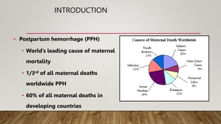
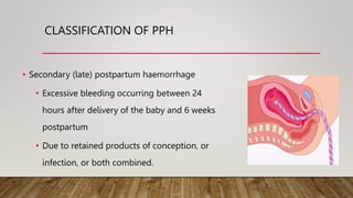
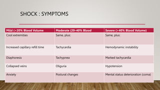
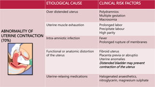
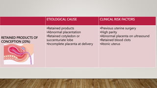
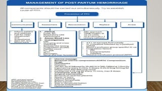
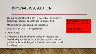
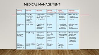
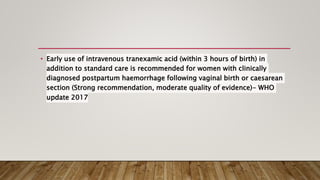
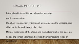
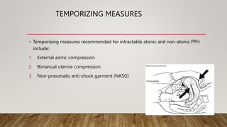
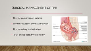
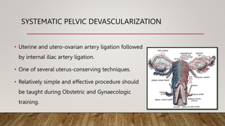
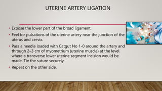
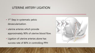
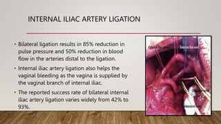
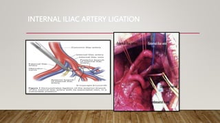
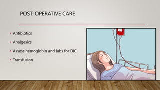
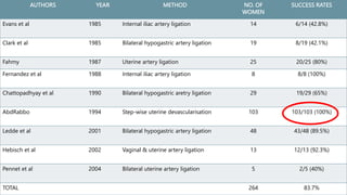
POST PARTUM
HEMORRHAGE
Dr Kuvaoga
INTRODUCTION
• Postpartum hemorrhage (PPH)
• World’s leading cause of maternal
mortality
• 1/3rd of all maternal deaths
worldwide PPH
• 60% of all maternal deaths in
developing countries
INTRODUCTION
• Majority of these deaths occur within 4
hours of delivery.
• A Practice Bulletin from the ACOG 2011,
places the estimate at 140,000 maternal
deaths per year or 1 woman every 4
minutes due to postpartum haemorrhage.
POST PARTUM HEMORRHAGE
• Blood loss
• Vaginal deliveries > 500mL (3-5%)
• Caesarean section deliveries >1000mL (5-
7%)
• For clinical purposes, any blood loss that has
the potential to produce hemodynamic
compromise should be considered a PPH.
CLASSIFICATION OF PPH
• Primary (immediate) postpartum
haemorrhage
• Excessive bleeding that occurs within the
first 24 hours after delivery
• 70% due to uterine atony (failure of the
uterus to contract adequately after the
child is born)
CLASSIFICATION OF PPH
• Secondary (late) postpartum haemorrhage
• Excessive bleeding occurring between 24
hours after delivery of the baby and 6 weeks
postpartum
• Due to retained products of conception, or
infection, or both combined.
SHOCK : SYMPTOMS
Mild (<20% Blood Volume Moderate (20–40% Blood Severe (>40% Blood Volume)
Cool extremities Same, plus: Same, plus:
Increased capillary refill time Tachycardia Hemodynamic instability
Diaphoresis Tachypnea Marked tachycardia
Collapsed veins Oliguria Hypotension
Anxiety Postural changes Mental status deterioration (coma)
CAUSES
4Ts
• TONE
• TEAR
• TISSUE
• THROMBUS
ETIOLOGICAL CAUSE CLINICAL RISK FACTORS
ABNORMALITY OF
UTERINE CONTRACTION
(70%)
Over distended uterus Polydramnios
Multiple gestation
Macrosomia
Uterine muscle exhaustion Prolonged labor
Precipitate labour
High parity
Intra-amniotic infection Fever
Prolonged rupture of membranes
Functional or anatomic distortion
of the uterus
Fibroid uterus
Placenta previa or abruptio
Uterine anomalies
Distended bladder may prevent
contraction of the uterus
Uterine-relaxing medications Halogenated anaesthetics,
nitroglycerin, magnesium sulphate
ETIOLOGICAL CAUSE CLINICAL RISK FACTORS
RETAINED PRODUCTS OF
CONCEPTION (20%)
•Retained products
•Abnormal placentation
•Retained cotyledon or
succenturiate lobe
•Incomplete placenta at delivery
•Previous uterine surgery
•High parity
•Abnormal placenta on ultrasound
•Retained blood clots
•Atonic uterus
ETIOLOGICAL CAUSE CLINICAL RISK FACTORS
GENITAL TRACT TRAUMA (10%)
•Tears (lacerations) of the cervix,
vagina, or perineum
•Ruptured vulval varicosities
•Precipitous delivery
•Operative delivery
•Mistimed or inappropriate use of
episiotomy
•Extensions, lacerations at
caesarean section
•Malposition
•Deep engagement
•Uterine rupture •Previous uterine surgery
•Uterine inversion •High parity
•Fundal placenta
ETIOLOGICAL CAUSE CLINICAL RISK FACTORS
ABNORMALITIES OF
COAGULATION (<1%)
Pre-existing states
- haemophilia A
-von Willebrand‘s disease1
History of hereditary
coagulopathies
• History of liver disease
Acquired in pregnancy
- Idiopathic thrombocytopenic
purpura2
- Thrombocytopenia with
preeclampsia
- Disseminated intravascular
coagulation
- Preeclampsia
- Dead foetus in utero
- Severe infection/sepsis
- Placental abruption
- Amniotic fluid embolus
•Bruising
•Elevated BP
•Elevated BP
•Foetal demise
•Fever
•Elevated white blood cells
•Antepartum hemorrhage
•Sudden collapse
Therapeutic anticoagulation History of thrombotic disease
LOCAL DATA
• Low parity, advanced maternal age, and antenatal hospitalisation were among the strongest risk
factors, with more modest associations for history of poor maternal or perinatal outcomes and
borderline anaemia at the time of booking. (Tsu et al,1993)
MANAGEMENT OF POSTPARTUM
HAEMORRHAGE
IMMEDIATE RESUSCITATION
• The primary treatment of PPH is to control the source of
bleeding as soon as possible and to replace fluid.
• Maintain airway, breathing and circulation
• Large bore IV Line: Fluid replacement
• O2 Inhalation
• Crystalloid is the first fluid of choice for resuscitation.
Immediately administer 2 L of isotonic sodium chloride
solution or lactated Ringer’s solution in response to shock
from blood loss
• Blood transfusion
Crystalloids restore
volume in a 3:1 ratio
Colloids restore
volume in a 1:1 ratio
MEDICAL MANAGEMENT
• Early use of intravenous tranexamic acid (within 3 hours of birth) in
addition to standard care is recommended for women with clinically
diagnosed postpartum haemorrhage following vaginal birth or caesarean
section (Strong recommendation, moderate quality of evidence)- WHO
update 2017
MANAGEMENT OF PPH
• External and internal bi-manual uterine massage
• Aortic compression
• Umbilical vein injection (injection of uterotonic into the umbilical cord
attached to the undelivered placenta)
• Manual exploration of the uterus and manual removal of the placenta
• Repair of perineal, vaginal and cervical trauma including repair of
episiotomy
TEMPORIZING MEASURES
• Temporizing measures recommended for intractable atonic and non-atonic PPH
include:
1. External aortic compression
2. Bimanual uterine compression
3. Non-pneumatic anti-shock garment (NASG)
SURGICAL MANAGEMENT OF PPH
• Uterine compression sutures
• Systematic pelvic devascularization
• Uterine artery embolization
• Total or sub-total hysterectomy
SYSTEMATIC PELVIC DEVASCULARIZATION
• Uterine and utero-ovarian artery ligation followed
by internal iliac artery ligation.
• One of several uterus-conserving techniques.
• Relatively simple and effective procedure should
be taught during Obstetric and Gynaecologic
training.
UTERINE ARTERY LIGATION
• Expose the lower part of the broad ligament.
• Feel for pulsations of the uterine artery near the junction of the
uterus and cervix.
• Pass a needle loaded with Catgut No 1-0 around the artery and
through 2–3 cm of myometrium (uterine muscle) at the level
where a transverse lower uterine segment incision would be
made. Tie the suture securely.
• Repeat on the other side.
UTERINE ARTERY LIGATION
• 1ST Step in systematic pelvic
devascularisation.
• uterine arteries which provide
approximately 90% of uterine blood flow.
• Ligation of uterine arteries alone has
success rate of 80% in controlling PPH
SITES FOR LIGATING UTERINE ARTERY
UTERO OVARIAN ANASTOMOSIS
• If the artery has been torn, clamp and tie the bleeding ends.
• Ligate the utero-ovarian artery just below the point where the ovarian
suspensory ligament joins the uterus.
• Repeat on the other side.
• Observe for continued bleeding or formation of hematoma.
• Close the abdomen in layers.
INTERNAL ILIAC ARTERY LIGATION
• Experiments in the 1960’s by Burchell,
ascertained that ligating the hypogastric
artery turned the pelvic circulation like a
venous system, thereby aiding clotting and
controlling PPH.
• It is effective in Uterine atony, midline
perforation, large broad ligament or lateral
pelvic wall haematoma, multiple cervical
tears and lower segment bleeding.
INTERNAL ILIAC ARTERY LIGATION
• Bilateral ligation results in 85% reduction in
pulse pressure and 50% reduction in blood
flow in the arteries distal to the ligation.
• Internal iliac artery ligation also helps the
vaginal bleeding as the vagina is supplied by
the vaginal branch of internal iliac.
• The reported success rate of bilateral internal
iliac artery ligation varies widely from 42% to
93%.
INTERNAL ILIAC ARTERY LIGATION
POST-OPERATIVE CARE
• Antibiotics
• Analgesics
• Assess hemoglobin and labs for DIC
• Transfusion
AUTHORS YEAR METHOD NO. OF
WOMEN
SUCCESS RATES
Evans et al 1985 Internal iliac artery ligation 14 6/14 (42.8%)
Clark et al 1985 Bilateral hypogastric artery ligation 19 8/19 (42.1%)
Fahmy 1987 Uterine artery ligation 25 20/25 (80%)
Fernandez et al 1988 Internal iliac artery ligation 8 8/8 (100%)
Chattopadhyay et al 1990 Bilateral hypogastric aretry ligation 29 19/29 (65%)
AbdRabbo 1994 Step-wise uterine devascularisation 103 103/103 (100%)
Ledde et al 2001 Bilateral hypogastric artery ligation 48 43/48 (89.5%)
Hebisch et al 2002 Vaginal & uterine artery ligation 13 12/13 (92.3%)
Pennet et al 2004 Bilateral uterine artery ligation 5 2/5 (40%)
TOTAL 264 83.7%
CONCLUSION
• Blood loss is consistently underestimated.
• Underestimation may result in inadequate treatment
resulting in complications or death.
• Ongoing trickling can lead to significant blood loss.
• Anaemia and other underlying health conditions may
profoundly affect a woman‘s ability to tolerate any amount
of blood loss.
CONCLUSION
• Systematic pelvic devascularisation is an effective and uterus
conserving surgical method to control PPH.
• All health care providers providing maternity care require to
learn this life-saving skill to enable them to make a significant
contribution to reducing maternal deaths and to promote safe
motherhood.
REFERENCES
• Countdown to 2015: Maternal, Newborn and Child Survival, WHO, 2012
• Lu M C, Fridman M, Korst L M. et al. Variations in the incidence of postpartum
hemorrhage across hospitals in California. Matern Child Health J. 2005;9:297–306.
• Sivan E, Spira M, Achiron R, Rimon U, Golan G, Mazaki-Tovi S, et al. Prophylactic
pelvic artery catheterization and embolization in women with placenta accreta:
can it prevent cesarean hysterectomy? Am J Perinatol 2010;27:455–61
• Harrisons Principle of Internal Medicine, 19th edition
• Global causes of maternal death: a WHO systematic analysis Lale et al.The Lancet
Global Health June 2014 , Volume 2 , Issue 6 , e323 - e333
• Iwata A, Murayama Y, Itakura A, Baba K, Seki H, Takeda S. Limitations of internal
iliac artery ligation for the reduction of intraoperative hemorrhage during
stepwisepelvicdevascularisation-180120130719.pptx

stepwisepelvicdevascularisation-180120130719.pptx

  • 1.
  • 2.
    INTRODUCTION • Postpartum hemorrhage(PPH) • World’s leading cause of maternal mortality • 1/3rd of all maternal deaths worldwide PPH • 60% of all maternal deaths in developing countries
  • 3.
    INTRODUCTION • Majority ofthese deaths occur within 4 hours of delivery. • A Practice Bulletin from the ACOG 2011, places the estimate at 140,000 maternal deaths per year or 1 woman every 4 minutes due to postpartum haemorrhage.
  • 4.
    POST PARTUM HEMORRHAGE •Blood loss • Vaginal deliveries > 500mL (3-5%) • Caesarean section deliveries >1000mL (5- 7%) • For clinical purposes, any blood loss that has the potential to produce hemodynamic compromise should be considered a PPH.
  • 5.
    CLASSIFICATION OF PPH •Primary (immediate) postpartum haemorrhage • Excessive bleeding that occurs within the first 24 hours after delivery • 70% due to uterine atony (failure of the uterus to contract adequately after the child is born)
  • 6.
    CLASSIFICATION OF PPH •Secondary (late) postpartum haemorrhage • Excessive bleeding occurring between 24 hours after delivery of the baby and 6 weeks postpartum • Due to retained products of conception, or infection, or both combined.
  • 7.
    SHOCK : SYMPTOMS Mild(<20% Blood Volume Moderate (20–40% Blood Severe (>40% Blood Volume) Cool extremities Same, plus: Same, plus: Increased capillary refill time Tachycardia Hemodynamic instability Diaphoresis Tachypnea Marked tachycardia Collapsed veins Oliguria Hypotension Anxiety Postural changes Mental status deterioration (coma)
  • 8.
  • 9.
    ETIOLOGICAL CAUSE CLINICALRISK FACTORS ABNORMALITY OF UTERINE CONTRACTION (70%) Over distended uterus Polydramnios Multiple gestation Macrosomia Uterine muscle exhaustion Prolonged labor Precipitate labour High parity Intra-amniotic infection Fever Prolonged rupture of membranes Functional or anatomic distortion of the uterus Fibroid uterus Placenta previa or abruptio Uterine anomalies Distended bladder may prevent contraction of the uterus Uterine-relaxing medications Halogenated anaesthetics, nitroglycerin, magnesium sulphate
  • 10.
    ETIOLOGICAL CAUSE CLINICALRISK FACTORS RETAINED PRODUCTS OF CONCEPTION (20%) •Retained products •Abnormal placentation •Retained cotyledon or succenturiate lobe •Incomplete placenta at delivery •Previous uterine surgery •High parity •Abnormal placenta on ultrasound •Retained blood clots •Atonic uterus
  • 11.
    ETIOLOGICAL CAUSE CLINICALRISK FACTORS GENITAL TRACT TRAUMA (10%) •Tears (lacerations) of the cervix, vagina, or perineum •Ruptured vulval varicosities •Precipitous delivery •Operative delivery •Mistimed or inappropriate use of episiotomy •Extensions, lacerations at caesarean section •Malposition •Deep engagement •Uterine rupture •Previous uterine surgery •Uterine inversion •High parity •Fundal placenta
  • 12.
    ETIOLOGICAL CAUSE CLINICALRISK FACTORS ABNORMALITIES OF COAGULATION (<1%) Pre-existing states - haemophilia A -von Willebrand‘s disease1 History of hereditary coagulopathies • History of liver disease Acquired in pregnancy - Idiopathic thrombocytopenic purpura2 - Thrombocytopenia with preeclampsia - Disseminated intravascular coagulation - Preeclampsia - Dead foetus in utero - Severe infection/sepsis - Placental abruption - Amniotic fluid embolus •Bruising •Elevated BP •Elevated BP •Foetal demise •Fever •Elevated white blood cells •Antepartum hemorrhage •Sudden collapse Therapeutic anticoagulation History of thrombotic disease
  • 13.
    LOCAL DATA • Lowparity, advanced maternal age, and antenatal hospitalisation were among the strongest risk factors, with more modest associations for history of poor maternal or perinatal outcomes and borderline anaemia at the time of booking. (Tsu et al,1993)
  • 14.
  • 16.
    IMMEDIATE RESUSCITATION • Theprimary treatment of PPH is to control the source of bleeding as soon as possible and to replace fluid. • Maintain airway, breathing and circulation • Large bore IV Line: Fluid replacement • O2 Inhalation • Crystalloid is the first fluid of choice for resuscitation. Immediately administer 2 L of isotonic sodium chloride solution or lactated Ringer’s solution in response to shock from blood loss • Blood transfusion Crystalloids restore volume in a 3:1 ratio Colloids restore volume in a 1:1 ratio
  • 17.
  • 18.
    • Early useof intravenous tranexamic acid (within 3 hours of birth) in addition to standard care is recommended for women with clinically diagnosed postpartum haemorrhage following vaginal birth or caesarean section (Strong recommendation, moderate quality of evidence)- WHO update 2017
  • 19.
    MANAGEMENT OF PPH •External and internal bi-manual uterine massage • Aortic compression • Umbilical vein injection (injection of uterotonic into the umbilical cord attached to the undelivered placenta) • Manual exploration of the uterus and manual removal of the placenta • Repair of perineal, vaginal and cervical trauma including repair of episiotomy
  • 20.
    TEMPORIZING MEASURES • Temporizingmeasures recommended for intractable atonic and non-atonic PPH include: 1. External aortic compression 2. Bimanual uterine compression 3. Non-pneumatic anti-shock garment (NASG)
  • 21.
    SURGICAL MANAGEMENT OFPPH • Uterine compression sutures • Systematic pelvic devascularization • Uterine artery embolization • Total or sub-total hysterectomy
  • 22.
    SYSTEMATIC PELVIC DEVASCULARIZATION •Uterine and utero-ovarian artery ligation followed by internal iliac artery ligation. • One of several uterus-conserving techniques. • Relatively simple and effective procedure should be taught during Obstetric and Gynaecologic training.
  • 23.
    UTERINE ARTERY LIGATION •Expose the lower part of the broad ligament. • Feel for pulsations of the uterine artery near the junction of the uterus and cervix. • Pass a needle loaded with Catgut No 1-0 around the artery and through 2–3 cm of myometrium (uterine muscle) at the level where a transverse lower uterine segment incision would be made. Tie the suture securely. • Repeat on the other side.
  • 24.
    UTERINE ARTERY LIGATION •1ST Step in systematic pelvic devascularisation. • uterine arteries which provide approximately 90% of uterine blood flow. • Ligation of uterine arteries alone has success rate of 80% in controlling PPH
  • 25.
    SITES FOR LIGATINGUTERINE ARTERY
  • 26.
    UTERO OVARIAN ANASTOMOSIS •If the artery has been torn, clamp and tie the bleeding ends. • Ligate the utero-ovarian artery just below the point where the ovarian suspensory ligament joins the uterus. • Repeat on the other side. • Observe for continued bleeding or formation of hematoma. • Close the abdomen in layers.
  • 27.
    INTERNAL ILIAC ARTERYLIGATION • Experiments in the 1960’s by Burchell, ascertained that ligating the hypogastric artery turned the pelvic circulation like a venous system, thereby aiding clotting and controlling PPH. • It is effective in Uterine atony, midline perforation, large broad ligament or lateral pelvic wall haematoma, multiple cervical tears and lower segment bleeding.
  • 28.
    INTERNAL ILIAC ARTERYLIGATION • Bilateral ligation results in 85% reduction in pulse pressure and 50% reduction in blood flow in the arteries distal to the ligation. • Internal iliac artery ligation also helps the vaginal bleeding as the vagina is supplied by the vaginal branch of internal iliac. • The reported success rate of bilateral internal iliac artery ligation varies widely from 42% to 93%.
  • 29.
  • 30.
    POST-OPERATIVE CARE • Antibiotics •Analgesics • Assess hemoglobin and labs for DIC • Transfusion
  • 31.
    AUTHORS YEAR METHODNO. OF WOMEN SUCCESS RATES Evans et al 1985 Internal iliac artery ligation 14 6/14 (42.8%) Clark et al 1985 Bilateral hypogastric artery ligation 19 8/19 (42.1%) Fahmy 1987 Uterine artery ligation 25 20/25 (80%) Fernandez et al 1988 Internal iliac artery ligation 8 8/8 (100%) Chattopadhyay et al 1990 Bilateral hypogastric aretry ligation 29 19/29 (65%) AbdRabbo 1994 Step-wise uterine devascularisation 103 103/103 (100%) Ledde et al 2001 Bilateral hypogastric artery ligation 48 43/48 (89.5%) Hebisch et al 2002 Vaginal & uterine artery ligation 13 12/13 (92.3%) Pennet et al 2004 Bilateral uterine artery ligation 5 2/5 (40%) TOTAL 264 83.7%
  • 32.
    CONCLUSION • Blood lossis consistently underestimated. • Underestimation may result in inadequate treatment resulting in complications or death. • Ongoing trickling can lead to significant blood loss. • Anaemia and other underlying health conditions may profoundly affect a woman‘s ability to tolerate any amount of blood loss.
  • 33.
    CONCLUSION • Systematic pelvicdevascularisation is an effective and uterus conserving surgical method to control PPH. • All health care providers providing maternity care require to learn this life-saving skill to enable them to make a significant contribution to reducing maternal deaths and to promote safe motherhood.
  • 34.
    REFERENCES • Countdown to2015: Maternal, Newborn and Child Survival, WHO, 2012 • Lu M C, Fridman M, Korst L M. et al. Variations in the incidence of postpartum hemorrhage across hospitals in California. Matern Child Health J. 2005;9:297–306. • Sivan E, Spira M, Achiron R, Rimon U, Golan G, Mazaki-Tovi S, et al. Prophylactic pelvic artery catheterization and embolization in women with placenta accreta: can it prevent cesarean hysterectomy? Am J Perinatol 2010;27:455–61 • Harrisons Principle of Internal Medicine, 19th edition • Global causes of maternal death: a WHO systematic analysis Lale et al.The Lancet Global Health June 2014 , Volume 2 , Issue 6 , e323 - e333 • Iwata A, Murayama Y, Itakura A, Baba K, Seki H, Takeda S. Limitations of internal iliac artery ligation for the reduction of intraoperative hemorrhage during