Downloaded 486 times


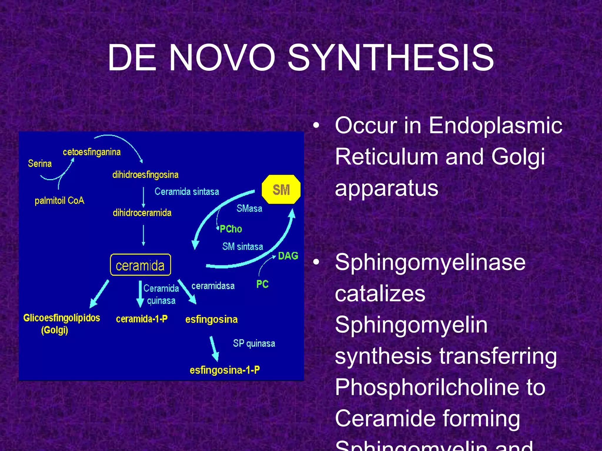

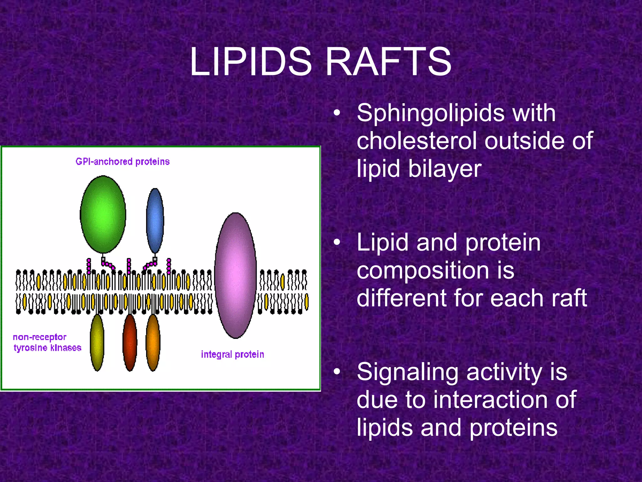

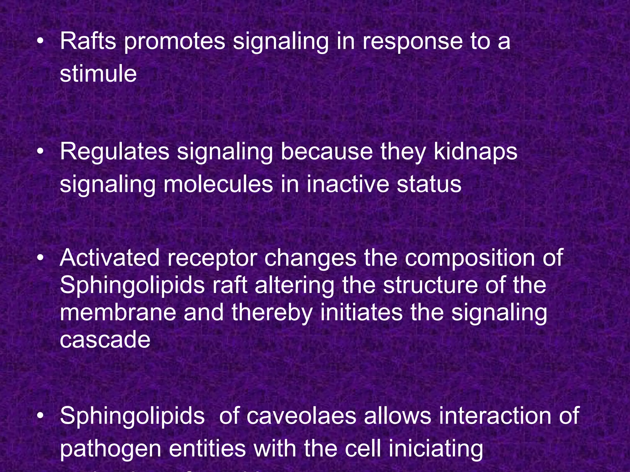


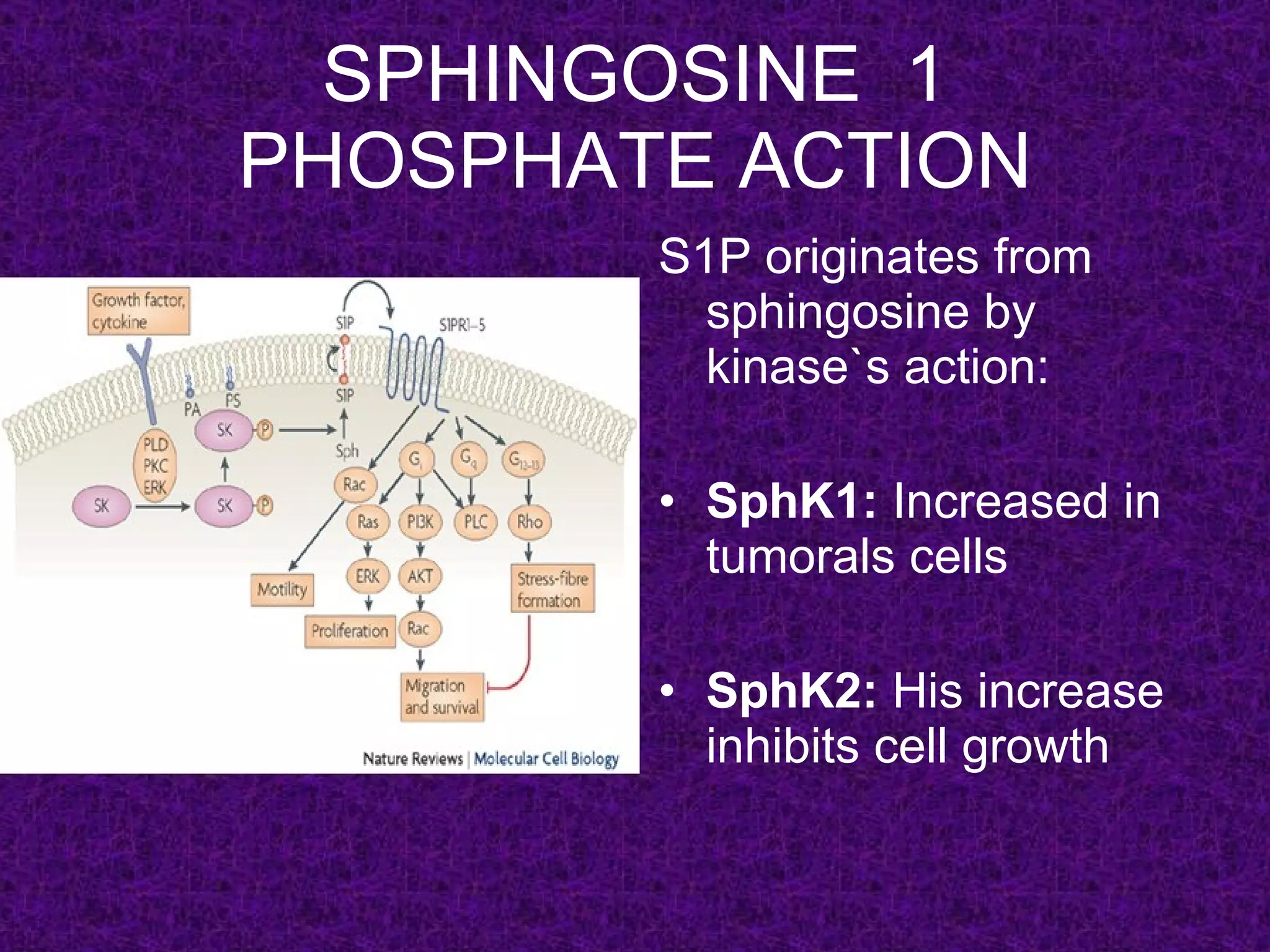


Sphingolipids are a class of lipids that include sphingomyelin, glycosphingolipids, and ceramide. They serve important structural and signaling functions in the cell membrane and participate in processes like cell growth, differentiation, and apoptosis. Sphingolipids are synthesized through de novo pathways or through the breakdown of sphingomyelin. Their metabolites, including ceramide, sphingosine-1-phosphate, and gangliosides act as second messengers and influence pathways such as PI3K/Akt and JNK. Sphingolipids are also components of lipid rafts and caveolae, which regulate protein activity and cellular signaling. Imbalances in sph


