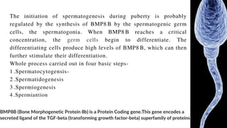
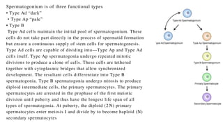
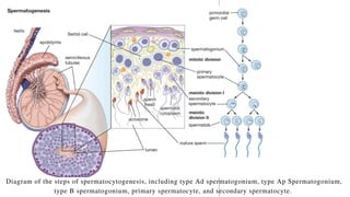
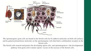
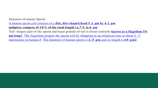
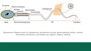
Presentation on .
DATTA MEGHE INSTITUTE OF HIGHER
EDUCATION AND RESEARCH,
SAHS.
Presented by-
Neeraj Vishwakarma
Spermatogenesis
Core Concepts
Fundamental Introduction of anatomy and
physiology of Male reproduction .
Male Gametogenesis
What is Spermatogenesis ?
Steps in Spermatogenesis
• Step 1: Spermatocytogenesis
• Step 2: Spermatidogenesis
• Step 3: Spermiogenesis
• Physiology of Spermatogenesis
Andrology
l
Fundamental Introduction of anatomy and physiology.
Understanding the fundamentals of anatomy and
physiology of male reproductive system is a key to
effective evaluation and treatment of male infertility. It
comprises of the hypothalamic-pituitary-testis axis,
epididymis, vas deferens, seminal vesicles, prostate and
urethra.
Short Discription of Anatomy of Male Reproductive
organ (at the time of Gustation period).
System Development -
The male urinary and reproductive systems share a common
developmental origin. The testes and extra testicular ducts arise from
three different tissues:
1 .Intermediate mesoderm- The intermediate mesoderm forms a
urogenital ridge that gives rise to testicular stroma and the
mesonephric (Wolffian) duct.
2 .Mesodermal epithelium- The mesodermal (coelomic) epithelium
gives rise to Sertoli cells and the paramesonephric duct.
3 .Primordial germ cells (PGC)- The primordial germ cells migrate
from yolk sac and give rise to the spermatagonia.
Male Gametogenesis.
Biology
1
Spermatozoa are the mature male gametes in many sexually reproducing organisms.
Spermatogenesis is the male version of gametogenesis and results in the formation of
spermatocytes possessing half the normal complement of genetic material.
In mammals, it occurs in the male testes and epididymis in a stepwise fashion that
takes approximately 64 days.
Spermatogenesis, essential for sexual reproduction is highly dependent upon optimal
conditions to occur correctly. DNA methylation and histone modification have been
implicated in the regulation of this process. It starts at puberty and usually continues
uninterrupted until death, although a slight decrease can be discerned in the quantity
of sperm produced with increase in age.
Testis
• 90% seminiferous tubules (Sertoli and germ cells).
• 10% interstitial (Leydig cells, connective tissue, blood supply).
• Two functions: – Spermatogenesis—in the seminiferous tubules.
Biosynthesis of testosterone (T)—Leydig cells.
=Synthesized from cholesterol
As Cholesterol is the parent compound of all steroid hormones, including testosterone.
It is synthesized in the liver and transported to the testicles by the blood. There, an
enzyme called Cytochrome P450 converts cholesterol into pregnenolone, which is
then converted into testosterone by the action of luteinizing hormone (LH) on the
Leydig cells.
High cholesterol levels may impair the blood flow and the testosterone production in
the testicles.
Sertoli cells
sertoli cells forms” blood testes barrier” and protect the
sperm from immune system of the body sertoli cells
function as endocrine gland of the body .i.e. scerete three
type of biochemicals
• AMH- Antimullarian hormone ~ It inhibit mullarian
duct
• Inhibin hormone - Gives negative feedback to the
Pitutary gland (mainly)and Hypothalamus .
• Androgen binding Protein - Conc. testesteron in Sertoli
Cells to aid Spermatogenesis
Defining
Spermatogenesis
~ Spermatogenesis is the production of Haploid spermatozoa
from the primordial germ cells. Once the vertebrate PGCs
arrive at the genital ridge of a male embryo, they become
incorporated into the sex cords. They remain there until
maturity, at which time the sex cords hollow out to form the
seminiferous tubules, and the mesodermal epithelium of the
tubules differentiates into the Sertoli cells.
BMP8B (Bone Morphogenetic Protein 8b) is a Protein Coding gene.This gene encodes a
secreted ligand of the TGF-beta (transforming growth factor-beta) superfamily of proteins
The initiation of spermatogenesis during puberty is probably
regulated by the synthesis of BMP8 B by the spermatogenic germ
cells, the spermatogonia. When BMP8 B reaches a critical
concentration, the germ cells begin to differentiate. The
differentiating cells produce high levels of BMP8 B, which can then
further stimulate their differentiation.
Whole process carried out in four basic steps-
1 .Spermatocytogensis-
2 .Spermatidogenesis
3 .Spermiogenesis
4 .Spermiantion
Spermatogonium is of three functional types
• Type Ad “dark”
• Type Ap “pale”
• Type B
Type Ad cells maintain the initial pool of spermatogonium. These
cells do not take part directly in the process of spermatid formation
but ensure a continuous supply of stem cells for spermatogenesis.
Type Ad cells are capable of dividing into—Type Ap and Type Ad
cells itself. Type Ap spermatogonia undergo repeated mitotic
divisions to produce a clone of cells. These cells are tethered
together with cytoplasmic bridges that allow synchronized
development. The resultant cells differentiate into Type B
spermatogonia. Type B spermatogonia undergo mitosis to produce
diploid intermediate cells, the primary spermatocytes. The primary
spermatocytes are arrested in the prophase of the first meiotic
division until puberty and thus have the longest life span of all
types of spermatogonia. At puberty, the diploid (2 N) primary
spermatocytes enter meiosis I and divide by to become haploid (N)
secondary spermatocytes
Biology
1
Diagram of the steps of spermatocytogenesis, including type Ad spermatogonium, type Ap Spermatogonium,
type B spermatogonium, primary spermatocyte, and secondary spermatocyte.
• Mitotic division of a diploid spermatogonium that resides in the basal
compartment of the seminiferous tubules, resulting in two diploid
intermediate cells called primary spermatocytes.
• Each primary spermatocyte then moves into the adluminal compartment
of the seminiferous tubules, duplicates its DNA, and subsequently
undergoes meiosis I to produce two haploid secondary spermatocytes.
• Secondary spermatocytes later divide into haploid spermatids.
Step 2 : Spermatidogenesis
The creation of spermatids from secondary
spermatocytes. Secondary spermatocytes produced
earlier rapidly enter meiosis II and divide to produce
haploid spermatids. The brevity of this stage means that
secondary spermatocytes are rarely seen in histological
preparations.
Step 3 : Spermiogenesis
At this stage, each spermatid begins to grow a tail and develop a
thickened midpiece where the mitochondria gather and form an
axoneme. Spermatid DNA also undergoes packaging, becoming
highly condensed.
The DNA is packaged with specific nuclear basic proteins,
which are subsequently replaced with protamines during
spermatid elongation. The resultant tightly packed chromatin is
transcriptionally inactive. The Golgi apparatus surrounds the
now condensed nucleus, becoming the acrosome.
One of the centrioles of the cell elongates to become the tail of
the sperm.
Biology
1
completed in 4 phase
-
• Golgi phase
• cap phase
• acrosomal phase
• maturation phase
Spermiation-
The non-motile spermatozoa are transported to the
epididymis in testicular fluid secreted by the Sertoli cells
with the aid of peristaltic contraction. While in the
epididymis, the spermatozoa gain motility and become
capable of fertilization.
However, transport of the mature spermatozoa through
the remainder of the male reproductive system is
achieved via muscle contraction rather than the
spermatozoon’s recently acquired motility.
Physiology of Spermatogenesis
Maturation takes place under the influence of testosterone, which removes
the remaining unnecessary cytoplasm and organelles. The excess
cytoplasm, known as residual bodies, is phagocytosed by surrounding
Sertoli cells in the testes. The resulting spermatozoa are now mature but
lack motility, rendering them sterile.
The mature spermatozoa are released from the protective Sertoli cells into
the lumen of the seminiferous tubule in a process called spermiation.
Factors affecting Spermatogenesis .
• Changes in temperature
• Deficiency in the diet
• Alcoholism
• Exposure to drugs
• Presence of disease
Spermatogenesis is highly sensitive to fluctuations in the environment,
particularly hormones and temperature.
The seminiferous epithelium is sensitive to elevated temperature in humans and
is adversely affected by temperatures as high as normal body temperature.
Consequently, the testes are located outside the body in a sack of skin called the
scrotum. The optimal temperature is maintained at 3 .4 °C below body
temperature in human males.
This is achieved by the regulation of blood flow and positioning towards and
away from the heat of the body by the cremaster muscle and the dartos smooth
muscle in the scrotum.
• Dietary deficiencies (such as vitamins B, E,
and A), anabolic steroids, metals (cadmium
and lead), x-ray exposure, dioxin, alcohol,
and infectious diseases will also adversely
affect the rate of spermatogenesis.
The spermatogenic germ cells are bound to the Sertoli cells by N-cadherin molecules on both cell surfaces
and by galactosyltransferase molecules on the spermatogenic cells that bind a carbohydrate receptor on the
Sertoli cells
The Sertoli cells nourish and protect the developing sperm cells, and spermatogenesis—the developmental
pathway from germ cell to mature sperm—occurs in the recesses of the Sertoli cells .
Structure of mature Sperm
A human sperm cell consists of a flat, disc-shaped head 5 .1 µm by 4 .1 µm
midpiece compres of 1 0 % of the total length i.e.7 .5 to 6 µm
Tail- longest part of the sperm and basal granule of tail is distal centriole known as a flagellum 5 0
µm long1. The flagellum propels the sperm cell by whipping in an elliptical cone at about 1 –3
mm/minute in humans1. The diameter of human sperm is 2 –5 μm and its length is 6 0 μm2.
Spermatozoon: Diagram of parts of a spermatozoon, including the acrosome, plasma membrane, nucleus, centriole,
mitochondria, terminal disc, axial filament, tail, endpiece, midpiece, and head.
Thanking you....

Spermatogenesis presentation.ppsx

  • 1.
    Presentation on . DATTAMEGHE INSTITUTE OF HIGHER EDUCATION AND RESEARCH, SAHS. Presented by- Neeraj Vishwakarma Spermatogenesis
  • 2.
    Core Concepts Fundamental Introductionof anatomy and physiology of Male reproduction . Male Gametogenesis What is Spermatogenesis ? Steps in Spermatogenesis • Step 1: Spermatocytogenesis • Step 2: Spermatidogenesis • Step 3: Spermiogenesis • Physiology of Spermatogenesis Andrology l
  • 3.
    Fundamental Introduction ofanatomy and physiology. Understanding the fundamentals of anatomy and physiology of male reproductive system is a key to effective evaluation and treatment of male infertility. It comprises of the hypothalamic-pituitary-testis axis, epididymis, vas deferens, seminal vesicles, prostate and urethra.
  • 4.
    Short Discription ofAnatomy of Male Reproductive organ (at the time of Gustation period). System Development - The male urinary and reproductive systems share a common developmental origin. The testes and extra testicular ducts arise from three different tissues: 1 .Intermediate mesoderm- The intermediate mesoderm forms a urogenital ridge that gives rise to testicular stroma and the mesonephric (Wolffian) duct. 2 .Mesodermal epithelium- The mesodermal (coelomic) epithelium gives rise to Sertoli cells and the paramesonephric duct. 3 .Primordial germ cells (PGC)- The primordial germ cells migrate from yolk sac and give rise to the spermatagonia.
  • 5.
    Male Gametogenesis. Biology 1 Spermatozoa arethe mature male gametes in many sexually reproducing organisms. Spermatogenesis is the male version of gametogenesis and results in the formation of spermatocytes possessing half the normal complement of genetic material. In mammals, it occurs in the male testes and epididymis in a stepwise fashion that takes approximately 64 days. Spermatogenesis, essential for sexual reproduction is highly dependent upon optimal conditions to occur correctly. DNA methylation and histone modification have been implicated in the regulation of this process. It starts at puberty and usually continues uninterrupted until death, although a slight decrease can be discerned in the quantity of sperm produced with increase in age.
  • 6.
    Testis • 90% seminiferoustubules (Sertoli and germ cells). • 10% interstitial (Leydig cells, connective tissue, blood supply). • Two functions: – Spermatogenesis—in the seminiferous tubules. Biosynthesis of testosterone (T)—Leydig cells. =Synthesized from cholesterol As Cholesterol is the parent compound of all steroid hormones, including testosterone. It is synthesized in the liver and transported to the testicles by the blood. There, an enzyme called Cytochrome P450 converts cholesterol into pregnenolone, which is then converted into testosterone by the action of luteinizing hormone (LH) on the Leydig cells. High cholesterol levels may impair the blood flow and the testosterone production in the testicles.
  • 7.
    Sertoli cells sertoli cellsforms” blood testes barrier” and protect the sperm from immune system of the body sertoli cells function as endocrine gland of the body .i.e. scerete three type of biochemicals • AMH- Antimullarian hormone ~ It inhibit mullarian duct • Inhibin hormone - Gives negative feedback to the Pitutary gland (mainly)and Hypothalamus . • Androgen binding Protein - Conc. testesteron in Sertoli Cells to aid Spermatogenesis
  • 8.
    Defining Spermatogenesis ~ Spermatogenesis isthe production of Haploid spermatozoa from the primordial germ cells. Once the vertebrate PGCs arrive at the genital ridge of a male embryo, they become incorporated into the sex cords. They remain there until maturity, at which time the sex cords hollow out to form the seminiferous tubules, and the mesodermal epithelium of the tubules differentiates into the Sertoli cells.
  • 9.
    BMP8B (Bone MorphogeneticProtein 8b) is a Protein Coding gene.This gene encodes a secreted ligand of the TGF-beta (transforming growth factor-beta) superfamily of proteins The initiation of spermatogenesis during puberty is probably regulated by the synthesis of BMP8 B by the spermatogenic germ cells, the spermatogonia. When BMP8 B reaches a critical concentration, the germ cells begin to differentiate. The differentiating cells produce high levels of BMP8 B, which can then further stimulate their differentiation. Whole process carried out in four basic steps- 1 .Spermatocytogensis- 2 .Spermatidogenesis 3 .Spermiogenesis 4 .Spermiantion
  • 10.
    Spermatogonium is ofthree functional types • Type Ad “dark” • Type Ap “pale” • Type B Type Ad cells maintain the initial pool of spermatogonium. These cells do not take part directly in the process of spermatid formation but ensure a continuous supply of stem cells for spermatogenesis. Type Ad cells are capable of dividing into—Type Ap and Type Ad cells itself. Type Ap spermatogonia undergo repeated mitotic divisions to produce a clone of cells. These cells are tethered together with cytoplasmic bridges that allow synchronized development. The resultant cells differentiate into Type B spermatogonia. Type B spermatogonia undergo mitosis to produce diploid intermediate cells, the primary spermatocytes. The primary spermatocytes are arrested in the prophase of the first meiotic division until puberty and thus have the longest life span of all types of spermatogonia. At puberty, the diploid (2 N) primary spermatocytes enter meiosis I and divide by to become haploid (N) secondary spermatocytes
  • 11.
    Biology 1 Diagram of thesteps of spermatocytogenesis, including type Ad spermatogonium, type Ap Spermatogonium, type B spermatogonium, primary spermatocyte, and secondary spermatocyte.
  • 12.
    • Mitotic divisionof a diploid spermatogonium that resides in the basal compartment of the seminiferous tubules, resulting in two diploid intermediate cells called primary spermatocytes. • Each primary spermatocyte then moves into the adluminal compartment of the seminiferous tubules, duplicates its DNA, and subsequently undergoes meiosis I to produce two haploid secondary spermatocytes. • Secondary spermatocytes later divide into haploid spermatids.
  • 13.
    Step 2 :Spermatidogenesis The creation of spermatids from secondary spermatocytes. Secondary spermatocytes produced earlier rapidly enter meiosis II and divide to produce haploid spermatids. The brevity of this stage means that secondary spermatocytes are rarely seen in histological preparations.
  • 14.
    Step 3 :Spermiogenesis At this stage, each spermatid begins to grow a tail and develop a thickened midpiece where the mitochondria gather and form an axoneme. Spermatid DNA also undergoes packaging, becoming highly condensed. The DNA is packaged with specific nuclear basic proteins, which are subsequently replaced with protamines during spermatid elongation. The resultant tightly packed chromatin is transcriptionally inactive. The Golgi apparatus surrounds the now condensed nucleus, becoming the acrosome. One of the centrioles of the cell elongates to become the tail of the sperm.
  • 15.
    Biology 1 completed in 4phase - • Golgi phase • cap phase • acrosomal phase • maturation phase
  • 16.
    Spermiation- The non-motile spermatozoaare transported to the epididymis in testicular fluid secreted by the Sertoli cells with the aid of peristaltic contraction. While in the epididymis, the spermatozoa gain motility and become capable of fertilization. However, transport of the mature spermatozoa through the remainder of the male reproductive system is achieved via muscle contraction rather than the spermatozoon’s recently acquired motility.
  • 17.
    Physiology of Spermatogenesis Maturationtakes place under the influence of testosterone, which removes the remaining unnecessary cytoplasm and organelles. The excess cytoplasm, known as residual bodies, is phagocytosed by surrounding Sertoli cells in the testes. The resulting spermatozoa are now mature but lack motility, rendering them sterile. The mature spermatozoa are released from the protective Sertoli cells into the lumen of the seminiferous tubule in a process called spermiation.
  • 18.
    Factors affecting Spermatogenesis. • Changes in temperature • Deficiency in the diet • Alcoholism • Exposure to drugs • Presence of disease
  • 19.
    Spermatogenesis is highlysensitive to fluctuations in the environment, particularly hormones and temperature. The seminiferous epithelium is sensitive to elevated temperature in humans and is adversely affected by temperatures as high as normal body temperature. Consequently, the testes are located outside the body in a sack of skin called the scrotum. The optimal temperature is maintained at 3 .4 °C below body temperature in human males. This is achieved by the regulation of blood flow and positioning towards and away from the heat of the body by the cremaster muscle and the dartos smooth muscle in the scrotum.
  • 20.
    • Dietary deficiencies(such as vitamins B, E, and A), anabolic steroids, metals (cadmium and lead), x-ray exposure, dioxin, alcohol, and infectious diseases will also adversely affect the rate of spermatogenesis.
  • 21.
    The spermatogenic germcells are bound to the Sertoli cells by N-cadherin molecules on both cell surfaces and by galactosyltransferase molecules on the spermatogenic cells that bind a carbohydrate receptor on the Sertoli cells The Sertoli cells nourish and protect the developing sperm cells, and spermatogenesis—the developmental pathway from germ cell to mature sperm—occurs in the recesses of the Sertoli cells .
  • 22.
    Structure of matureSperm A human sperm cell consists of a flat, disc-shaped head 5 .1 µm by 4 .1 µm midpiece compres of 1 0 % of the total length i.e.7 .5 to 6 µm Tail- longest part of the sperm and basal granule of tail is distal centriole known as a flagellum 5 0 µm long1. The flagellum propels the sperm cell by whipping in an elliptical cone at about 1 –3 mm/minute in humans1. The diameter of human sperm is 2 –5 μm and its length is 6 0 μm2.
  • 23.
    Spermatozoon: Diagram ofparts of a spermatozoon, including the acrosome, plasma membrane, nucleus, centriole, mitochondria, terminal disc, axial filament, tail, endpiece, midpiece, and head.
  • 24.