SlideShare a Scribd company logo
1 of 8
CASE REVIEW                                                       SIMPLE BONE CYST WITH FRACTURE MERCURY IMAGING INSTITUTE  SCO 172-173 SEC 9C  CHANDIGARH MERCURY IMAGING CENTRE  SCO 16-17 SEC 20D CHANDIGARH 24 Yr old  Female  H/o fall and pain in the Rt hip joint .
Altered MR signal is appreciated in the Rt femur - subcapitate , neck , interochanteric and proximal shaft region. The lesion has following morphological characters : Central / expansible  lesion with Lobulated  contour. Narrow zone of the transition. Primarily involving medullary cavity with associated thinning of the cortex . Size : 82mm x 39mmx 38mm ( longitudinal x anteroposterior x transverse ). MR signal character : Intermediate on T1, heterogeneously hyperintense on t2/ stir,  with interspread bloom On GRE . Features are corroborative with fluid  and interspread haemorrhage in the core of the lesion.  Cortical break appreciated along the medial aspect of the proximal femoral shaft (just below the lesser trochanter ) . Reactionary edema / hematoma appreciated in the adjacent myofascial planes ( primarily vastusintermedius involved ) . MR  Bilateral hip joints done
HETEROGENOUSLY HYPERINTENSE ON T2 INTERMEDIATE ON T1W HETEROGENOUSLY HYPERINTENSE  ON FATSAT INTERSPREAD BLOOM ON GRE  S/O HAEMORRHAGE
INTERSPREAD BLOOM ON GRE SUGGESTIVE OF HAEMORRHAGE  IN THE LESION.
HYPERINTENSE SIGNAL OF THE LESION  ON FAT SATURATION- SEQUENCE CORTICAL BREAK
ALTERED MR SIGNAL  IN RT PROXIMAL FEMORAL SHAFT WITH INTERSPREAD HETEROGENOUSITY . APPRECIATE : NARROW ZONE OF THE TRANSITION .  EXPANSILE  NATURE  CENTRAL LOCATION  CORTICAL BREAK
SHARP ZONE OF TRANSITION .      LESION LIMITED BY THE PHYSEAL SCAR SMOOTH HOMOGENOUS INTERMEDIATE SIGNAL ON T1 W
Simple bone cyst / unicameral bone cyst  a brief .................................... Unicameral / simple bone cyst . 5 % of primary bone tumors Etiology                                                  ? Trauma                                         ? Vascular anomaly  3 to 19 years  Occurs during active phase of growth Asymptomatic unless fractured. Usually in proximal femur , proximal humerus Intramedullary centric metaphyseal lesion adjacent to epiphyseal cartilage  migrating to diaphysis  with growth.   2 to 3 cm radiolucent with long axis parallel to the long axis of the bone .  Fine sclerotic boundary . Fallen fragment sign if fractured                                                           (  centrally  dislodged fragment  falls into the dependant position) .

More Related Content

What's hot

Presentation1, radiological imaging of chest wall tumour.
Presentation1, radiological imaging of chest wall tumour.Presentation1, radiological imaging of chest wall tumour.
Presentation1, radiological imaging of chest wall tumour.Abdellah Nazeer
 
Volkmann contracture
Volkmann contractureVolkmann contracture
Volkmann contracturePraveen RK
 
Presentation1, radiological application of diffusion weighted imges in neuror...
Presentation1, radiological application of diffusion weighted imges in neuror...Presentation1, radiological application of diffusion weighted imges in neuror...
Presentation1, radiological application of diffusion weighted imges in neuror...Abdellah Nazeer
 
Presentation1, artifacts and pitfalls of the wrist and elbow joints.
Presentation1, artifacts and pitfalls of the wrist and elbow joints.Presentation1, artifacts and pitfalls of the wrist and elbow joints.
Presentation1, artifacts and pitfalls of the wrist and elbow joints.Abdellah Nazeer
 
Presentation1.pptx, radiological classical signs and appearances in neuroradi...
Presentation1.pptx, radiological classical signs and appearances in neuroradi...Presentation1.pptx, radiological classical signs and appearances in neuroradi...
Presentation1.pptx, radiological classical signs and appearances in neuroradi...Abdellah Nazeer
 
15 cerebellopontine angle masses on magnetic resonance imaging
15 cerebellopontine angle masses on magnetic resonance imaging15 cerebellopontine angle masses on magnetic resonance imaging
15 cerebellopontine angle masses on magnetic resonance imagingDr. Muhammad Bin Zulfiqar
 
Presentation2, radiological imaging of phakomatosis.
Presentation2, radiological imaging of phakomatosis.Presentation2, radiological imaging of phakomatosis.
Presentation2, radiological imaging of phakomatosis.Abdellah Nazeer
 
Intramedullary vs extramedullary spinal cord lesions
Intramedullary vs extramedullary spinal cord lesionsIntramedullary vs extramedullary spinal cord lesions
Intramedullary vs extramedullary spinal cord lesionsDr. Yagnik Chhotala
 
The Hand Anatomy Part 2
The Hand Anatomy Part 2The Hand Anatomy Part 2
The Hand Anatomy Part 2nihattt
 
Presentation1, radiological imaging of corpus callosum lesios.
Presentation1, radiological imaging of corpus callosum lesios.Presentation1, radiological imaging of corpus callosum lesios.
Presentation1, radiological imaging of corpus callosum lesios.Abdellah Nazeer
 
Salter Harris Type 5 Fracture
Salter Harris Type 5 FractureSalter Harris Type 5 Fracture
Salter Harris Type 5 FractureTodd Peterson
 
Brachial plexus injury and Thoracic outlet syndromes
Brachial plexus injury and Thoracic outlet syndromesBrachial plexus injury and Thoracic outlet syndromes
Brachial plexus injury and Thoracic outlet syndromesBhavin Mandowara
 
Presentation1.pptx, radiological imaging of keinbok,s disease
Presentation1.pptx, radiological imaging of keinbok,s diseasePresentation1.pptx, radiological imaging of keinbok,s disease
Presentation1.pptx, radiological imaging of keinbok,s diseaseAbdellah Nazeer
 

What's hot (18)

Presentation1, radiological imaging of chest wall tumour.
Presentation1, radiological imaging of chest wall tumour.Presentation1, radiological imaging of chest wall tumour.
Presentation1, radiological imaging of chest wall tumour.
 
Volkmann contracture
Volkmann contractureVolkmann contracture
Volkmann contracture
 
Presentation1, radiological application of diffusion weighted imges in neuror...
Presentation1, radiological application of diffusion weighted imges in neuror...Presentation1, radiological application of diffusion weighted imges in neuror...
Presentation1, radiological application of diffusion weighted imges in neuror...
 
Presentation1, artifacts and pitfalls of the wrist and elbow joints.
Presentation1, artifacts and pitfalls of the wrist and elbow joints.Presentation1, artifacts and pitfalls of the wrist and elbow joints.
Presentation1, artifacts and pitfalls of the wrist and elbow joints.
 
Presentation1.pptx, radiological classical signs and appearances in neuroradi...
Presentation1.pptx, radiological classical signs and appearances in neuroradi...Presentation1.pptx, radiological classical signs and appearances in neuroradi...
Presentation1.pptx, radiological classical signs and appearances in neuroradi...
 
Vic
VicVic
Vic
 
15 cerebellopontine angle masses on magnetic resonance imaging
15 cerebellopontine angle masses on magnetic resonance imaging15 cerebellopontine angle masses on magnetic resonance imaging
15 cerebellopontine angle masses on magnetic resonance imaging
 
Anatomy of cavernous sinus
Anatomy of cavernous sinusAnatomy of cavernous sinus
Anatomy of cavernous sinus
 
Presentation2, radiological imaging of phakomatosis.
Presentation2, radiological imaging of phakomatosis.Presentation2, radiological imaging of phakomatosis.
Presentation2, radiological imaging of phakomatosis.
 
Intramedullary vs extramedullary spinal cord lesions
Intramedullary vs extramedullary spinal cord lesionsIntramedullary vs extramedullary spinal cord lesions
Intramedullary vs extramedullary spinal cord lesions
 
The Hand Anatomy Part 2
The Hand Anatomy Part 2The Hand Anatomy Part 2
The Hand Anatomy Part 2
 
Presentation1, radiological imaging of corpus callosum lesios.
Presentation1, radiological imaging of corpus callosum lesios.Presentation1, radiological imaging of corpus callosum lesios.
Presentation1, radiological imaging of corpus callosum lesios.
 
Salter Harris Type 5 Fracture
Salter Harris Type 5 FractureSalter Harris Type 5 Fracture
Salter Harris Type 5 Fracture
 
Barton’s fracture
Barton’s fractureBarton’s fracture
Barton’s fracture
 
Venous malformation hand
Venous malformation handVenous malformation hand
Venous malformation hand
 
Ganglion & Bursitis.
Ganglion & Bursitis.Ganglion & Bursitis.
Ganglion & Bursitis.
 
Brachial plexus injury and Thoracic outlet syndromes
Brachial plexus injury and Thoracic outlet syndromesBrachial plexus injury and Thoracic outlet syndromes
Brachial plexus injury and Thoracic outlet syndromes
 
Presentation1.pptx, radiological imaging of keinbok,s disease
Presentation1.pptx, radiological imaging of keinbok,s diseasePresentation1.pptx, radiological imaging of keinbok,s disease
Presentation1.pptx, radiological imaging of keinbok,s disease
 

Viewers also liked

Retinoblastoma in short - By :Mayank Kesharwani (KGMU)
Retinoblastoma in short - By :Mayank Kesharwani (KGMU)Retinoblastoma in short - By :Mayank Kesharwani (KGMU)
Retinoblastoma in short - By :Mayank Kesharwani (KGMU)Mayank Kesharwani
 
Powerpoint final case study presentation
Powerpoint final case study presentationPowerpoint final case study presentation
Powerpoint final case study presentationJLUM13
 
Evaluation Of Pachymetry,Progression Index And Back Difference Elevation Valu...
Evaluation Of Pachymetry,Progression Index And Back Difference Elevation Valu...Evaluation Of Pachymetry,Progression Index And Back Difference Elevation Valu...
Evaluation Of Pachymetry,Progression Index And Back Difference Elevation Valu...Dr. Jagannath Boramani
 

Viewers also liked (7)

Intramedullary tumors
Intramedullary tumorsIntramedullary tumors
Intramedullary tumors
 
Retinoblastoma in short - By :Mayank Kesharwani (KGMU)
Retinoblastoma in short - By :Mayank Kesharwani (KGMU)Retinoblastoma in short - By :Mayank Kesharwani (KGMU)
Retinoblastoma in short - By :Mayank Kesharwani (KGMU)
 
Goldenhar Syndrome-A Case Report
Goldenhar Syndrome-A Case Report Goldenhar Syndrome-A Case Report
Goldenhar Syndrome-A Case Report
 
Powerpoint final case study presentation
Powerpoint final case study presentationPowerpoint final case study presentation
Powerpoint final case study presentation
 
Patient Case Presentation
Patient Case PresentationPatient Case Presentation
Patient Case Presentation
 
Evaluation Of Pachymetry,Progression Index And Back Difference Elevation Valu...
Evaluation Of Pachymetry,Progression Index And Back Difference Elevation Valu...Evaluation Of Pachymetry,Progression Index And Back Difference Elevation Valu...
Evaluation Of Pachymetry,Progression Index And Back Difference Elevation Valu...
 
A Case Of Endophytic Retinoblastoma
A Case Of Endophytic RetinoblastomaA Case Of Endophytic Retinoblastoma
A Case Of Endophytic Retinoblastoma
 

Similar to Simple bone cyst

Foot pathology MRI part 1.shorouk zaki
Foot pathology MRI part 1.shorouk zakiFoot pathology MRI part 1.shorouk zaki
Foot pathology MRI part 1.shorouk zakiShorouk Zaky
 
Mr knee orthopaedic perspective
Mr knee orthopaedic perspectiveMr knee orthopaedic perspective
Mr knee orthopaedic perspectiveRitesh Mahajan
 
Benign bone tumors imaging
Benign bone tumors imagingBenign bone tumors imaging
Benign bone tumors imagingRiyaz Ahmed
 
Fracture of humerus
Fracture of humerusFracture of humerus
Fracture of humerusRam Kumar
 
Benign bone disease spotters
Benign bone disease spottersBenign bone disease spotters
Benign bone disease spottersAGRAWAL14
 
Humerus fracture
Humerus fractureHumerus fracture
Humerus fracturevaruntandra
 
Vertebral hemangiomas.pptx
Vertebral hemangiomas.pptxVertebral hemangiomas.pptx
Vertebral hemangiomas.pptxssuser0aca5c
 
INFRATEMPORAL FOSSA AND PTERYGOPALATINE FOSSA NEW.pptx
INFRATEMPORAL FOSSA AND PTERYGOPALATINE FOSSA NEW.pptxINFRATEMPORAL FOSSA AND PTERYGOPALATINE FOSSA NEW.pptx
INFRATEMPORAL FOSSA AND PTERYGOPALATINE FOSSA NEW.pptxSudin Kayastha
 
Temporo mandibular joint
Temporo mandibular jointTemporo mandibular joint
Temporo mandibular jointnitya Krishna
 
Mr imaging for diagnosis of meniscal tears
Mr imaging for diagnosis of meniscal tearsMr imaging for diagnosis of meniscal tears
Mr imaging for diagnosis of meniscal tearsSumiya Arshad
 
SURGICAL ANATOMY OF SALIVARY GLANDS
SURGICAL ANATOMY OF SALIVARY GLANDSSURGICAL ANATOMY OF SALIVARY GLANDS
SURGICAL ANATOMY OF SALIVARY GLANDSDr SAHYA S LAL
 
Temporomandibular joint 1
Temporomandibular joint 1Temporomandibular joint 1
Temporomandibular joint 1ANIL KUMAR
 
LIGAMENTOUS ANATOMY OF KNEE
LIGAMENTOUS ANATOMY OF KNEELIGAMENTOUS ANATOMY OF KNEE
LIGAMENTOUS ANATOMY OF KNEETamilSelvan470
 
Presentation1.pptx, ultrasound of the hand and fingers.
Presentation1.pptx, ultrasound of the hand and fingers.Presentation1.pptx, ultrasound of the hand and fingers.
Presentation1.pptx, ultrasound of the hand and fingers.Abdellah Nazeer
 

Similar to Simple bone cyst (20)

Foot pathology MRI part 1.shorouk zaki
Foot pathology MRI part 1.shorouk zakiFoot pathology MRI part 1.shorouk zaki
Foot pathology MRI part 1.shorouk zaki
 
Mr knee orthopaedic perspective
Mr knee orthopaedic perspectiveMr knee orthopaedic perspective
Mr knee orthopaedic perspective
 
Benign bone tumors imaging
Benign bone tumors imagingBenign bone tumors imaging
Benign bone tumors imaging
 
Gct
GctGct
Gct
 
Fracture of humerus
Fracture of humerusFracture of humerus
Fracture of humerus
 
Benign bone disease spotters
Benign bone disease spottersBenign bone disease spotters
Benign bone disease spotters
 
Humerus fracture
Humerus fractureHumerus fracture
Humerus fracture
 
Vertebral hemangiomas.pptx
Vertebral hemangiomas.pptxVertebral hemangiomas.pptx
Vertebral hemangiomas.pptx
 
Tmj
TmjTmj
Tmj
 
Benign bone tumors
Benign bone tumorsBenign bone tumors
Benign bone tumors
 
INFRATEMPORAL FOSSA AND PTERYGOPALATINE FOSSA NEW.pptx
INFRATEMPORAL FOSSA AND PTERYGOPALATINE FOSSA NEW.pptxINFRATEMPORAL FOSSA AND PTERYGOPALATINE FOSSA NEW.pptx
INFRATEMPORAL FOSSA AND PTERYGOPALATINE FOSSA NEW.pptx
 
Temporo mandibular joint
Temporo mandibular jointTemporo mandibular joint
Temporo mandibular joint
 
Mr imaging for diagnosis of meniscal tears
Mr imaging for diagnosis of meniscal tearsMr imaging for diagnosis of meniscal tears
Mr imaging for diagnosis of meniscal tears
 
SURGICAL ANATOMY OF SALIVARY GLANDS
SURGICAL ANATOMY OF SALIVARY GLANDSSURGICAL ANATOMY OF SALIVARY GLANDS
SURGICAL ANATOMY OF SALIVARY GLANDS
 
Tmj prostho
Tmj prosthoTmj prostho
Tmj prostho
 
Temporomandibular joint 1
Temporomandibular joint 1Temporomandibular joint 1
Temporomandibular joint 1
 
Femoral triangle
Femoral triangleFemoral triangle
Femoral triangle
 
LIGAMENTOUS ANATOMY OF KNEE
LIGAMENTOUS ANATOMY OF KNEELIGAMENTOUS ANATOMY OF KNEE
LIGAMENTOUS ANATOMY OF KNEE
 
Mri of knee
Mri of kneeMri of knee
Mri of knee
 
Presentation1.pptx, ultrasound of the hand and fingers.
Presentation1.pptx, ultrasound of the hand and fingers.Presentation1.pptx, ultrasound of the hand and fingers.
Presentation1.pptx, ultrasound of the hand and fingers.
 

More from Ritesh Mahajan

Ultrasound november 17
Ultrasound november 17Ultrasound november 17
Ultrasound november 17Ritesh Mahajan
 
Usg image gallery nov 16
Usg image gallery nov 16Usg image gallery nov 16
Usg image gallery nov 16Ritesh Mahajan
 
Usg image gallery advanced usg lounge
Usg image gallery  advanced usg loungeUsg image gallery  advanced usg lounge
Usg image gallery advanced usg loungeRitesh Mahajan
 
Ultrasound image gallery 2016 (2)
Ultrasound image gallery 2016 (2)Ultrasound image gallery 2016 (2)
Ultrasound image gallery 2016 (2)Ritesh Mahajan
 
Ultrasound image gallery 2016
Ultrasound image gallery 2016Ultrasound image gallery 2016
Ultrasound image gallery 2016Ritesh Mahajan
 
Ultrasound image gallery
Ultrasound image galleryUltrasound image gallery
Ultrasound image galleryRitesh Mahajan
 
3 d power doppler angiogram
3 d power doppler  angiogram3 d power doppler  angiogram
3 d power doppler angiogramRitesh Mahajan
 
Anterior ectopic anus advanced usg lounge
Anterior  ectopic anus  advanced usg loungeAnterior  ectopic anus  advanced usg lounge
Anterior ectopic anus advanced usg loungeRitesh Mahajan
 
Fetal anomaly archive advanced usg lounge
Fetal anomaly archive advanced usg loungeFetal anomaly archive advanced usg lounge
Fetal anomaly archive advanced usg loungeRitesh Mahajan
 
Transcranial doppler advanced usg lounge
Transcranial doppler advanced usg loungeTranscranial doppler advanced usg lounge
Transcranial doppler advanced usg loungeRitesh Mahajan
 
Advanced usg lounge fetal pulmonary sequestration
Advanced usg lounge fetal pulmonary sequestrationAdvanced usg lounge fetal pulmonary sequestration
Advanced usg lounge fetal pulmonary sequestrationRitesh Mahajan
 
Advanced usg lounge 3 d image gallery
Advanced usg lounge  3 d image galleryAdvanced usg lounge  3 d image gallery
Advanced usg lounge 3 d image galleryRitesh Mahajan
 
New microsoft office power point presentation
New microsoft office power point presentationNew microsoft office power point presentation
New microsoft office power point presentationRitesh Mahajan
 
Fetal anomaly series prune belly syndrome.
Fetal anomaly series prune  belly syndrome.Fetal anomaly series prune  belly syndrome.
Fetal anomaly series prune belly syndrome.Ritesh Mahajan
 
ADVANCED USG LOUNGE FETAL FEET
ADVANCED USG LOUNGE  FETAL FEET ADVANCED USG LOUNGE  FETAL FEET
ADVANCED USG LOUNGE FETAL FEET Ritesh Mahajan
 
Advanced usg lounge gynaecology imaging series
Advanced usg lounge gynaecology imaging seriesAdvanced usg lounge gynaecology imaging series
Advanced usg lounge gynaecology imaging seriesRitesh Mahajan
 

More from Ritesh Mahajan (20)

Ultrasound november 17
Ultrasound november 17Ultrasound november 17
Ultrasound november 17
 
Usg image gallery nov 16
Usg image gallery nov 16Usg image gallery nov 16
Usg image gallery nov 16
 
Usg image gallery advanced usg lounge
Usg image gallery  advanced usg loungeUsg image gallery  advanced usg lounge
Usg image gallery advanced usg lounge
 
Ultrasound image gallery 2016 (2)
Ultrasound image gallery 2016 (2)Ultrasound image gallery 2016 (2)
Ultrasound image gallery 2016 (2)
 
Ultrasound image gallery 2016
Ultrasound image gallery 2016Ultrasound image gallery 2016
Ultrasound image gallery 2016
 
Ultrasound image gallery
Ultrasound image galleryUltrasound image gallery
Ultrasound image gallery
 
3 d power doppler angiogram
3 d power doppler  angiogram3 d power doppler  angiogram
3 d power doppler angiogram
 
Anterior ectopic anus advanced usg lounge
Anterior  ectopic anus  advanced usg loungeAnterior  ectopic anus  advanced usg lounge
Anterior ectopic anus advanced usg lounge
 
Fetal anomaly archive advanced usg lounge
Fetal anomaly archive advanced usg loungeFetal anomaly archive advanced usg lounge
Fetal anomaly archive advanced usg lounge
 
Transcranial doppler advanced usg lounge
Transcranial doppler advanced usg loungeTranscranial doppler advanced usg lounge
Transcranial doppler advanced usg lounge
 
Advanced usg lounge fetal pulmonary sequestration
Advanced usg lounge fetal pulmonary sequestrationAdvanced usg lounge fetal pulmonary sequestration
Advanced usg lounge fetal pulmonary sequestration
 
Advanced usg lounge 3 d image gallery
Advanced usg lounge  3 d image galleryAdvanced usg lounge  3 d image gallery
Advanced usg lounge 3 d image gallery
 
New microsoft office power point presentation
New microsoft office power point presentationNew microsoft office power point presentation
New microsoft office power point presentation
 
Fetal anomaly series prune belly syndrome.
Fetal anomaly series prune  belly syndrome.Fetal anomaly series prune  belly syndrome.
Fetal anomaly series prune belly syndrome.
 
ADVANCED USG LOUNGE FETAL FEET
ADVANCED USG LOUNGE  FETAL FEET ADVANCED USG LOUNGE  FETAL FEET
ADVANCED USG LOUNGE FETAL FEET
 
Advanced usg lounge gynaecology imaging series
Advanced usg lounge gynaecology imaging seriesAdvanced usg lounge gynaecology imaging series
Advanced usg lounge gynaecology imaging series
 
Torsion testis final
Torsion testis finalTorsion testis final
Torsion testis final
 
Acute tenosynovitis
Acute tenosynovitisAcute tenosynovitis
Acute tenosynovitis
 
Anencephaly
AnencephalyAnencephaly
Anencephaly
 
Pedicle artery sign
Pedicle  artery signPedicle  artery sign
Pedicle artery sign
 

Simple bone cyst

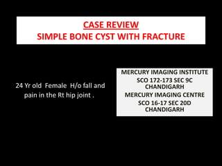
  • 1. CASE REVIEW SIMPLE BONE CYST WITH FRACTURE MERCURY IMAGING INSTITUTE SCO 172-173 SEC 9C CHANDIGARH MERCURY IMAGING CENTRE SCO 16-17 SEC 20D CHANDIGARH 24 Yr old Female H/o fall and pain in the Rt hip joint .
  • 2. Altered MR signal is appreciated in the Rt femur - subcapitate , neck , interochanteric and proximal shaft region. The lesion has following morphological characters : Central / expansible lesion with Lobulated contour. Narrow zone of the transition. Primarily involving medullary cavity with associated thinning of the cortex . Size : 82mm x 39mmx 38mm ( longitudinal x anteroposterior x transverse ). MR signal character : Intermediate on T1, heterogeneously hyperintense on t2/ stir, with interspread bloom On GRE . Features are corroborative with fluid and interspread haemorrhage in the core of the lesion. Cortical break appreciated along the medial aspect of the proximal femoral shaft (just below the lesser trochanter ) . Reactionary edema / hematoma appreciated in the adjacent myofascial planes ( primarily vastusintermedius involved ) . MR Bilateral hip joints done
  • 3. HETEROGENOUSLY HYPERINTENSE ON T2 INTERMEDIATE ON T1W HETEROGENOUSLY HYPERINTENSE ON FATSAT INTERSPREAD BLOOM ON GRE S/O HAEMORRHAGE
  • 4. INTERSPREAD BLOOM ON GRE SUGGESTIVE OF HAEMORRHAGE IN THE LESION.
  • 5. HYPERINTENSE SIGNAL OF THE LESION ON FAT SATURATION- SEQUENCE CORTICAL BREAK
  • 6. ALTERED MR SIGNAL IN RT PROXIMAL FEMORAL SHAFT WITH INTERSPREAD HETEROGENOUSITY . APPRECIATE : NARROW ZONE OF THE TRANSITION . EXPANSILE NATURE CENTRAL LOCATION CORTICAL BREAK
  • 7. SHARP ZONE OF TRANSITION . LESION LIMITED BY THE PHYSEAL SCAR SMOOTH HOMOGENOUS INTERMEDIATE SIGNAL ON T1 W
  • 8. Simple bone cyst / unicameral bone cyst a brief .................................... Unicameral / simple bone cyst . 5 % of primary bone tumors Etiology ? Trauma ? Vascular anomaly 3 to 19 years Occurs during active phase of growth Asymptomatic unless fractured. Usually in proximal femur , proximal humerus Intramedullary centric metaphyseal lesion adjacent to epiphyseal cartilage migrating to diaphysis with growth. 2 to 3 cm radiolucent with long axis parallel to the long axis of the bone . Fine sclerotic boundary . Fallen fragment sign if fractured ( centrally dislodged fragment falls into the dependant position) .