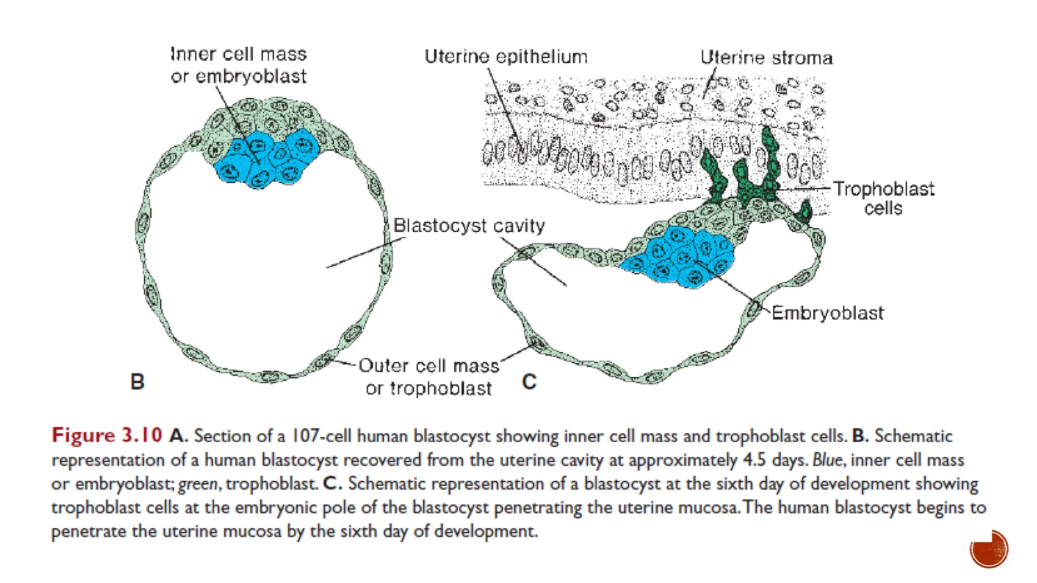
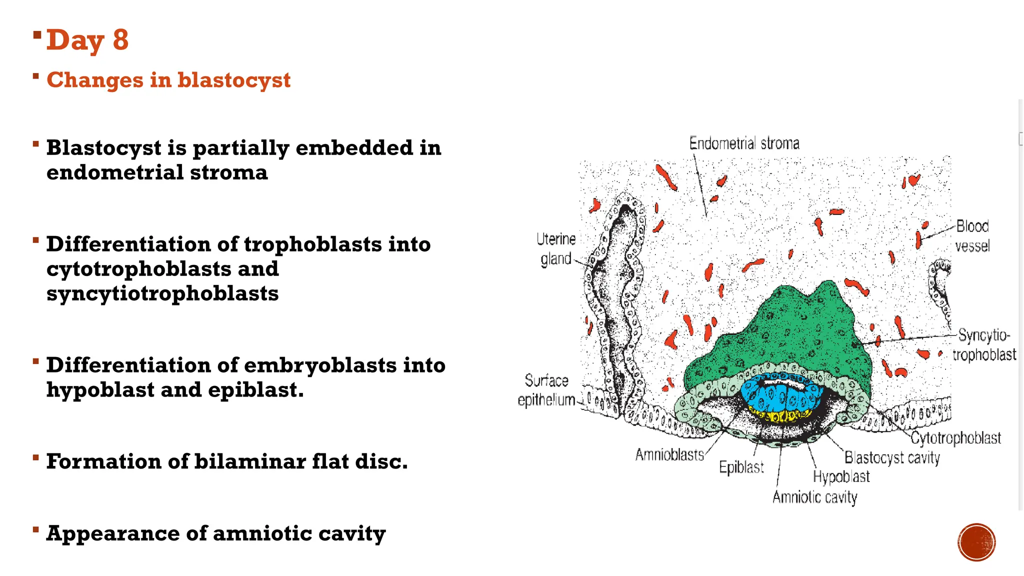
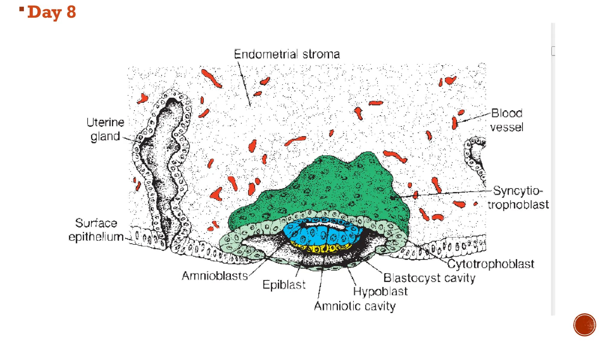
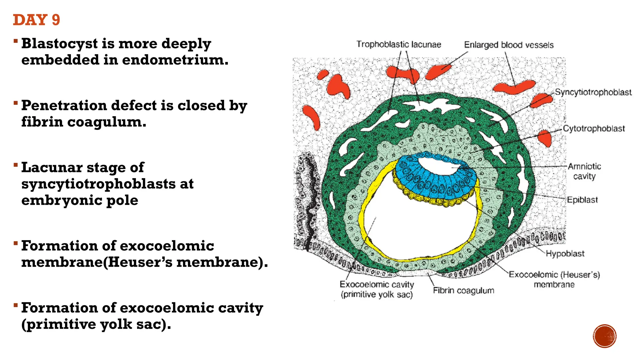
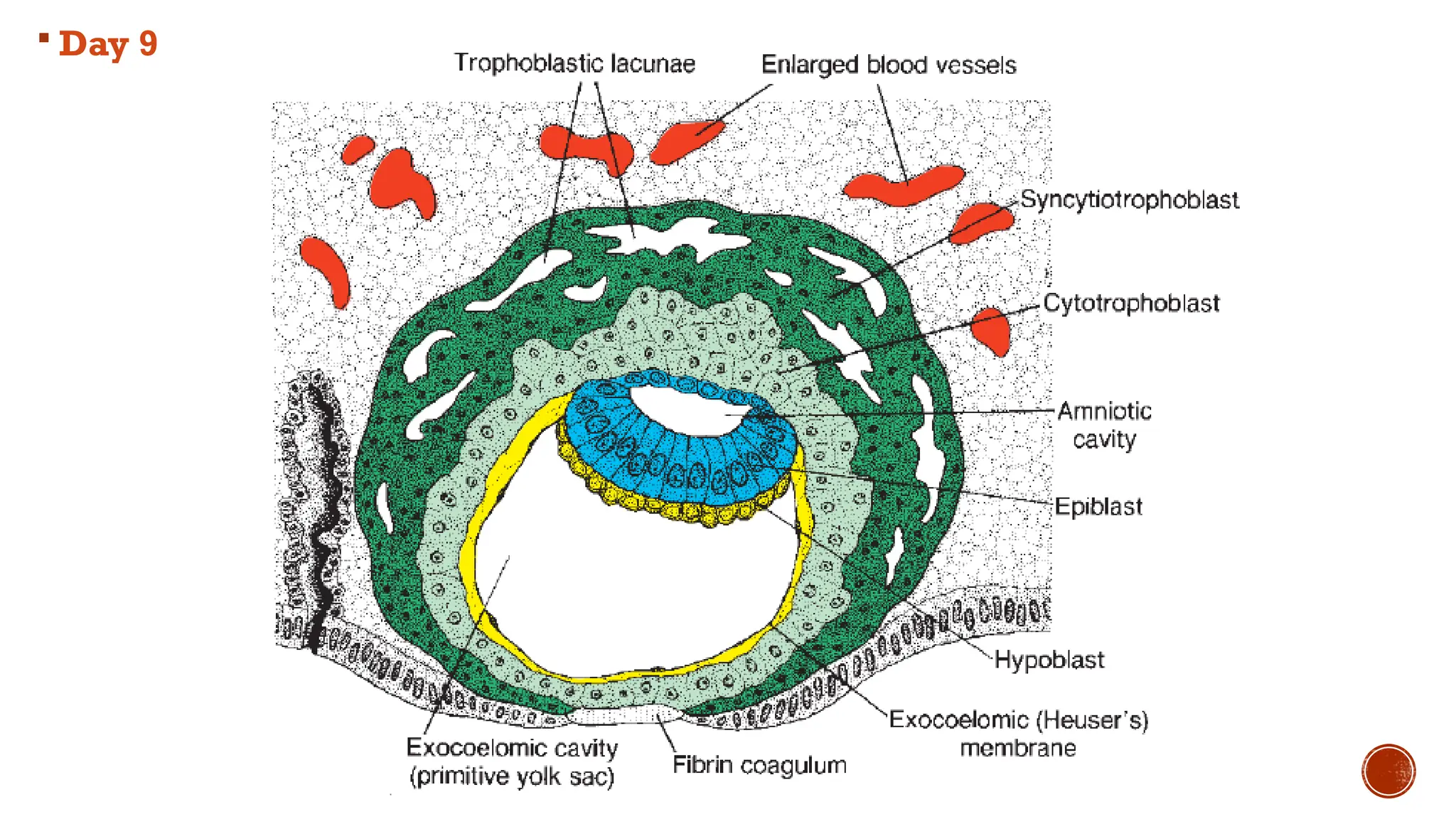
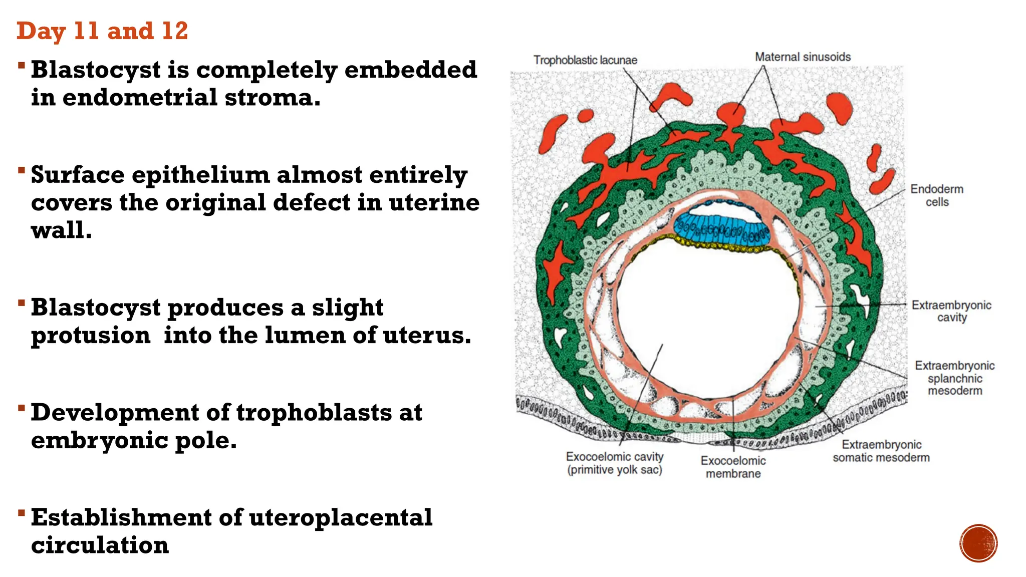
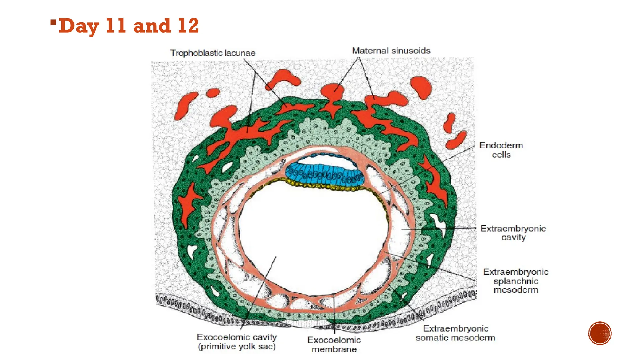
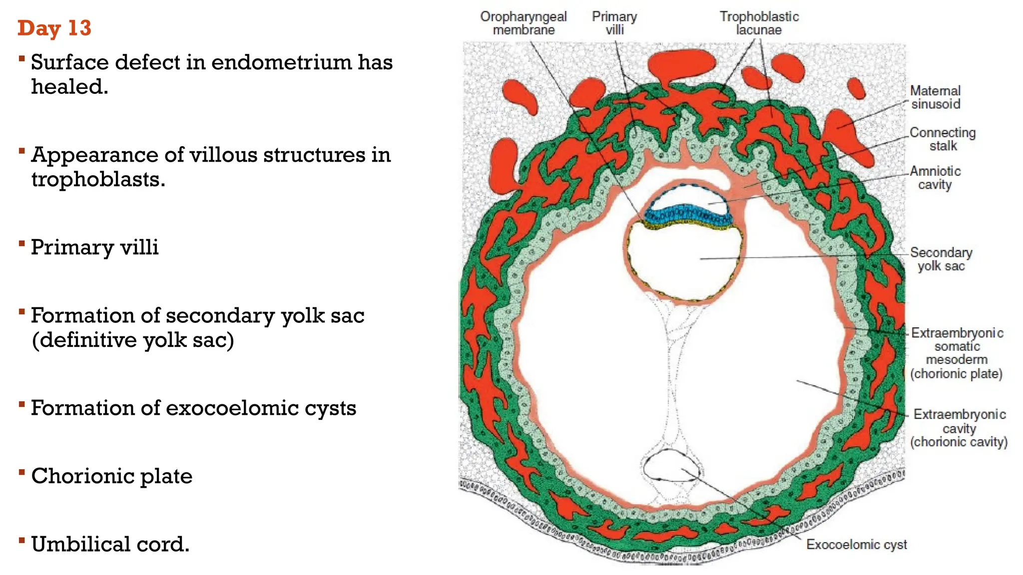
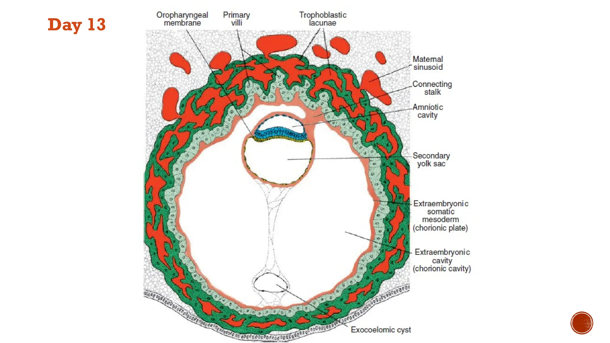
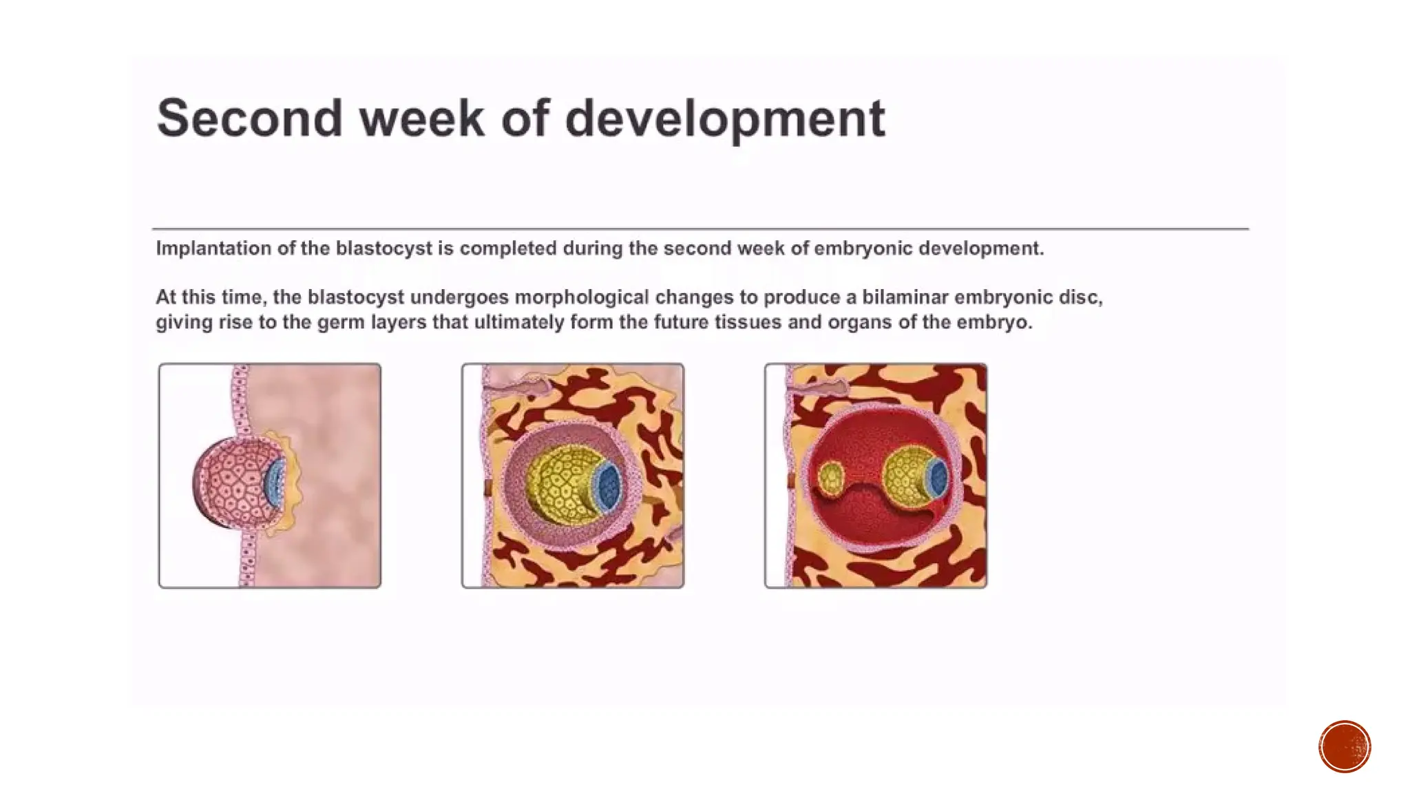
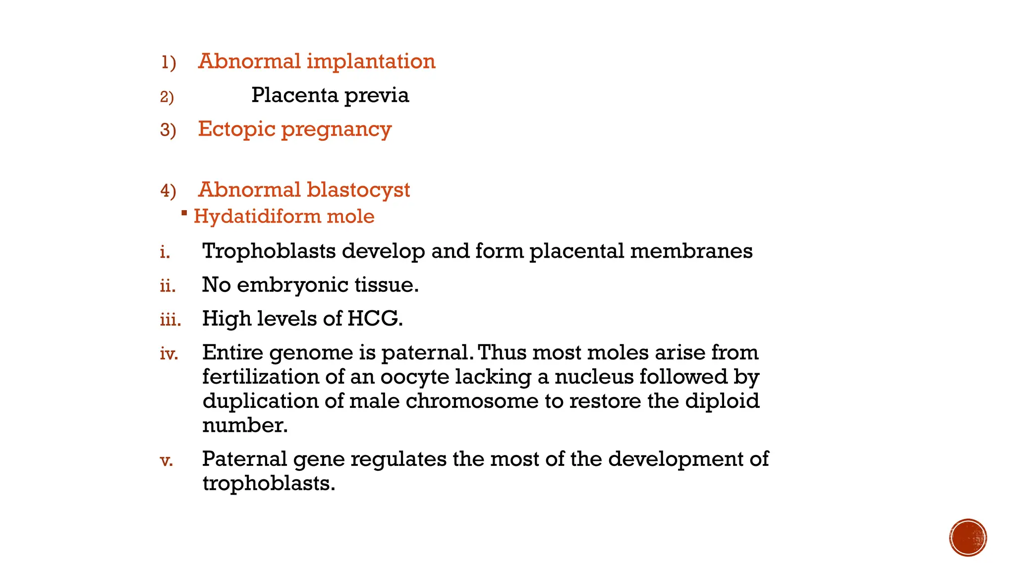
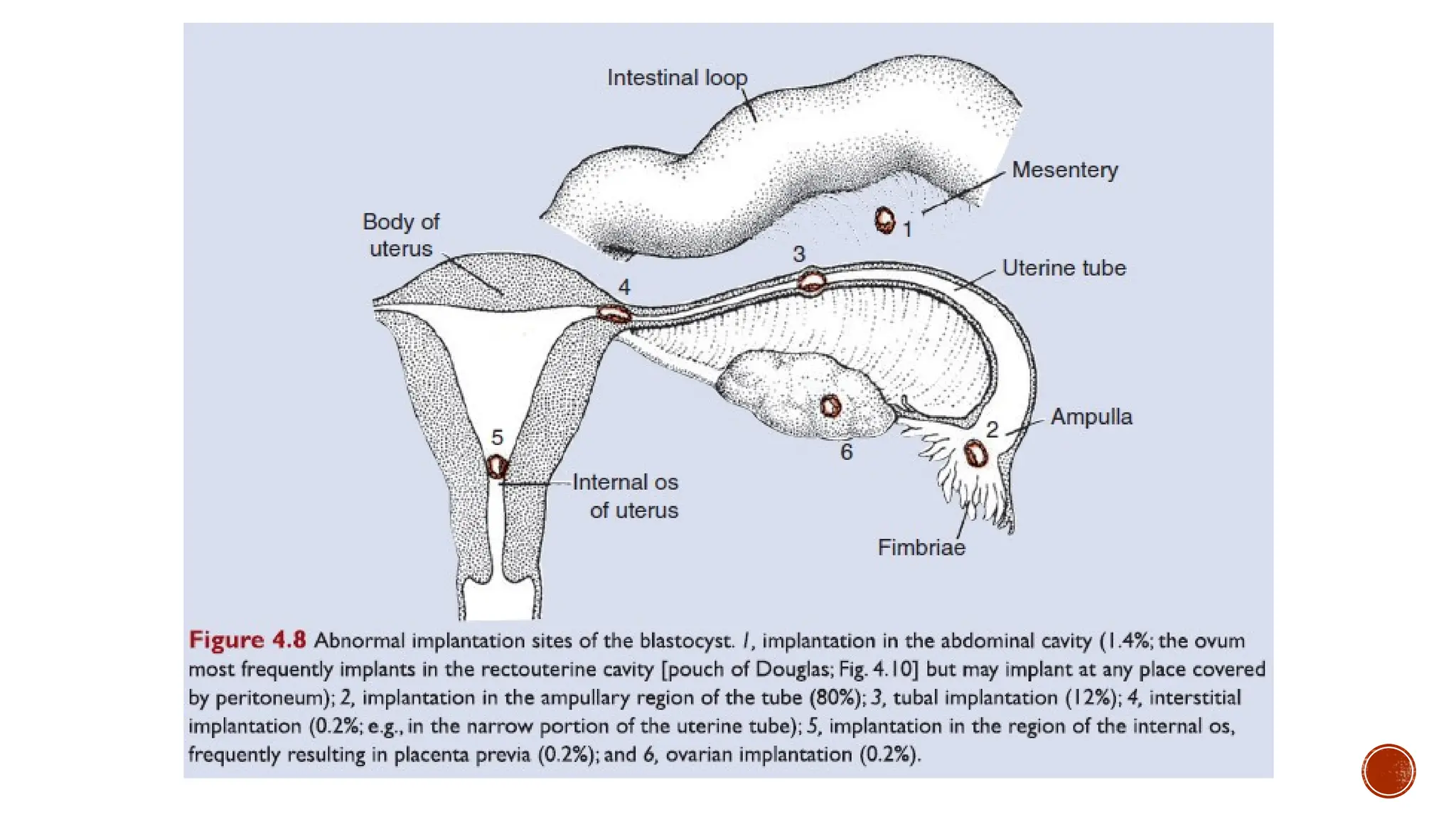
2ND
WEEK OF DEVELOPMENT
Dr Anber Saleem
Day 8
 Changes in blastocyst
 Blastocyst is partially embedded in
endometrial stroma
 Differentiation of trophoblasts into
cytotrophoblasts and
syncytiotrophoblasts
 Differentiation of embryoblasts into
hypoblast and epiblast.
 Formation of bilaminar flat disc.
 Appearance of amniotic cavity
Day 8
2. Changes in endometrial stroma
Becomes edematous and highly vascular
Large and tortuous glands secrete abundant
glycogen and mucus
DAY 9
 Blastocyst is more deeply
embedded in endometrium.
 Penetration defect is closed by
fibrin coagulum.
 Lacunar stage of
syncytiotrophoblasts at
embryonic pole
 Formation of exocoelomic
membrane(Heuser’s membrane).
 Formation of exocoelomic cavity
(primitive yolk sac).
 Day 9
Day 11 and 12
 Blastocyst is completely embedded
in endometrial stroma.
 Surface epithelium almost entirely
covers the original defect in uterine
wall.
 Blastocyst produces a slight
protusion into the lumen of uterus.
 Development of trophoblasts at
embryonic pole.
 Establishment of uteroplacental
circulation
Day 11 and 12
Formation of extraembryonic mesoderm.
Formation of extraembyonic cavity (chorionic cavity).
Formation of Extraembryonic somatic and splanchnic
mesoderm.
Growth of bilaminar germ disc in comparison with
growth of trophoblasts.
Decidua reaction in endometrium
Day 13
 Surface defect in endometrium has
healed.
 Appearance of villous structures in
trophoblasts.
 Primary villi
 Formation of secondary yolk sac
(definitive yolk sac)
 Formation of exocoelomic cysts
 Chorionic plate
 Umbilical cord.
Day 13
Week of 2’s
Trophoblasts differentiate into 2 layers
1. Cytotrophoblasts
2. Syncytiotrophoblasts
Embryoblasts forms 2 layers;
3. epiblasts
4. hypoblasts
Extraembryonic mesoderm splits into 2 layers
5. Somatic
6. Splanchnic
2 cavities form
7. Amniotic
8. Yolk sac
CLINICALS
1) Abnormal implantation
2) Placenta previa
3) Ectopic pregnancy
4) Abnormal blastocyst
 Hydatidiform mole
i. Trophoblasts develop and form placental membranes
ii. No embryonic tissue.
iii. High levels of HCG.
iv. Entire genome is paternal.Thus most moles arise from
fertilization of an oocyte lacking a nucleus followed by
duplication of male chromosome to restore the diploid
number.
v. Paternal gene regulates the most of the development of
trophoblasts.
JAZAKALLAH

second week of development pathology.pptx

  • 2.
  • 4.
    Day 8  Changesin blastocyst  Blastocyst is partially embedded in endometrial stroma  Differentiation of trophoblasts into cytotrophoblasts and syncytiotrophoblasts  Differentiation of embryoblasts into hypoblast and epiblast.  Formation of bilaminar flat disc.  Appearance of amniotic cavity
  • 5.
  • 6.
    2. Changes inendometrial stroma Becomes edematous and highly vascular Large and tortuous glands secrete abundant glycogen and mucus
  • 7.
    DAY 9  Blastocystis more deeply embedded in endometrium.  Penetration defect is closed by fibrin coagulum.  Lacunar stage of syncytiotrophoblasts at embryonic pole  Formation of exocoelomic membrane(Heuser’s membrane).  Formation of exocoelomic cavity (primitive yolk sac).
  • 8.
  • 9.
    Day 11 and12  Blastocyst is completely embedded in endometrial stroma.  Surface epithelium almost entirely covers the original defect in uterine wall.  Blastocyst produces a slight protusion into the lumen of uterus.  Development of trophoblasts at embryonic pole.  Establishment of uteroplacental circulation
  • 10.
  • 11.
    Formation of extraembryonicmesoderm. Formation of extraembyonic cavity (chorionic cavity). Formation of Extraembryonic somatic and splanchnic mesoderm. Growth of bilaminar germ disc in comparison with growth of trophoblasts. Decidua reaction in endometrium
  • 12.
    Day 13  Surfacedefect in endometrium has healed.  Appearance of villous structures in trophoblasts.  Primary villi  Formation of secondary yolk sac (definitive yolk sac)  Formation of exocoelomic cysts  Chorionic plate  Umbilical cord.
  • 13.
  • 14.
    Week of 2’s Trophoblastsdifferentiate into 2 layers 1. Cytotrophoblasts 2. Syncytiotrophoblasts Embryoblasts forms 2 layers; 3. epiblasts 4. hypoblasts Extraembryonic mesoderm splits into 2 layers 5. Somatic 6. Splanchnic 2 cavities form 7. Amniotic 8. Yolk sac
  • 16.
  • 17.
    1) Abnormal implantation 2)Placenta previa 3) Ectopic pregnancy 4) Abnormal blastocyst  Hydatidiform mole i. Trophoblasts develop and form placental membranes ii. No embryonic tissue. iii. High levels of HCG. iv. Entire genome is paternal.Thus most moles arise from fertilization of an oocyte lacking a nucleus followed by duplication of male chromosome to restore the diploid number. v. Paternal gene regulates the most of the development of trophoblasts.
  • 20.