Rickets and Osteomalacia: Types, Causes, Symptoms and Treatment
•Download as PPTX, PDF•
6 likes•2,392 views

Rickets and osteomalacia

Recommended
More Related Content
What's hot
What's hot (20)
Similar to Rickets and Osteomalacia: Types, Causes, Symptoms and Treatment
Similar to Rickets and Osteomalacia: Types, Causes, Symptoms and Treatment (20)
Recently uploaded
Recently uploaded (20)
Rickets and Osteomalacia: Types, Causes, Symptoms and Treatment
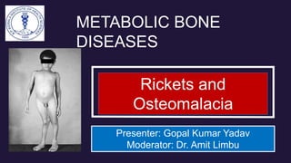
- 1. Rickets and Osteomalacia Presenter: Gopal Kumar Yadav Moderator: Dr. Amit Limbu METABOLIC BONE DISEASES
- 2. OBJECTIVES 1. Introduction and Definition 2. Causes of Rickets and Osteomalacia 3. Rickets 4. Osteomalacia
- 3. Introduction •Osteomalacic diseases: These are diseases characterized by an increase in the ratio of the organic fraction to the mineralized fraction, i.e. the available organic matter is undermineralized. •Rickets: Lack of adequate mineralization of growing bones. It occurs before closure of growth plates. •Osteomalacia: Lack of adequate mineralization of trabecular bone.
- 5. RICKETS: Types, Clinical features, Investigations, Diagnosis and Treatment
- 6. Types of Rickets (1/2) Classification I • Fetal rickets-osteomalacic mothers. • Infantile rickets-6mths to 3 yrs ,most common form, nutritional. • Late rickets or rachitis tarda-late onset , familial, vitamin D resistant.
- 7. Types of Rickets (2/2) Classification II Type I Rickets: Dietary deficiency or defects in the metabolism of vitamin D. Type II Rickets: Low serum phosphorus due to dietary deficiency or defective renal tubular resorption
- 8. Clinical Features (1/5) •A – Abdomen protuberant due to muscular hypotonia •B – Bowing of bones (on weight bearing)
- 9. Clinical Features (2/5) • C – Costochondral Junction prominent - (Rosary), Craniotabes (soft skull)
- 10. Clinical Features (3/5) • D – Diaphragm pull - Harrisons groove (lateral indentation of chest due to pull of diaphragm on ribs)/Double malleolus d/t metaphyseal hyperplasia
- 11. Clinical Features (4/5) • E – Enamel defect of teeth and delayed dentition • F – Forward sternum - Pigeon chest (Pectus carinatum) • G – Growth plate - widening
- 12. Clinical Features (5/5) • H – Hypocalcemia causing Hyper PtH • I – Irritability • J – Joint deformities - Genu valgum/genu varum/coxa vara • K – Kyphosis • L – Loosers zones • M – Milestone delayed Muscle weakness
- 13. Q. In which condition is Windswept deformity seen? some books say RA, others say Rickets? American authors have published RA, British have published Rickets...which one to pick sir? Ans. Both diseases have wind swept deformity Rickets at knees and at hand in RA but classically it has been named for rickets. It is called as tackle deformity also.
- 14. Investigations (1/2) •Laboratory: Serum Ca2+, Serum PO4-, Serum ALP
- 15. Investigations (2/2) • Radiological Features: X-ray of both wrist and knees- AP views
- 16. Diagnosis: • Is based on the combination of a history of poor vitamin D intake and risk factors for decreased cutaneous synthesis, radiographic changes consistent with rickets, and typical laboratory findings .
- 17. Treatment: Medical (1/4) 1.Nutritional rickets: Two strategies for administration of vitamin D. Stoss therapy, 300,000–600,000 I.U. of vitamin D are administered orally or intramuscularly. The alternative is daily high dose vitamin D, 2000– 5000 I.U./day over 4–6 week. Followed by 400 I.U. Vit D/Day and supplements of calcium for 2–4 months
- 18. Treatment: Medical (2/4) 2. Hypophosphatemic Rickets Oral phosphate and Vit D supplements Joule’s Solution—Dibasic sodium phosphate, phosphoric acid is given in hypophosphatemic Rickets. 3. VDDR I: Calcitriol, calcium, phosphate supplements 4. VDDR II: Treatment not satisfactory large doses
- 19. Treatment: Medical (3/4) 5. Renal tubular acidosis Bicarbonate supplements (Shohl’s solution— Citric acid, sodium citrate) and phosphate supplementation. 6. CRF Calcitriol, calcium supplements and phosphate restriction.
- 20. Treatment: Orthopedic (4/4) Conservative Methods: Designed splints (mermaid splints) or orthopedic shoes for correction of knee deformities Operative methods: Corrective Osteotomies, after 6 months of starting the medical treatment
- 21. OSTEOMALACIA: Clinical features, Investigations and Treatment
- 22. Clinical features: Early stages: Non-specific Bone pains: Backache to diffuse pains Muscle weakness: Waddling gait, Carpopedal spasm and facial twitching Spontaneous fractures: usually in spine
- 23. Investigations Radiological Examination: Looser’s zone (pseudo-fractures) , Triradiate pelvis and Protrusio-acetabuli Bone biopsy: from iliac crest confirms the diagnosis (Excessive uncalcified osteoid) Serum: Low serum calcium and phosphates level, and high ALP level
- 24. Looser’s Zone ( Milkman’s Pseudofractures ) Pathognomonic
- 25. Trefoil Pelvis
- 26. Treatment of Osteomalacia Identify the cause. Vit. D 50000 IU / week X 3-12 weeks, Followed by maintenance 400-800IU /day Along with elemental Calcium 1.5 to 2 g / day
- 27. References
- 28. Thank You! • To feel Low or to doubt in yourself is to commit mental suicide which is more catastrophic than physical suicide because mental suicide is chronic incident that melts you daily on your voyage! –GK Yadav
Editor's Notes
- There are four types of metabolic bone diseases. • Osteopenic diseases: These diseases are characterized by a generalized decrease in bone mass (i.e. loss of bone matrix), though whatever bone is there, is normally mineralized (e.g. osteoporosis). • Osteosclerotic diseases: There are diseases characterized by an increase in bone mass (e.g. fluorosis). • Osteomalacic diseases: These are diseases characterized by an increase in the ratio of the organic fraction to the mineralized fraction, i.e. the available organic matter is undemineralized. • Mixed diseases: These are diseases that are a combination of osteopenia and osteomalacia (e.g. hyperparathyroidism).
- DMP1: Dentin Matrix Protein 1 PHEX: phosphate-regulating gene with homologies to endopeptidases on the X chromosome HYPOPHOSPHATASIA—DIFFERENT FROM HYPOPHOSPHATEMIA! • Genetic error in synthesis of ALP • Normal calcium and phosphate levels • Low ALP • Autososmal recessive • Phosphoethanolamine in urine/serum • Changes early in life • Delayed dentition • Genu valgum/varum • Mortality 50–70% • Treatment is supportive
- A – Abdomen protuberant B – Bowing of bones (on weight bearing) C – Costochondral Junction prominent - (Rosary), Craniotabes (open fontanelles) D – Diaphragm pull - Harrisons groove (lateral indentation of chest due to pull of diaphragm on ribs)/Double malleolus E – Enamel defect of teeth and delayed dentition F – Forward sternum - Pigeon chest (Pectus carinatum) G – Growth plate - widening H – Hypocalcemia causing Hyper PtH I – Irritability J – Joint deformities - Genu valgum/genu varum/coxa vara K – Kyphosis L – Loosers zones M – Milestone delayed Muscle weakness R – Rickets
- A – Abdomen protuberant B – Bowing of bones (on weight bearing) C – Costochondral Junction prominent - (Rosary), Craniotabes (open fontanelles) D – Diaphragm pull - Harrisons groove (lateral indentation of chest due to pull of diaphragm on ribs)/Double malleolus E – Enamel defect of teeth and delayed dentition F – Forward sternum - Pigeon chest (Pectus carinatum) G – Growth plate - widening H – Hypocalcemia causing Hyper PtH I – Irritability J – Joint deformities - Genu valgum/genu varum/coxa vara K – Kyphosis L – Loosers zones M – Milestone delayed Muscle weakness R – Rickets
- A – Abdomen protuberant B – Bowing of bones (on weight bearing) C – Costochondral Junction prominent - (Rosary), Craniotabes (open fontanelles) D – Diaphragm pull - Harrisons groove (lateral indentation of chest due to pull of diaphragm on ribs)/Double malleolus E – Enamel defect of teeth and delayed dentition F – Forward sternum - Pigeon chest (Pectus carinatum) G – Growth plate - widening H – Hypocalcemia causing Hyper PtH I – Irritability J – Joint deformities - Genu valgum/genu varum/coxa vara K – Kyphosis L – Loosers zones M – Milestone delayed Muscle weakness R – Rickets
- A – Abdomen protuberant B – Bowing of bones (on weight bearing) C – Costochondral Junction prominent - (Rosary), Craniotabes (open fontanelles) D – Diaphragm pull - Harrisons groove (lateral indentation of chest due to pull of diaphragm on ribs)/Double malleolus E – Enamel defect of teeth and delayed dentition F – Forward sternum - Pigeon chest (Pectus carinatum) G – Growth plate - widening H – Hypocalcemia causing Hyper PtH I – Irritability J – Joint deformities - Genu valgum/genu varum/coxa vara K – Kyphosis L – Loosers zones M – Milestone delayed Muscle weakness R – Rickets
- A – Abdomen protuberant B – Bowing of bones (on weight bearing) C – Costochondral Junction prominent - (Rosary), Craniotabes (open fontanelles) D – Diaphragm pull - Harrisons groove (lateral indentation of chest due to pull of diaphragm on ribs)/Double malleolus E – Enamel defect of teeth and delayed dentition F – Forward sternum - Pigeon chest (Pectus carinatum) G – Growth plate - widening H – Hypocalcemia causing Hyper PtH I – Irritability J – Joint deformities - Genu valgum/genu varum/coxa vara K – Kyphosis L – Loosers zones M – Milestone delayed Muscle weakness R – Rickets
- Urinalysis is useful for detecting the glycosuria and aminoaciduria (positive dipstick for protein) seen with Fanconi syndrome. Evaluation of urinary excretion of calcium (24 hr collection for calcium or calcium-creatinine ratio) is helpful if hereditary hypophosphatemic rickets with hypercalciuria or Fanconi syndrome is suspected. Direct measurement of other fat-soluble vitamins (A, E, and K) or indirect assessment of deficiency (prothrombin time for vitamin K deficiency) is appropriate if malabsorption is a consideration. Bone Biopsy: Osteoid seams --wider and extensive. Tetracycline labelling shows mineralisation defective
- 1. Nutritional rickets • Two strategies for administration of vitamin D. Stoss therapy, 300,000–600,000 I.U. of vitamin D are administered orally or intramuscularly. The alternative is daily high dose vitamin D, 2000–5000 I.U./day over 4–6 week. Followed by 400 I.U. Vit D/Day and supplements of calcium for 2–4 months 2. Hypophosphatemic Rickets • Oral phosphate and Vit D supplements • Joule’s Solution—Dibasic sodium phosphate, phosphoric acid is given in hypophosphatemic Rickets. 3. VDDR I • Calcitriol, calcium, phosphate supplements 4. VDDR II • Treatment not satisfactory large doses of calcitriol and calcium, phosphate supplements. 5. Renal tubular acidosis • Bicarbonate supplements (Shohl’s solution—Citric acid, sodium citrate) and phosphate supplementation. 6. CRF • Calcitriol, calcium supplements and phosphate restriction.
- Protrusio acetabular (acetabulum protrudes into pelvis due to bone softening when bilateral called as Otto Pelvis),
- What is the deformity? B/L Genu Varum 3 D/Ds of bowing of legs? Physiological Rickets Blount’s disease What is the commonest inheritance pattern in hypophosphatemic rickets? X-linked dominant Other features of X-Linked Hypophosphatemic Rickets? Short Stature Frequent Tooth abscesses How will you treat? Oral Phosphorus + Calcitriol