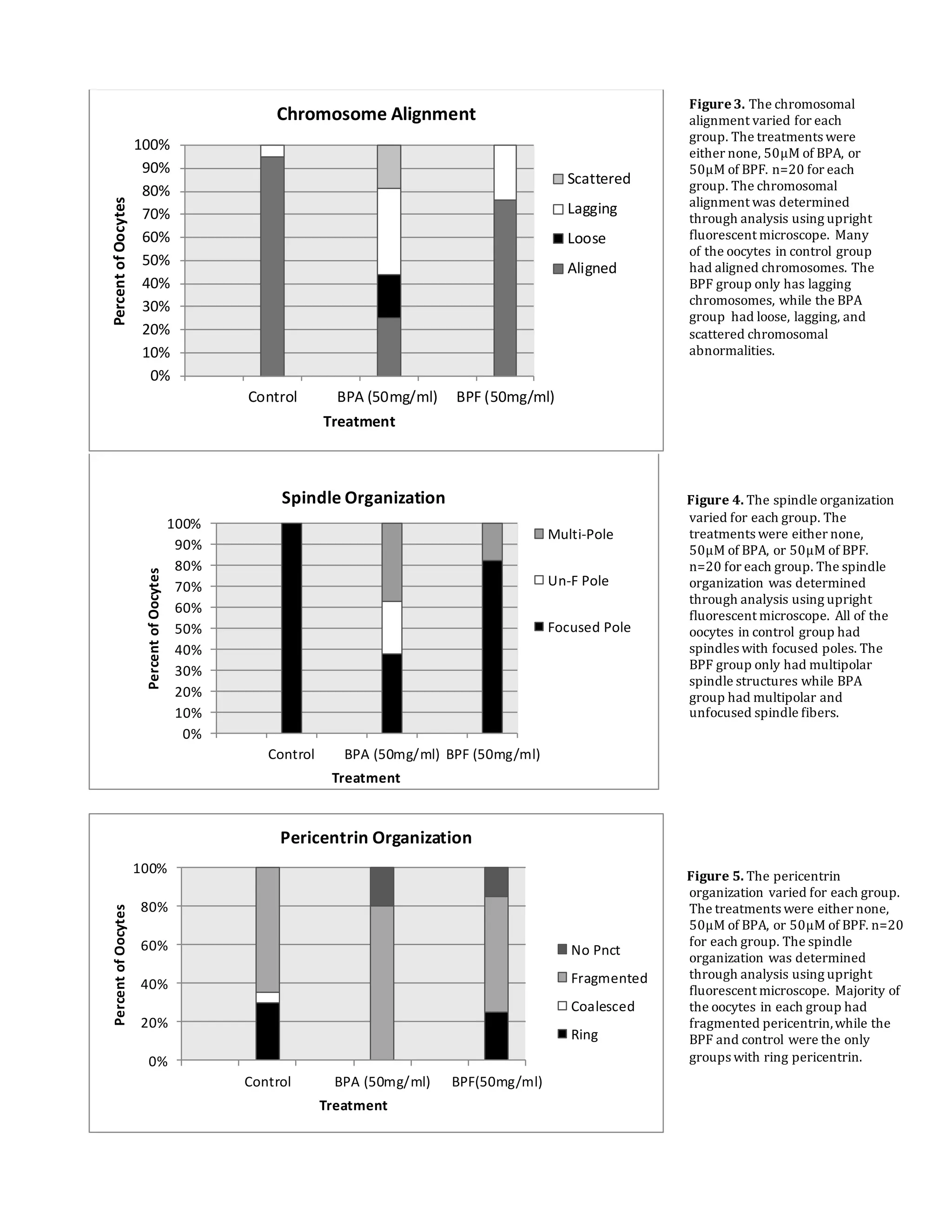
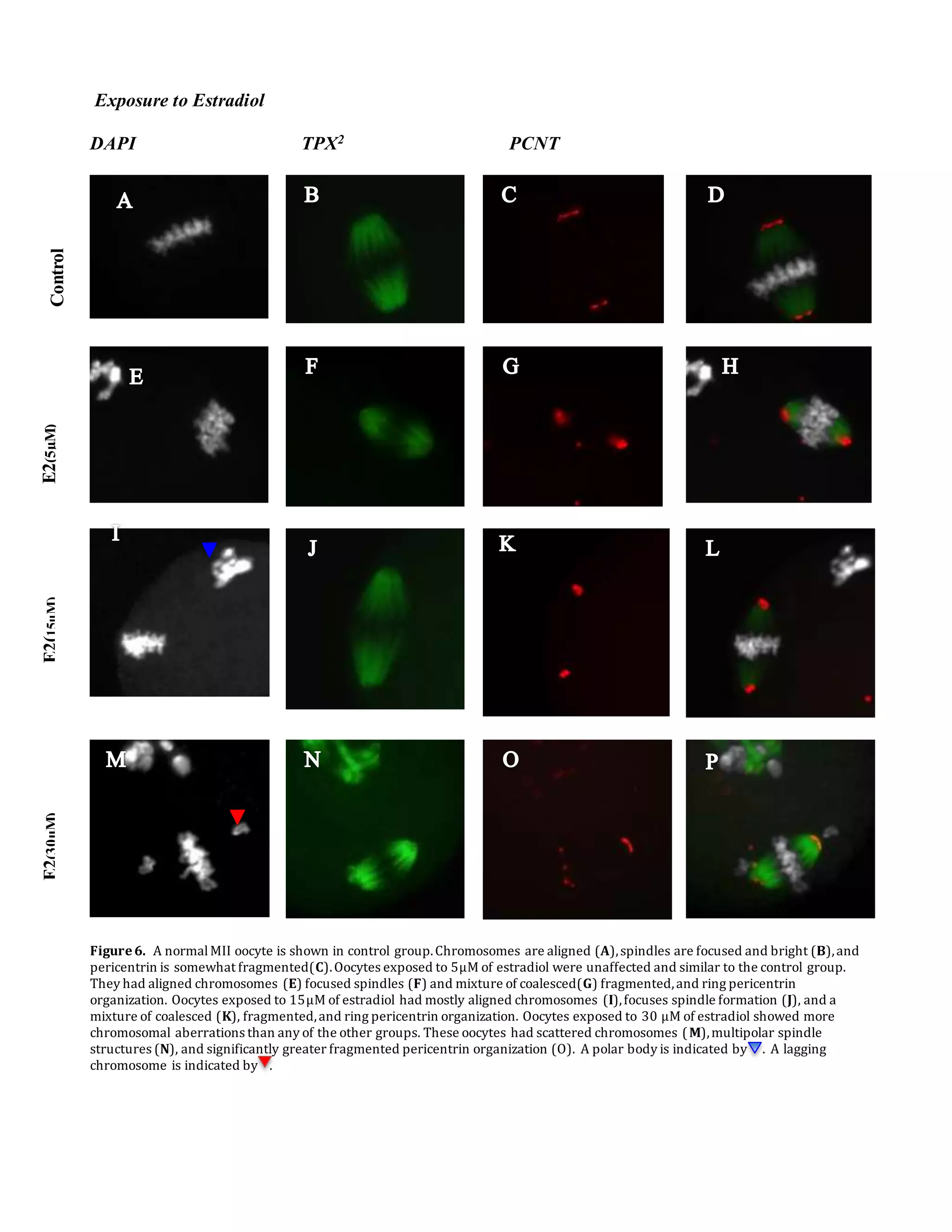
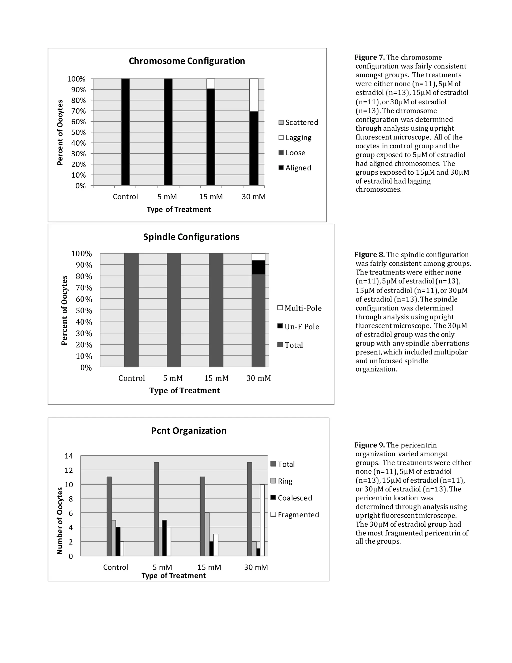
This document summarizes a study that examined the effects of bisphenol A (BPA), bisphenol F (BPF), and high levels of estrogen on oocyte maturation and quality. Mouse oocytes were exposed to varying concentrations of BPA, BPF, and estrogen. The oocytes were then analyzed for spindle formation, centrosome position, and chromosome alignment. Oocytes exposed to BPA, BPF, and high estrogen showed lower maturation rates and more chromosome abnormalities compared to the control group. BPA exposure resulted in more chromosomal errors than BPF exposure. The findings suggest that exposure to synthetic estrogens like BPA and BPF can negatively impact oocyte development and potentially cause birth defects.
Epigenetics is the study, in the field of genetics, of cellular and physiological phenotypic trait variations that are caused by external or environmental factors that switch genes on and off and affect how cells read genes instead of being caused by changes in the DNA sequence. -Wikipedia
PowerPoint giving a summary on research in stem cells (brief historical overview), and the explanatory component of the papers which changed the game of stem cell research Yamanka's Nuclear Reprogramming.
Pregnancy outcome following swim up preparation of both fresh and cryopreserv...lukeman Joseph Ade shittu
This study was designed to assess the impact of swim up preparation of both fresh and cryopreserved sperm on the pregnancy outcome in a private fertility centre in Lagos. A cross-sectional prospective analysis of 34 asthenozoospermic semen samples of men whose wives were undergoing assisted reproduction was studied. The basic semen parameters comprising of the volume, count, and motility of the sperm before and after swim up preparations with pregnancy outcome were measured. For fresh semen (n = 28, mean age = 37.0 ± 1.1 years, mean volume = 2.16 ± 0.1 ml), the sperm count decreased significantly (p<0.01)><0.01)><0.01)><0.01) from 25.1 ± 4.01 to 32.8 ± 6.18%. The pregnancy outcome of cryopreserved was 30%. The pregnancy outcome was higher with fresh than the cryopreserved semen. However, the motility was a significant indicator for the successful outcome. Swim up procedure improve the motility of both cryopreserved and fresh semen with a better pregnancy outcome in this study.
In conclusion this work supports the hypothesis that FtMt contributes to male fertility ensuring ATP energy level of spermatozoa. However the mechanisms by which FtMt acts remain to be elucidated.
Epigenetics, Pedigree Analysis, Twins studies, Genetic Counseling, Prenatal testing, Linked genes, Chemical Nature of the Gene, History of Genetics, DNA Structure, RNA Structure and DNA Analysis.
The Potential Impact of Preimplantation Genetic Diagnosis on Discrimination o...blaine_5
The argument that selection against specific genetic traits will lead to increased discrimination is both compelling and troubling. Indeed, it is reasonable to conclude that if a large number of people use PGD to select against traits they consider to be disabilities then the probability of increased discrimination and marginalization would be greatly increased. However, as this Note argues, most participants in the PGD disability debate overlook important limitations of both trait selection and large-scale PGD adoption that will likely mitigate the negative potentially negative impact of PGD technology.
Ridge IVF is palce for Infertility Treatment in Delhi/NCR with team of best Infertility Specialist. Female infertility treatment Delhi, male infertility treatment Delhi, Infertility Specialist in Delhi, In Vitro Fertility, IVF Doctors Delhi, IVF Specialist Delhi, infertility hospital Delhi.Our Clinic is North India's leading chain of fertility centers providing comprehensive male and female infertility treatment "
We provide a wide variety of infertility treatments, from basic infertility care to advanced infertility treatment at our state of the art centers in india. We have three tertiary care infertifilty treatment centers in India, conveniently located in North and South Delhi.
Detection of misfolded aβ oligomers for sensitive biochemical diagnosis of Al...José Luis Moreno Garvayo
El equipo del Dr. Claudio Soto de la Universidad de Texas ha conseguido demostrar que son capaces de detectar pequeños fragmentos de proteínas mal plegadas, los precursores de las placas seniles, que pueden estar circulando por nuestro cuerpo durante años o décadas antes de que surjan los primeros síntomas de la enfermedad de Alzheimer
1. The Effect of BPA on Oocyte Maturation and Quality
Dr. Maria Viveiros
Department of Physiology and Pharmacology
College of Veterinary Medicine
viveiros@uga.edu
706.542.5869
Madison Kathleen Cook
393 Oconee Street Athens, GA, 30601
mkc29463@uga.edu
770.331.5298
University of Georgia
Fall 2016
VPHY 4960 H
2. Objective
To determine whether BPA, BPF, or high levels of estrogen affect oocyte maturity or quality through
spindle disruption, changes in centrosome position, and chromosome misalignment.
Abstract
Synthetic estrogens are present in many products throughout the world and can cause meiotic errors in
germ cell division. Errors in meiosis, through unfocused spindle formation or chromosome
misalignment, can potentially cause aneuploidy, which is one of the leading causes of birth defects.
BPA is an estrogen-mimicking compound used in plastic products, canned goods, and thermal receipts.
Past experiments have shown relevant concentrations of BPA can cause spindle disruption and
chromosome misalignment. BPF, an analogue of BPA, is its number one substitute. The effects of BPF
on meiosis is less clear, however, it has been shown to cause similar negative effects on germ cells.
This experiment tested whether varying levels of estrogen, whether it is natural or synthetic, affects
meiotic division of female mouse germ cells. Oocytes were collected from mice that were injected with
5IU PMSG (stimulates follicle growth). The oocytes were then placed in a culture with various
concentrations ranging from 5-50M of Bisphenol A (BPA), Bisphenol F(BPF), and estradiol(E2).
Next, the oocytes were immunostained using primary and secondary antibodies that tagged
microtubulin and microtubule organizing centers (centrosomes). DAPI was then used as a
chromosomal counterstain. Oocytes were analyzed using an upright fluorescent microscope. Oocytes
exposed to both BPA and BPF showed lower maturation rates and more chromosome disruptions than
the control. BPA, however, showed significantly more chromosomal errors than BPF. Oocytes exposed
to high levels of estradiol also showed some chromosomal abnormalities. Thus, it can be concluded
that high estrogen levels including those from estrogen mimicking compounds, can alter oocyte quality
and potentially lead to birth defects.
3. Introduction:
Meiosis is a cellular division that takes place in sex cells. It reduces the number of
chromosomes of the parent cell to half of the original number and creates genetic diversity. It involves
two meiotic divisions. The first meiotic division reduces the amount of chromosomes to one and the
amount of chromatids to two. After the second meiotic division, the one chromosome is reduced to one
chromatid. The purpose of the first meiotic division is to separate homologous chromosomes. The
second meiotic division occurs only at fertilization to produce a haploid cell ready to combine with
another haploid sex cell, to produce a zygote (De la Fuente et al, 2011) The one chromosome lost in
the first meiotic division is released in the form of a polar body. A chromatid is released in a second
polar body in the second meiotic division. Oocytes are first arrested in GV-stage (prophase 1) until
ovulation is induced by luteinizing hormone and follicle stimulating hormone. The oocyte then goes on
to its first meiotic division and second if it is fertilized. In the experiment, the focus will be on the first
meiotic division because this is where most errors are known to occur. Any errors in the first division
of meiosis could lead to an abnormal number of chromosomes. Ensuring a proper first meiotic division
is crucial for normal embryonic development.
Two main components of meiotic division are spindle formation and centrosome location.
Centrosomes assemble the spindles and the protein pericentrin, found in centrosomes, helps the spindle
fibers anchor to the centrosome (Can et al, 2005). Spindle fibers are made of microtubule protein
fibers. These spindles attach to the chromosomes at the kinetochore region and to the centrosomes at
the end of each oocyte. Spindles ensure the proper alignment and equal separation of the chromosomes
(Compton 2000). A spindle assembly checkpoint (SAC) corrects any errors by stabilizing
chromosome and microtubule interactions before proceeding to the second meiotic division. However,
oocytes with spindle errors sometimes still proceed onto the second meiotic division (De la Fuente et
al, 2011). Abnormal spindle formation could lead to an unequal separation of sister chromatids and
thus aneuploidy. Aneuploidy, where an abnormal amount of chromosomes is present, is one of the
4. main causes of birth defects in older women and is also a main characteristic of cancerous tumors
(Marchetti et al, 2009). Other factors besides age can affect the first meiotic division, including
environmental toxins.
BPA is a type of endocrine disrupting compound that is found in many plastic products,
including aluminum cans and thermal receipts. It has been increasingly used throughout the world
since its introduction to the plastic industry in 1940. Over 10 million tons are used per year (Santangeli
et al, 2016). The most common route of exposure is through ingestion, due to the leaking of epoxy
resins from BPA into food sources (metallic food cans). BPA was found in over 90% of people
according to the National Health and Nutrition Survey (Machtinger et al, 2013). It has been found in
human urine, blood, placental tissue, and breast milk (Chen, et al 2016).
BPA is a weak mimic of estrogen and has been shown to cause defects in germ cell division.
Estrogen is crucial for cell growth including processes such as meiosis and DNA replication. However
overexposure to estrogen can cause adverse effects, which makes the increasing presence of estrogen
mimicking compounds disconcerting (Can et al, 2005). BPA has shown negative effects on spindle
formation, centrosome position, and chromosome alignment in mouse and human oocytes (Nakano,
2015). Rodents exposed to normal doses of BPA experienced abnormalities in chromosome
segregation in the first meiotic division that led to aneuploidy in subsequent oocytes (Chen et al, 2016).
BPA also has shown affects on oocyte maturation, preventing the cells form undergoing the first and
second divisions of meiosis (Ferris 2015). Past studies showed that women with higher concentrations
of BPA exposure undergoing IVF treatment had less oocytes and less mature oocytes(MII). Studies
have also shown abnormal spindle formation including multipolarity in oocytes exposed to relevant
concentrations of BPA (Peretz et al, 2014). Overall, BPA has shown adverse affects on oocyte quality
in human and mouse species at concentrations similar to normal exposure levels.
Due to studies showing adverse affects of BPA on human health, new alternatives are being
utilized. Many of these are analogues of BPA and are structurally similar (Figure 1). There are 16 total
5. bisphenol analogues present, but only a few are being used in mass industrial production. BPF is one
of the more popular replacements for BPA because it has a wide range of applications including
adhesives, plastics, lacquers, and food packaging (Chen et al, 2016). BPF (bisphenol F, 4,4′-
dihydroxydiphenylmethane) is produced at the same high levels as BPA. Unlike BPA, BPF use is not
regulated. Human exposure has increased vastly over the past five years (Maćczak et al, 2015). BPF
was found in a variety of food products in the United States (Chen et el, 2016). In vivo studies showed
that BPF has estrogenic, androgenic, and thyroidgenic characteristics. In vitro studies showed cellular
dysfunction, DNA damage, and chromosomal abnormalities due to BPF exposure. In comparison with
BPA, studies have shown that BPF is just as potent if not more potent in regards to estrogenic activity
(Rochester et al, 2015). BPF may be equally or more detrimental than BPA to human health, making it
a poor substitute. Consequently, “BPA-free” may not explicitly indicate that a product is a safer option.
Figure1. The structures of BPA and BPF are very similar.
BPF lacks the two methyl groups present in BPA. Estradiol
is very different structurally than BPA and BPF. It has one
phenolgroup and 2 cyclohexane rings as well as one
cyclopentane ring with an OH group. Estradiol is a form of
estrogen.
6. Methods
Mouse Hormone Treatment and Oocyte Collection
Mouse oocytes were used for all experiments in this study. The specific mouse strain used is referred
to as B6D2F1 mice (C57BL/6J females x DBA/2J males). To collect pre-ovulatory stage oocytes, 21
to 23-day old females were injected with 5IU PMSG (EMD Biosciences) to stimulate follicle
development. For experiments requiring mature metaphase-II (MII) oocytes the females were
subsequently treated with 5 IU hCG (EMD Biosciences) to promote ovulation as previously described
(Ma and Viveiros, 2014). For collection of prophase-I arrested (GV-stage) oocytes, the ovaries were
surgically removed from female mice approximately 44h after PMSG treatment. The ovarian follicles
were punctured using 27 gauge needles to release the cumulus cell-oocyte complexes (COCs) into
Minimal Essential Medium (MEM) supplemented with 3 mg/ml crystallized bovine serum albumin
(BSA, from Sigma). Ovulated, MII-stage oocytes, were collected from the oviducts of super-ovulated
females, approximately 16 hours after treatment with 5 IU hCG.
Oocyte Culture and Exposure to Synthetic Estrogens
To test the influence of synthetic estrogens during meiotic maturation, COCs were placed in culture for
17 hours with high concentrations (50g/ml) of BPA and BPF. The control oocytes were cultured in
media alone. To test the influence of estrogen on the mature MII-stage, ovulated oocytes were
recovered from the oviducts and the surrounding cumulus cells were removed by a brief exposure to
hyaluronidase (Sigma). The denuded oocytes were then cultured for 4 hours with 30M Estradiol (E2).
The control oocytes were cultured in media alone. All oocytes were cultured in MEM supplemented
with 3 mg/ml BSA and 10% fetal calf serum (Hyclone). Cultures were maintained at 37°C with 5%
CO2, 5% O2 and 90% N2 (Ma and Viveiros, 2014). At the end of culture, each group of oocytes was
placed in fixative (4% PFA in PBS, supplemented with 0.25% Triton X) for 30 minutes at 37°C and
then washed 3 times (10 minute washes) in wash buffer (10 ml PBS, 500μl of 5% serum, 500μl
7. antibiotic/antimycotic 100x solution). The detergent (Triton-X) permeabilizes the cell membrane,
allowing for the antibodies to enter the oocytes. The oocytes were blocked at 4°C overnight in wash
buffer supplemented with 5% serum.
Immunostaining
After blocking, the oocytes were immunostained as previously described (Ma and Viveiros, 2014. The
primary antibody solution was prepared: 1l of rabbit anti-pericentrin, and 1l of mouse anti-
acetylated-tubulin primary antibody (mouse) was added to 1ml of immunowash buffer for a 1:1000
dilution of each antibody. It is important to use antibodies from two different species for double
staining immunofluorescence, when the antibodies are added together. The Pericentrin antibody labels
the centrosomes of the chromosome while the Ac-Tub antibody binds to the microtubules.
Immunostaining was undertaken in 96 well plates as previously described (Baumann and Viveiros,
2015. The oocytes were incubated in the primary antibody solution for 45 minutes at 37°C, then
serially transferred for 4 washes in wash buffer solution for 15-20 minute intervals each. The washes
ensure that no residual or excess antibody is present. The secondary antibody solution (1/1000
dilutions) was prepared using goat anti-rabbit AF555 and goat anti-mouse AF488 that bind to the
primary antibodies. The oocytes were incubated in the secondary antibody solution for 45 minutes at
37°C. The oocytes were then washed 4 times for 15-20 minute intervals with wash buffer once again.
After the fourth wash, the oocytes were mounted onto a slide. All of the oocytes were picked up using
a pipette (which already had some immunowash buffer from the previous well in it) and deposited in
the middle of a clean slide. Quickly, any excess wash buffer was aspirated and 6 μl of DAPI was
placed onto the oocytes. DAPI (4',6-diamidino-2-phenylindole) binds to the DNA and emits a blue
fluorescent tag. A coverslip was then slowly administered on top of the oocytes.
8. Results (General summary of your observations)
Exposure to BPA and BPF
DAPI TPX2 PCNT
V
Figure2. 20 oocytes were in each group. Most of the oocytes in the control group were in MII stage (A), had ring pericentrin(C)
and bright,focused spindles(B).The oocytes exposed to BPA displayed chromosomal misalignment, including scattered(E),loose,
and lagging chromosomes. The pericentrin was very fragmented in BPA oocytes(G) and spindles were bright but unfocused and
wide(F). Figures (I-L) show a polar view of an oocyte (rather than side). Oocytes exposed to BPF at 50µg/mlonly had
chromosomal errors in the form of lagging chromosomes(I). Lagging chromosomes often results in multipolarity of spindle
fibers(J) which is was a common error in BPF oocytes. Pericentrin was overwhelmingly more present in the cytoplasm for BPA
and BPF oocytes(K, G) than the control group (C).
The BPA and BPF group had more MI oocytes than MII oocytes and also had oocytes stuck in
the GV stage unlike the control group. The maturation rates for BPA and BPF groups were very
similar. The control group had many more MII oocytes than the BPA and BPF groups. It can be
concluded that BPA and BPF affect oocyte maturation.
ControlBPA
(50µg/ml)
BPF
(50µg/ml)
9. Chromosome alignment varied in BPA and BPF groups. Oocytes exposed to BPA showed
more chromosomal abnormalities, the most common being a lagging chromosome. Scattered (Figure
2E) and loose chromosome alignments were also present in the BPA group. The oocytes exposed to
BPF had less chromosomal abnormalities, only having lagging chromosomes(Figure 2I). The control
group showed no chromosomal misalignments (Figure 2A).
Spindle structures varied in the BPA and BPF groups. Oocytes in the BPA group had
multipolar and unfocused spindle formations (Figure 2F). Oocytes exposed to BPF had multipolar
spindle formations, which usually coincided with lagging chromosomes (Figure 2C), but no unfocused
spindle formations. Most of the oocytes (80%) in the BPF group had focused, bright spindle structures.
The oocytes in the control group had normal spindle formations. The brightness of the spindles did not
vary significantly amongst any of the three groups. Overall the oocytes in BPA group experienced the
most spindle abnormalities, the most predominate being multipolar (over 35%).
Pericentrin organization varied in every group. Majority of the oocytes in the BPA, BPF, and
control groups all had fragmented pericentrin. About 30% of oocytes in the control and BPF group had
ring pericentrin organization while the BPA group had no ring pericentrin formation. No pericentrin
corresponds to the oocytes in the GV stage.
0
10
20
30
40
50
60
70
80
Control BPA
(50mg/ml)
BPF
(50mg/ml)
PercentOocytes
Treatment
Maturation Rates
%GV
%MI
%MII
%Anaphase
Figure2. The maturation rates
varied for each group. The
treatments were either none,
50µM of BPA, or 50µM of BPF.
n=20 for each group. The
maturation rates were
determined through analysis
using upright fluorescent
microscope. Many of the
oocytes in control group
proceeded to MII stage while
many of the oocytes in BPA
and BPF group were arrested
in MI stage of meiosis.
10. 0%
10%
20%
30%
40%
50%
60%
70%
80%
90%
100%
Control BPA (50mg/ml) BPF (50mg/ml)
PercentofOocytes
Treatment
Chromosome Alignment
Scattered
Lagging
Loose
Aligned
0%
10%
20%
30%
40%
50%
60%
70%
80%
90%
100%
Control BPA (50mg/ml) BPF (50mg/ml)
PercentofOocytes
Treatment
Spindle Organization
Multi-Pole
Un-F Pole
Focused Pole
0%
20%
40%
60%
80%
100%
Control BPA (50mg/ml) BPF(50mg/ml)
PercentofOocytes
Treatment
Pericentrin Organization
No Pnct
Fragmented
Coalesced
Ring
Figure3. The chromosomal
alignment varied for each
group. The treatments were
either none, 50µM of BPA, or
50µM of BPF. n=20 for each
group. The chromosomal
alignment was determined
through analysis using upright
fluorescent microscope. Many
of the oocytes in control group
had aligned chromosomes. The
BPF group only has lagging
chromosomes, while the BPA
group had loose, lagging, and
scattered chromosomal
abnormalities.
Figure 4. The spindle organization
varied for each group. The
treatments were either none,
50µM of BPA, or 50µM of BPF.
n=20 for each group. The spindle
organization was determined
through analysis using upright
fluorescent microscope. All of the
oocytes in control group had
spindles with focused poles. The
BPF group only had multipolar
spindle structures while BPA
group had multipolar and
unfocused spindle fibers.
Figure 5. The pericentrin
organization varied for each group.
The treatments were either none,
50µM of BPA, or 50µM of BPF. n=20
for each group. The spindle
organization was determined
through analysis using upright
fluorescent microscope. Majority of
the oocytes in each group had
fragmented pericentrin,while the
BPF and control were the only
groups with ring pericentrin.
11. Exposure to Estradiol
DAPI TPX2 PCNT
Figure6. A normalMII oocyte is shown in control group.Chromosomes are aligned (A),spindles are focused and bright (B),and
pericentrin is somewhat fragmented(C).Oocytes exposed to 5µM of estradiol were unaffected and similar to the control group.
They had aligned chromosomes (E) focused spindles (F) and mixture of coalesced(G) fragmented,and ring pericentrin
organization. Oocytes exposed to 15µM of estradiol had mostly aligned chromosomes (I),focuses spindle formation (J), and a
mixture of coalesced (K), fragmented,and ring pericentrin organization. Oocytes exposed to 30 µM of estradiol showed more
chromosomal aberrations than any of the other groups. These oocytes had scattered chromosomes (M),multipolar spindle
structures (N), and significantly greater fragmented pericentrin organization (O). A polar body is indicated by . A lagging
chromosome is indicated by .
ControlE2(15µM)E2(5µM)E2(30µM)
12. At concentrations of 5µM of estradiol, oocytes showed no differences from the control group.
The oocytes all had aligned chromosomes and focused spindle formations (Figure 6E-F). The
pericentrin however varied from ring, fragmented, and coalesced organization. All were present in
relatively equal amounts of the population, however there were slightly more with coalesced
pericentrin.
At concentrations of 15µM of estradiol, oocytes had normal spindle formations (Figure 6J),
however 20% of the population had chromosomal abnormalities. These chromosomal aberrations were
limited to the presence of lagging chromosomes. The pericentrin organizations once again ranged from
ring, coalesced (Figure 6E) and fragmented. More than half of the oocytes had ring pericentrin.
At concentrations of 30µM of estradiol, oocytes had more chromosomal and spindle variations.
30% of the group had lagging chromosomes (Figure 6M), while 10% of spindle formations were
multipolar (Figure 6N) and 20% were unfocused at poles. The pericentrin was once again organized in
various forms, however a significant amount (77%) of the oocytes had fragmented pericentrin (Figure
6O).
Overall, only oocytes exposed to higher concentrations of estradiol showed any negative side
effects, including multipolar spindle formations and lagging chromosomes. Pericentrin formation
varied throughout the four different groups, although the group exposed to the highest concentration of
estradiol had a greater amount of fragmented pericentrin than the rest of the groups. The oocytes were
all ovulated at the MII stage, so maturation rates were not compared.
13. 0%
10%
20%
30%
40%
50%
60%
70%
80%
90%
100%
Control 5 mM 15 mM 30 mM
PercentofOocytes
Type of Treatment
Chromosome Configuration
Scattered
Lagging
Loose
Aligned
0%
10%
20%
30%
40%
50%
60%
70%
80%
90%
100%
Control 5 mM 15 mM 30 mM
PercentofOocytes
Type of Treatment
Spindle Configurations
Multi-Pole
Un-F Pole
Total
0
2
4
6
8
10
12
14
Control 5 mM 15 mM 30 mM
NumberofOocytes
Type of Treatment
Pcnt Organization
Total
Ring
Coalesced
Fragmented
Figure 7. The chromosome
configuration was fairly consistent
amongst groups. The treatments
were either none (n=11),5µM of
estradiol (n=13),15µM of estradiol
(n=11),or 30µM of estradiol
(n=13).The chromosome
configuration was determined
through analysis using upright
fluorescent microscope. All of the
oocytes in control group and the
group exposed to 5µM of estradiol
had aligned chromosomes. The
groups exposed to 15µM and 30µM
of estradiol had lagging
chromosomes.
Figure 9. The pericentrin
organization varied amongst
groups. The treatments were either
none (n=11),5µM of estradiol
(n=13),15µM of estradiol(n=11),
or 30µM of estradiol (n=13).The
pericentrin location was
determined through analysis using
upright fluorescent microscope.
The 30µM of estradiol group had
the most fragmented pericentrin of
all the groups.
Figure 8. The spindle configuration
was fairly consistent among groups.
The treatments were either none
(n=11),5µM of estradiol(n=13),
15µM of estradiol (n=11),or 30µM
of estradiol (n=13).The spindle
configuration was determined
through analysis using upright
fluorescent microscope. The 30µM
of estradiol group was the only
group with any spindle aberrations
present,which included multipolar
and unfocused spindle
organization.
14. Discussion
BPA and BPF promote MI and GV arrest in ooctyes
Most of the oocytes exposed to BPA and BPF were in the MI stage. 20% of the oocytes in the
BPA group were in the GV stage, where as 15% of the oocytes were in the GV stage in the BPF group.
The control group had no GV stage oocytes and 65% were in the MII stage, while over 65% of oocytes
in BPA and BPF groups were MI. This shows that BPA and BPF have potential to arrest oocytes in
GV and MI stage thus inhibiting maturation. A similar study done in 2009 exposed oocytes to .0001,
.001, 1, and 100 µM of BPA. The oocytes exposed to 100 µM BPA showed significantly less
maturation than the control group. 30% less of the oocytes went on to MII stage compared to the
control and 10% more of the oocytes were in GV stage after BPA exposure. The study tested another
phenol derivative, MP, which is used in skin care products, and the results were similar to that of BPA
exposed oocytes (Mlynarčíková et al, 2009). More oocytes were arrested in the GV and MI stage in
this experiment but the pattern is congruent with past studies. A larger sample size and higher
concentration of BPA was used in the 2009 experiment than this one, which could have explained
these differences. If a larger sample size were used, more accurate patterns would have been observed.
An experiment done in 2005 also showed oocytes arrested in the MI phase and GV phase after 30 µM
of BPA exposure (Can et al, 2005).
BPA causes chromosomal abnormality in oocytes
Compared to the control, the oocytes in the BPA group had significantly greater chromosome
misalignments with more than half of the oocytes showing a chromosomal error. Majority of the
abnormalities were due to lagging chromosomes. 20% of the oocytes in the BPF group had lagging
chromosomes but the rest showed normal, aligned structures. In this experiment, BPA had greater
influence on chromosome alignment than BPF. An experiment completed in 2015 using bovine
oocytes showed BPA exposure induced chromosome misalignment as well (Ferris et al, 2015). After
being accidentally exposed to BPA, chromosomal errors were observed in several mouse oocytes in an
15. experiment done in 2003. The most common errors from this experiment were scattered chromosomes,
which made up about 20% of the oocyte population in our experiment (Hunt et al, 2003). It can be
concluded that BPA has the potential to cause various chromosomal abnormalities in multiple species
of oocytes.
BPA and BPF cause spindle aberration and changes in pericentrin location in oocytes
Oocytes in the BPF and BPA groups had wider spindles that were less focused at singular
poles. A past experiment in 2005 showed that BPA exposure caused less focused, wider spindles
which were also observed in this experiment as seen in Figure 2F. A compressed spindle was also
observed in the experiment using bovine oocytes (Ferris et al, 2015). The experiment in 2005 also
showed more cytoplasmic pericentrin and less pericentrin around the spindle poles in the oocytes
exposed to BPA. This was also reflected in the experiment as seen in Figure 2K. The experiment in
2005 compared BPA effect on mitotic vs meiotic division, concluding that BPA has a greater influence
on gamete cells versus somatic cells (Can et al, 2005). A study in 2008 revealed that BPA induces
multipolar spindle structures in sea urchin oocytes and predicted that this was the mechanism of BPA
that would later cause aneuploidy and chromosomal errors. (George et al, 2008). 30% of the oocytes
exposed to BPA had multipolar spindle structures while 20% of the oocytes exposed to BPF showed
multipolar spindles. A majority of the multipolar spindles corresponded to lagging chromosomes
(Figure 2L). A relationship between chromosomal errors and spindle errors was observed.
High concentrations of Estradiol cause chromosome abnormalities and changes in pericentrin
organization
Estrogen is necessary for meiosis and reproduction. Many women use estradiol, which is the
most common form of estrogen found in the body, to increases successful IVF treatments. There is
controversy surrounding estradiol concentrations and oocyte quality. While many experiments show no
affect on oocytes, some experiments show negative consequences of estradiol on oocyte quality and
embryonic implantation(Bianco et al, 2009, Valbuena et al, 2001). Only higher concentrations of
16. estradiol (30 µM) caused chromosomal errors and spindle aberrations(Figure 6P) in oocytes in our
experiment. Fragmented pericentrin organization was greater as concentration of estradiol increased,
but no other patterns regarding pericentrin formation was observed. Further experiments on estradiol
would need to be conducted to determine more conclusive effects on oocyte quality. Only ovulated
MII oocytes were used in this experiment, so the effect of estradiol on maturation rates could not be
observed. However past studies show that estradiol inhibits oocyte maturation (Beker et al, 2002).
17. References:
Baumann C., and Viveiros (2015) Meiotic spindle assessment in mouse oocytes by siRNA-mediated
silencing. Journal of Visualized Experiments. Oct 11;(104). doi: 10.3791/53586.
Beker AR, Colenbrander B, Bevers MM: Effect of 17beta-estradiol on the in vitro maturation of
bovine oocytes. Theriogenology. 2002, 58: 1663-1673. 10.1016/S0093-691X(02)01082-8.
Bianco, K., Mahutte, N. G., Arici, A., Sakkas, D., & Taylor, H. S. (2009). Effect of estradiol on oocyte
development. International Journal of Gynaecology and Obstetrics: The Official Organ of the
International Federation of Gynaecology and Obstetrics, 104(3), 230–232.
http://doi.org/10.1016/j.ijgo.2008.10.015
Can, A., Semiz, O., & Cinar, O. (2005). Bisphenol-A induces cell cycle delay and alters centrosome
and spindle microtubular organization in oocytes during meiosis. Molecular Human
Reproduction, 11(5-6), 389-396.
Chen, D., Kannan, K., Tan, H., Zheng, Z., Feng, Y., Wu, Y., & Widelka, M. (2016). Bisphenol
Analogues Other Than BPA: Environmental Occurrence, Human Exposure, and Toxicity-A
Review. Environmental Science & Technology, 50(11), 5438-5453.
doi:10.1021/acs.est.5b05387
Chen, Y., Shu, L., Qiu, Z., Lee, D. Y., Settle, S. J., Que Hee, S., & ... Allard, P. (2016). Exposure
to the BPA-Substitute Bisphenol S Causes Unique Alterations of Germline Function. Plos
Genetics, 12(7), 1-22.doi:10.1371/journal.pgen.1006223
Compton, D. (2000). Spindle assembly in animal cells. Annual Review Of Biochemistry, 6995-114.
De La Fuente, R., Baumann, C., & Viveiros, M. M. (2011). Role of ATRX in chromatin structure and
function: implications for chromosome instability and human disease. Reproduction
(Cambridge, England), 142(2), 221–234. http://doi.org/10.1530/REP-10-0380
18. Ferris, J., Favetta, L., & King, W. (2015). Bisphenol A Exposure during Oocyte Maturation in vitro
Results in Spindle Abnormalities and Chromosome Misalignment in Bos taurus. Cytogenetic
And Genome Research, 145(1), 50-58.
George, O., Bryant, B., Chinnasamy, R., Corona, C., Arterburn, J., & Shuster, C. (2008). Bisphenol a
directly targets tubulin to disrupt spindle organization in embryonic and somatic cells. Acs
Chemical Biology, 3(3), 167-179.
Hunt PA, Koehler KE, Susiarjo M, Hodges CA, Ilagan A, Voigt RC, Thomas S, Thomas BF, Hassold
TJ (2003) Bisphenol a exposure causes meiotic aneuploidy in the female mouse. Curr Biol
13:546– 553
Ma W., and Viveiros M.M. (2014). Depletion of pericentrin in mouse oocytes disrupts microtubule
organizing center function and meiotic spindle organization. Molecular Reproduction and
Development 81, 1019-1029.
Maćczak, A., Bukowska, B., & Michałowicz, J. (2015). Comparative study of the effect of BPA and
its selected analogues on hemoglobin oxidation, morphological alterations and hemolytic
changes in human erythrocytes. Comparative Biochemistry And Physiology, Part C, 176-
17762-70. doi:10.1016/j.cbpc.2015.07.008
Machtinger, R., & Orvieto, R. (2014). Review: Bisphenol A, oocyte maturation, implantation, and IVF
outcome: review of animal and human data. Reproductive Biomedicine Online, 29404-410.
doi:10.1016/j.rbmo.2014.06.013
Marchetti, F., & Venkatachalam, S. (2009). The multiple roles of Bub1 in chromosome segregation
during mitosis and meiosis. Proceedings Of The National Academy Of Sciences (PNAS), (1).
Mlynarčíková, A., Nagyová, E., Ficková, M., & Scsuková, S. (2009). Effects of selected endocrine
disruptors on meiotic maturation, cumulus expansion, synthesis of hyaluronan and progesterone
by porcine oocyte–cumulus complexes. Toxicology In Vitro, 23371-377.
doi:10.1016/j.tiv.2008.12.017
19. Nakano, K., Nishio, M., Kobayashi, N., Hiradate, Y., Hoshino, Y., Sato, E., & Tanemura, K.
(2016). Comparison of the effects of BPA and BPAF on oocyte spindle assembly and polar
body release in mice. Zygote (Cambridge, England), 24(2), 172-180.
doi:10.1017/S0967199415000027
Peretz, J., Vrooman, L., Ricke, W. A., Hunt, P. A., Ehrlich, S., Hauser, R., & ... Flaws, J. A.
(2014). Bisphenol A and Reproductive Health: Update of Experimental and Human
Evidence, 2007–2013. Environmental Health Perspectives, 122(8), 775-786.
doi:10.1289/ehp.1307728
Rochester JR, Bolden AL. 2015. Bisphenol S and F: a systematic review and comparison of the
hormonal activity of bisphenol A substitutes. Environ Health Perspect 123:643–
650; http://dx.doi.org/10.1289/ehp.1408989
Santangeli, S., Maradonna, F., Olivotto, I., Piccinetti, C. C., Gioacchini, G., & Carnevali, O.
(2016). Research paper: Effects of BPA on female reproductive function: The involvement of
epigenetic mechanism. General And Comparative Endocrinology,
doi:10.1016/j.ygcen.2016.08.010
Valbuena D, Martin J, de Pablo JL, Remohi J, Pellicer A, Simon C. Increasing levels of estradiol are
deleterious to embryonic implantation because they directly affect the embryo. Fertil
Steril.2001;76(5):962–968.