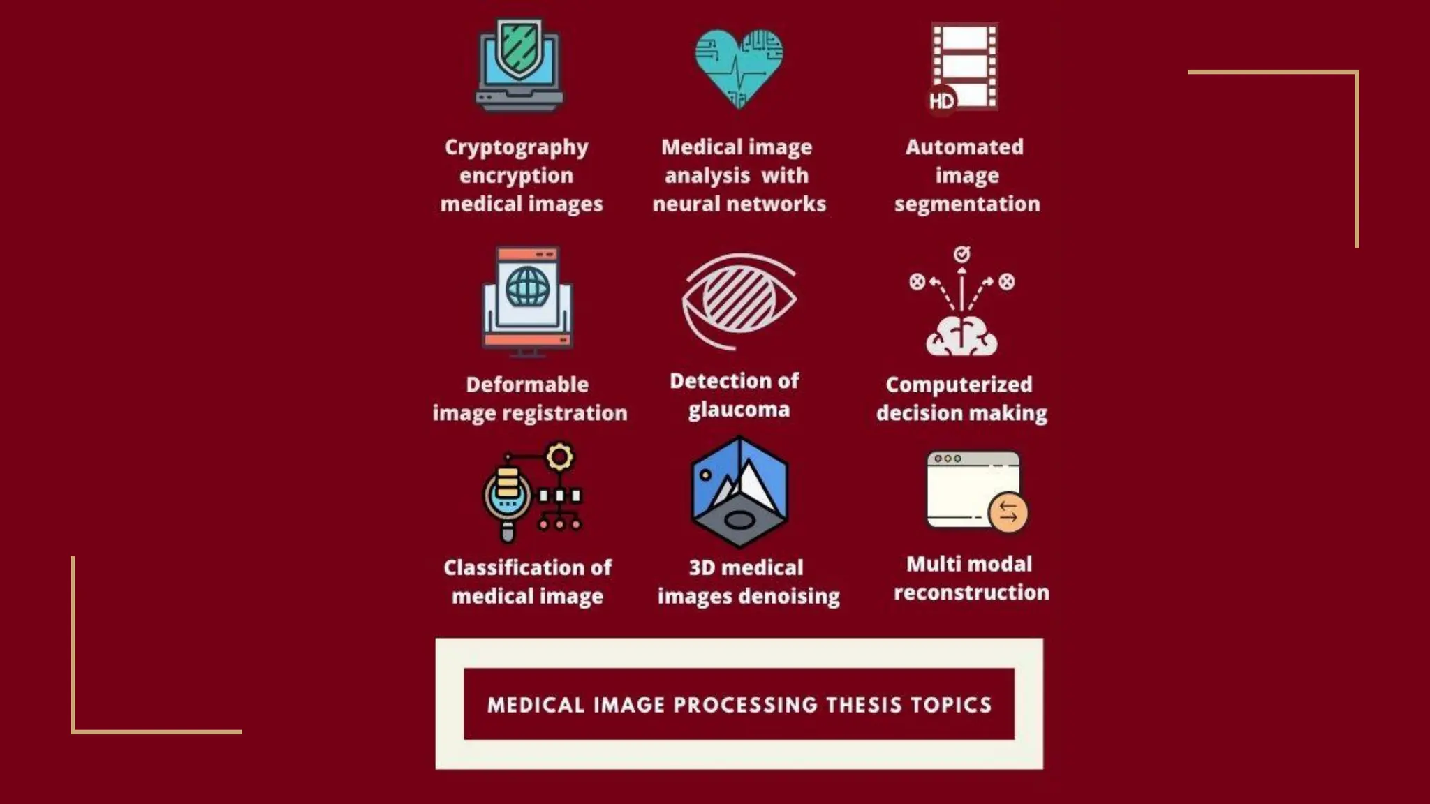
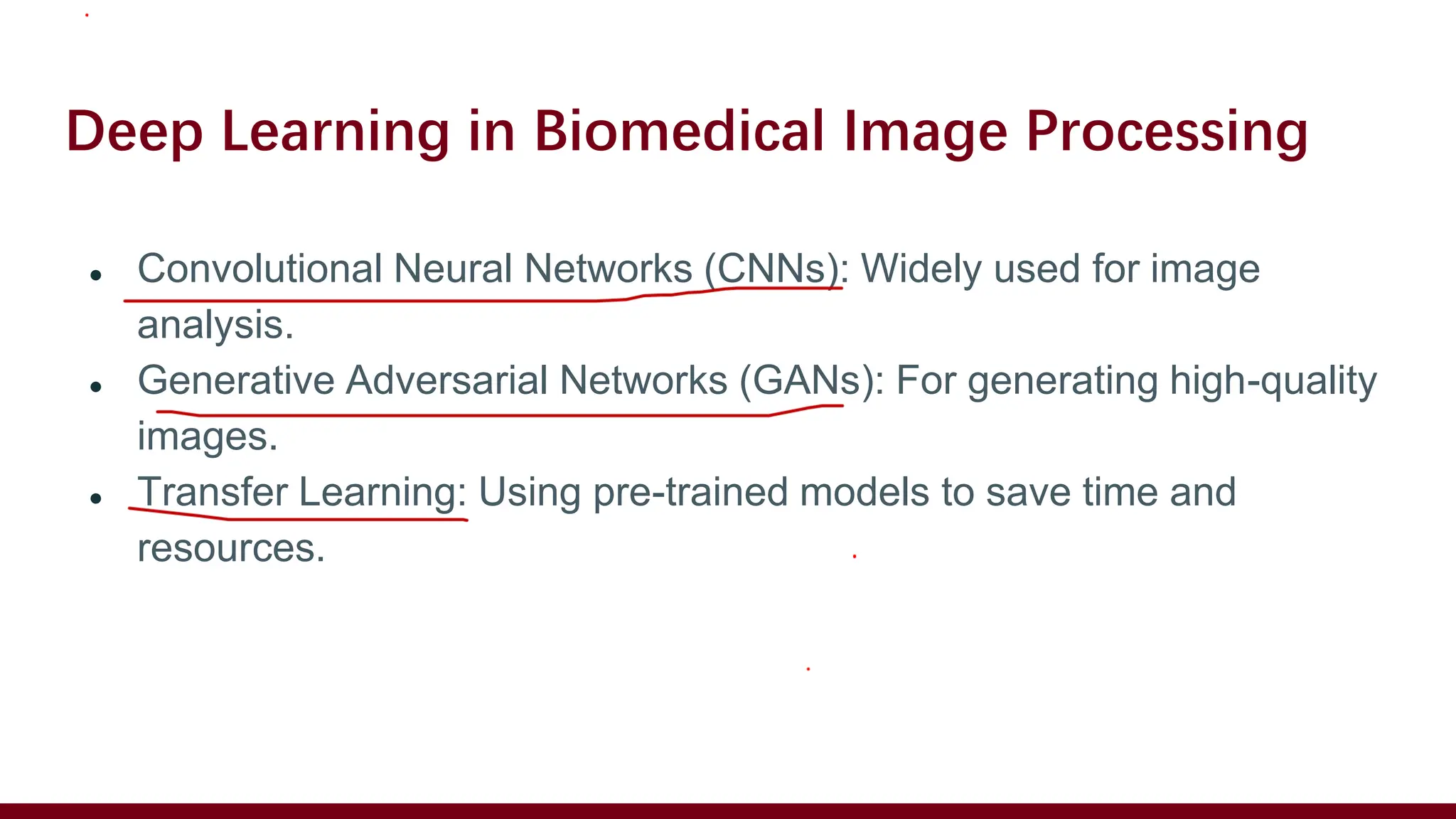
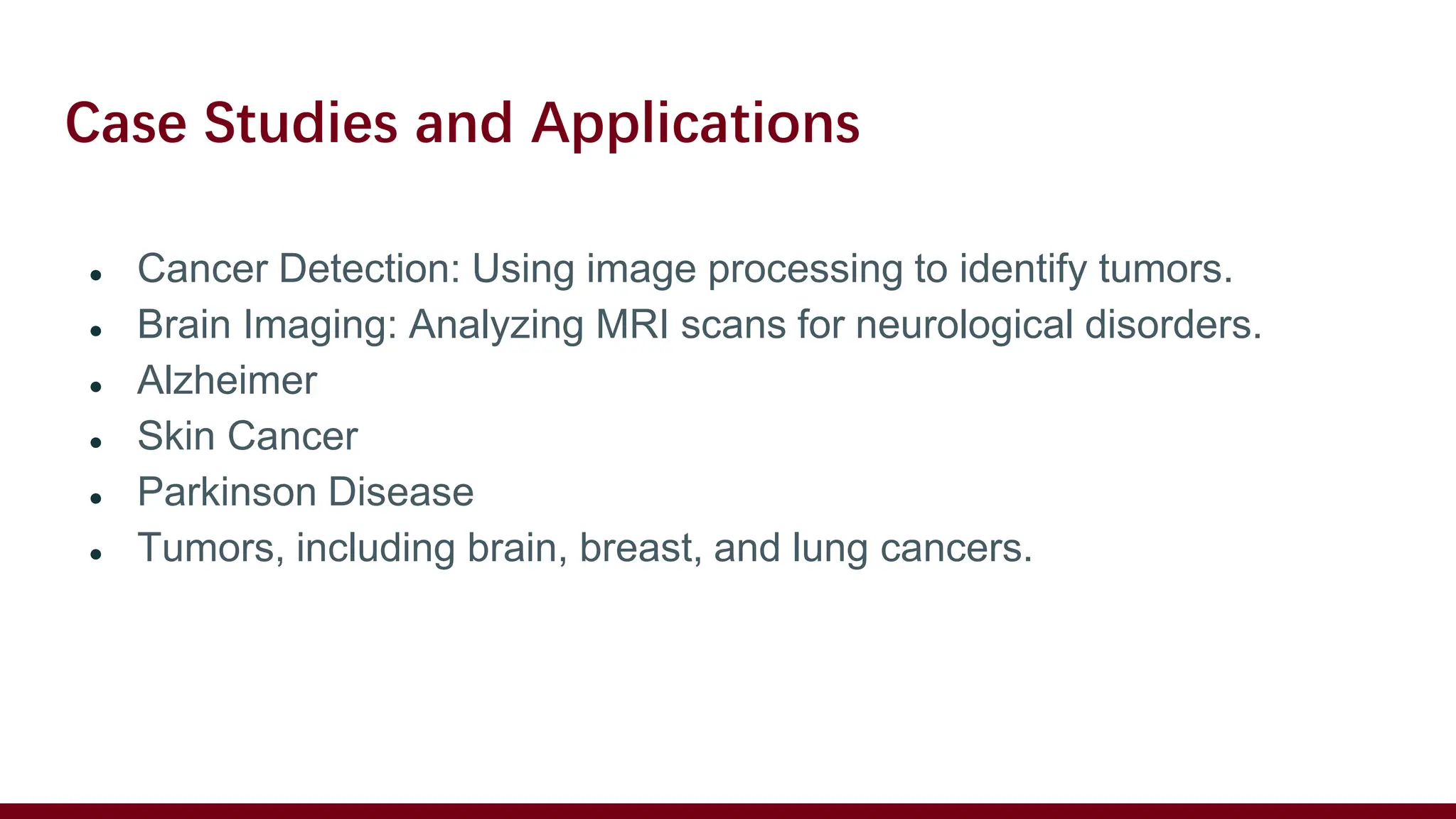
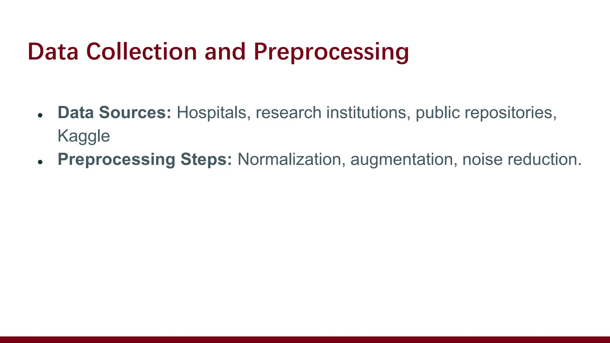
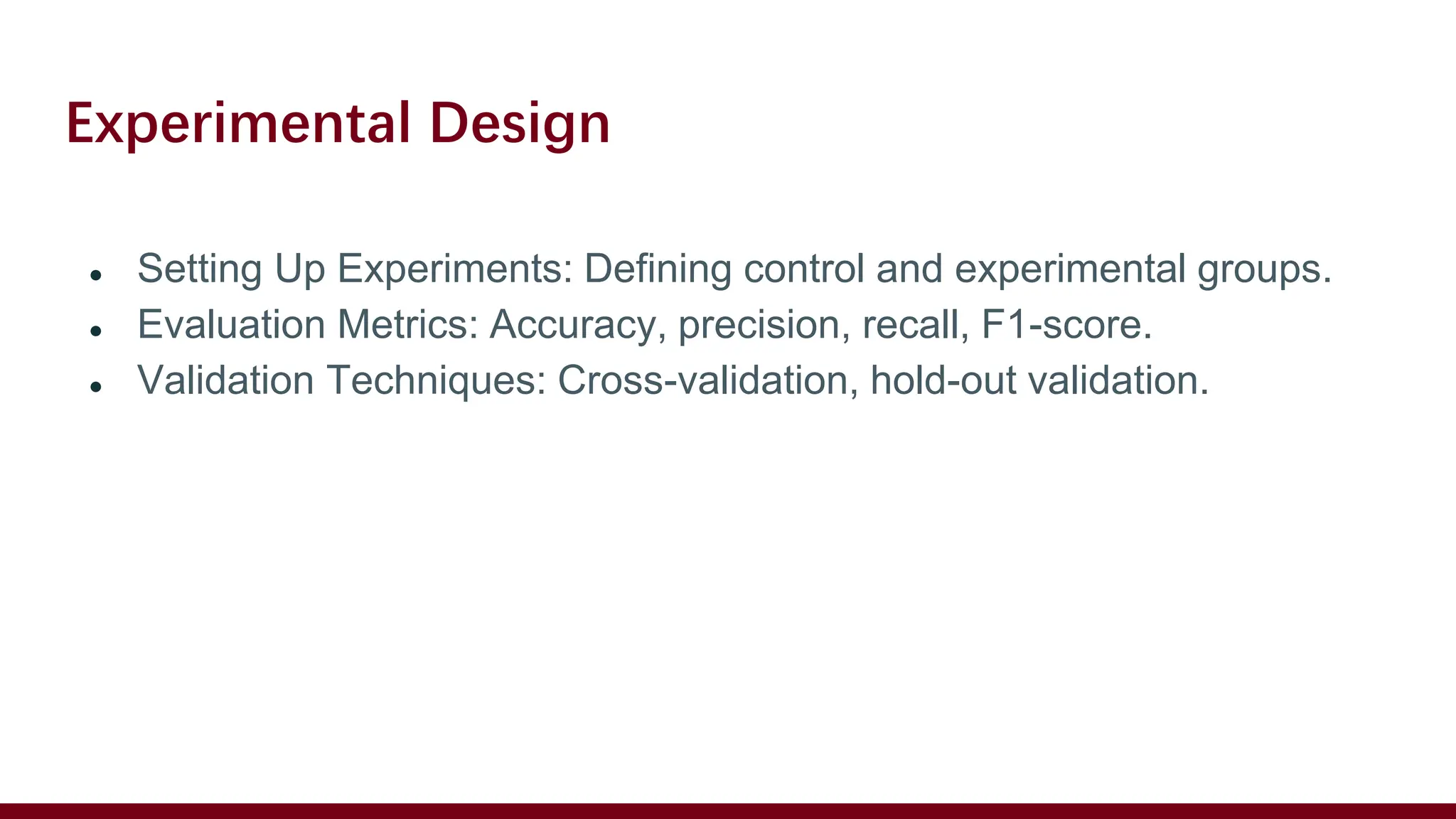
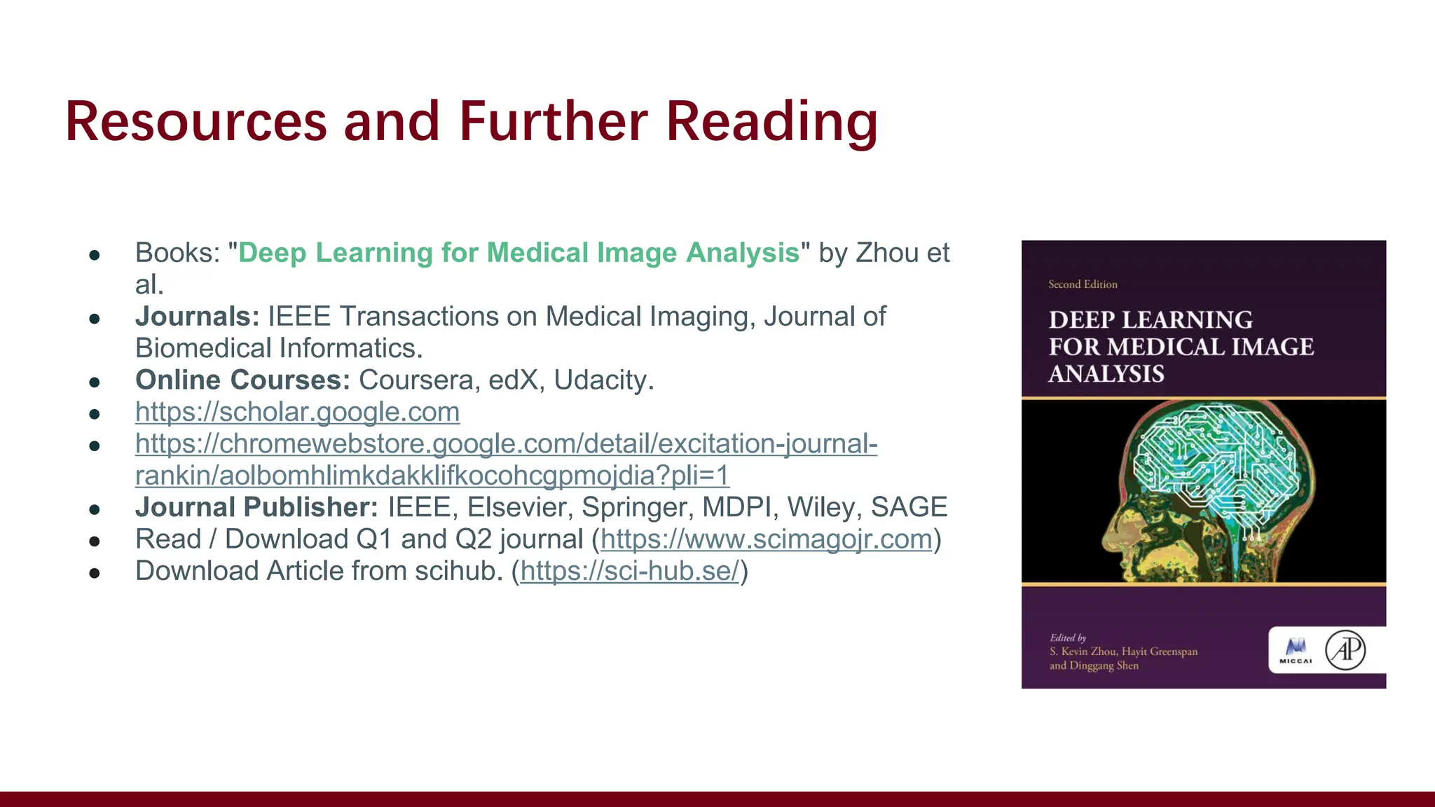
RESEARCH
Introduction
Introduction
● Creating new ideas or improving efficiency of any previous Model performance.
Understanding Biomedical Image Processing
● Biomedical image processing
involves the analysis and
manipulation of medical
images to improve their quality
and extract meaningful
information.
● Applications include
diagnostics, treatment
planning, and research.
Key Areas of Research
● Image Acquisition: Techniques for capturing medical images (e.g.,
MRI, CT, Ultrasound).
● Image Enhancement: Improving image quality for better
visualization.
● Image Segmentation: Partitioning images into meaningful regions.
● Feature Extraction: Identifying and quantifying key features.
● Image Classification: Categorizing images based on extracted
features.
Getting Started with Research
● Literature Review: Understanding current state-of-the-art methods
and technologies.
● Identify a Research Gap: Find areas that need improvement or new
approaches.
● Define Objectives: Clear goals and hypotheses for your research.
Choosing the Right Tools and Techniques
● Software: MATLAB, Python (with libraries like OpenCV, TensorFlow,
Keras).
● Techniques: Machine Learning, Deep Learning, Classical Image
Processing Methods.
● Datasets: Publicly available datasets (e.g., BraTS, ImageNet).
Deep Learning in Biomedical Image Processing
● Convolutional Neural Networks (CNNs): Widely used for image
analysis.
● Generative Adversarial Networks (GANs): For generating high-quality
images.
● Transfer Learning: Using pre-trained models to save time and
resources.
Case Studies and Applications
● Cancer Detection: Using image processing to identify tumors.
● Brain Imaging: Analyzing MRI scans for neurological disorders.
● Alzheimer
● Skin Cancer
● Parkinson Disease
● Tumors, including brain, breast, and lung cancers.
Data Collection and Preprocessing
● Data Sources: Hospitals, research institutions, public repositories,
Kaggle
● Preprocessing Steps: Normalization, augmentation, noise reduction.
Experimental Design
● Setting Up Experiments: Defining control and experimental groups.
● Evaluation Metrics: Accuracy, precision, recall, F1-score.
● Validation Techniques: Cross-validation, hold-out validation.
Challenges and Considerations
● Data Privacy: Ensuring patient confidentiality.
● Computational Resources: High-performance computing needs.
● Interdisciplinary Collaboration: Working with healthcare professionals.
Future Directions
● AI and Machine Learning: Continued integration in medical imaging.
● Real-time Processing: Enhancing speed and efficiency.
● Personalized Medicine: Tailoring treatments based on image analysis.
Resources and Further Reading
● Books: "Deep Learning for Medical Image Analysis" by Zhou et
al.
● Journals: IEEE Transactions on Medical Imaging, Journal of
Biomedical Informatics.
● Online Courses: Coursera, edX, Udacity.
● https://scholar.google.com
● https://chromewebstore.google.com/detail/excitation-journal-
rankin/aolbomhlimkdakklifkocohcgpmojdia?pli=1
● Journal Publisher: IEEE, Elsevier, Springer, MDPI, Wiley, SAGE
● Read / Download Q1 and Q2 journal (https://www.scimagojr.com)
● Download Article from scihub. (https://sci-hub.se/)
Research Outline on Biomedical Image Processing .pdf

Research Outline on Biomedical Image Processing .pdf

  • 1.
  • 3.
    Introduction ● Creating newideas or improving efficiency of any previous Model performance.
  • 4.
    Understanding Biomedical ImageProcessing ● Biomedical image processing involves the analysis and manipulation of medical images to improve their quality and extract meaningful information. ● Applications include diagnostics, treatment planning, and research.
  • 5.
    Key Areas ofResearch ● Image Acquisition: Techniques for capturing medical images (e.g., MRI, CT, Ultrasound). ● Image Enhancement: Improving image quality for better visualization. ● Image Segmentation: Partitioning images into meaningful regions. ● Feature Extraction: Identifying and quantifying key features. ● Image Classification: Categorizing images based on extracted features.
  • 6.
    Getting Started withResearch ● Literature Review: Understanding current state-of-the-art methods and technologies. ● Identify a Research Gap: Find areas that need improvement or new approaches. ● Define Objectives: Clear goals and hypotheses for your research.
  • 7.
    Choosing the RightTools and Techniques ● Software: MATLAB, Python (with libraries like OpenCV, TensorFlow, Keras). ● Techniques: Machine Learning, Deep Learning, Classical Image Processing Methods. ● Datasets: Publicly available datasets (e.g., BraTS, ImageNet).
  • 8.
    Deep Learning inBiomedical Image Processing ● Convolutional Neural Networks (CNNs): Widely used for image analysis. ● Generative Adversarial Networks (GANs): For generating high-quality images. ● Transfer Learning: Using pre-trained models to save time and resources.
  • 9.
    Case Studies andApplications ● Cancer Detection: Using image processing to identify tumors. ● Brain Imaging: Analyzing MRI scans for neurological disorders. ● Alzheimer ● Skin Cancer ● Parkinson Disease ● Tumors, including brain, breast, and lung cancers.
  • 10.
    Data Collection andPreprocessing ● Data Sources: Hospitals, research institutions, public repositories, Kaggle ● Preprocessing Steps: Normalization, augmentation, noise reduction.
  • 11.
    Experimental Design ● SettingUp Experiments: Defining control and experimental groups. ● Evaluation Metrics: Accuracy, precision, recall, F1-score. ● Validation Techniques: Cross-validation, hold-out validation.
  • 12.
    Challenges and Considerations ●Data Privacy: Ensuring patient confidentiality. ● Computational Resources: High-performance computing needs. ● Interdisciplinary Collaboration: Working with healthcare professionals.
  • 13.
    Future Directions ● AIand Machine Learning: Continued integration in medical imaging. ● Real-time Processing: Enhancing speed and efficiency. ● Personalized Medicine: Tailoring treatments based on image analysis.
  • 14.
    Resources and FurtherReading ● Books: "Deep Learning for Medical Image Analysis" by Zhou et al. ● Journals: IEEE Transactions on Medical Imaging, Journal of Biomedical Informatics. ● Online Courses: Coursera, edX, Udacity. ● https://scholar.google.com ● https://chromewebstore.google.com/detail/excitation-journal- rankin/aolbomhlimkdakklifkocohcgpmojdia?pli=1 ● Journal Publisher: IEEE, Elsevier, Springer, MDPI, Wiley, SAGE ● Read / Download Q1 and Q2 journal (https://www.scimagojr.com) ● Download Article from scihub. (https://sci-hub.se/)