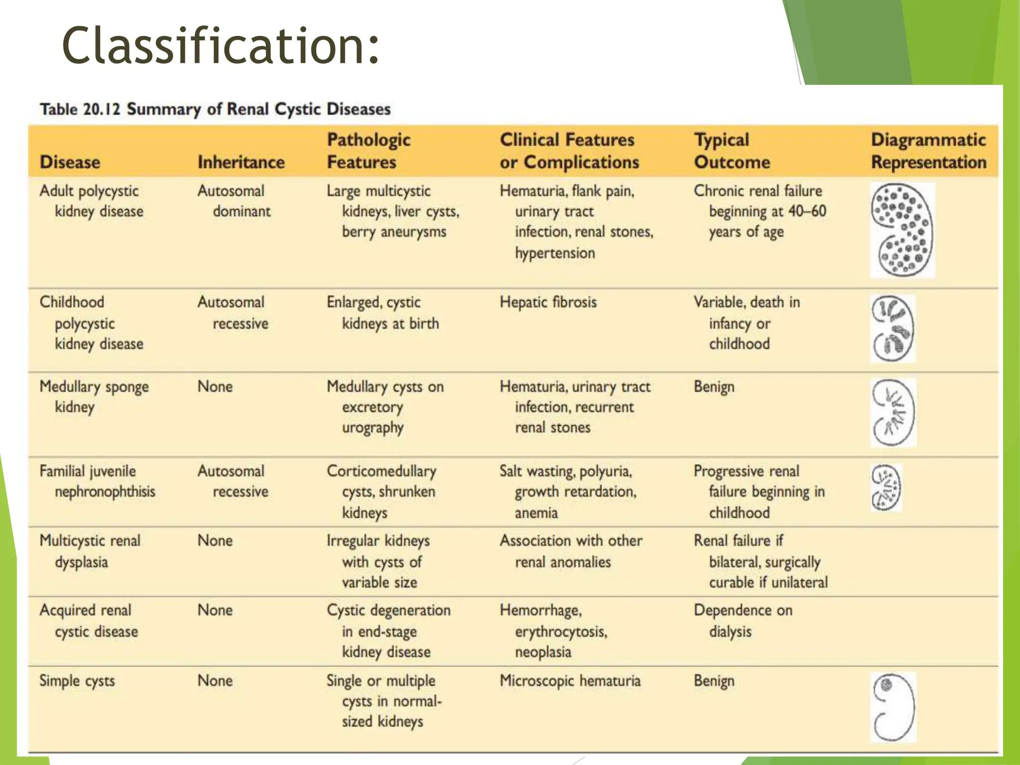
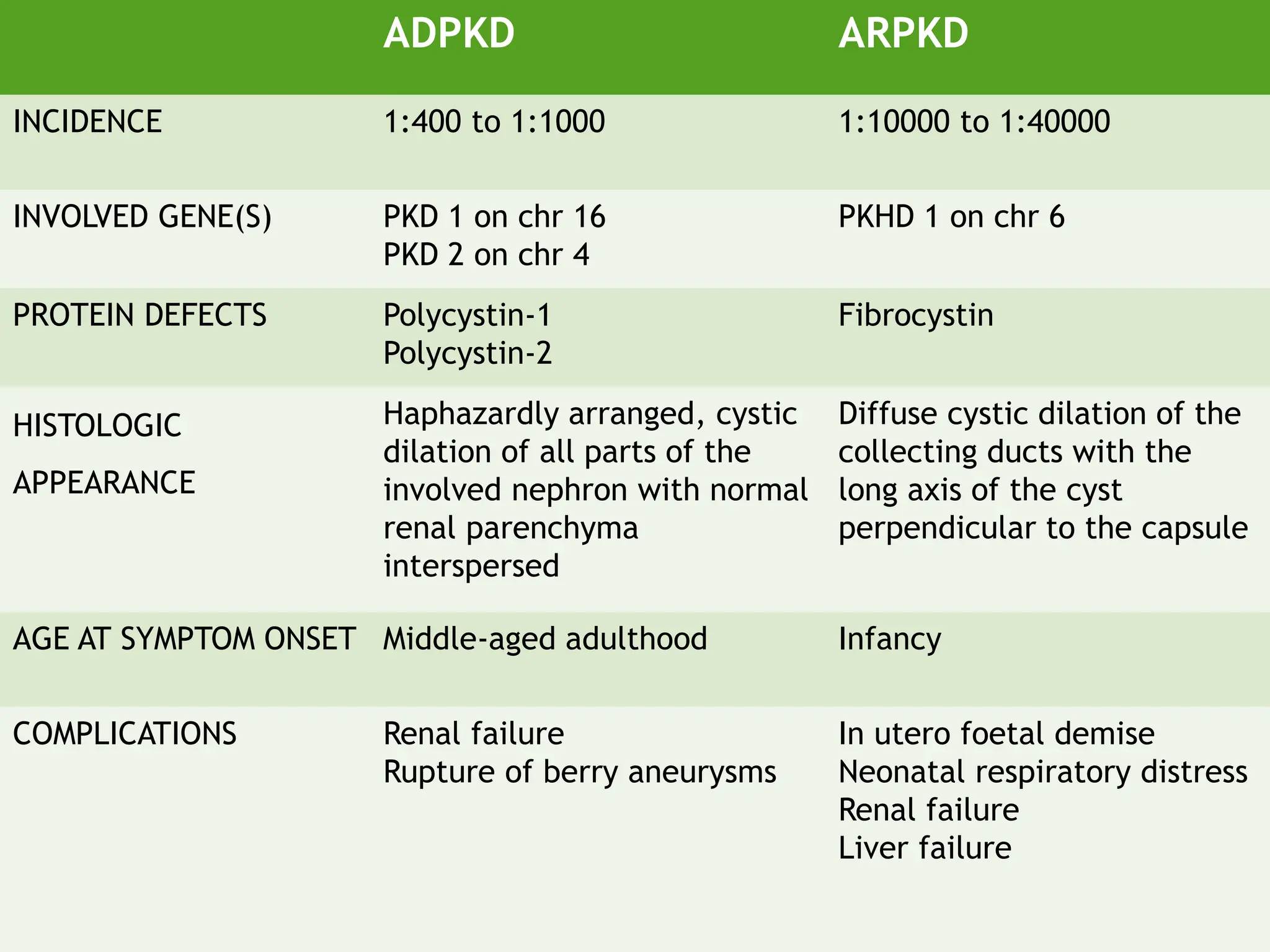
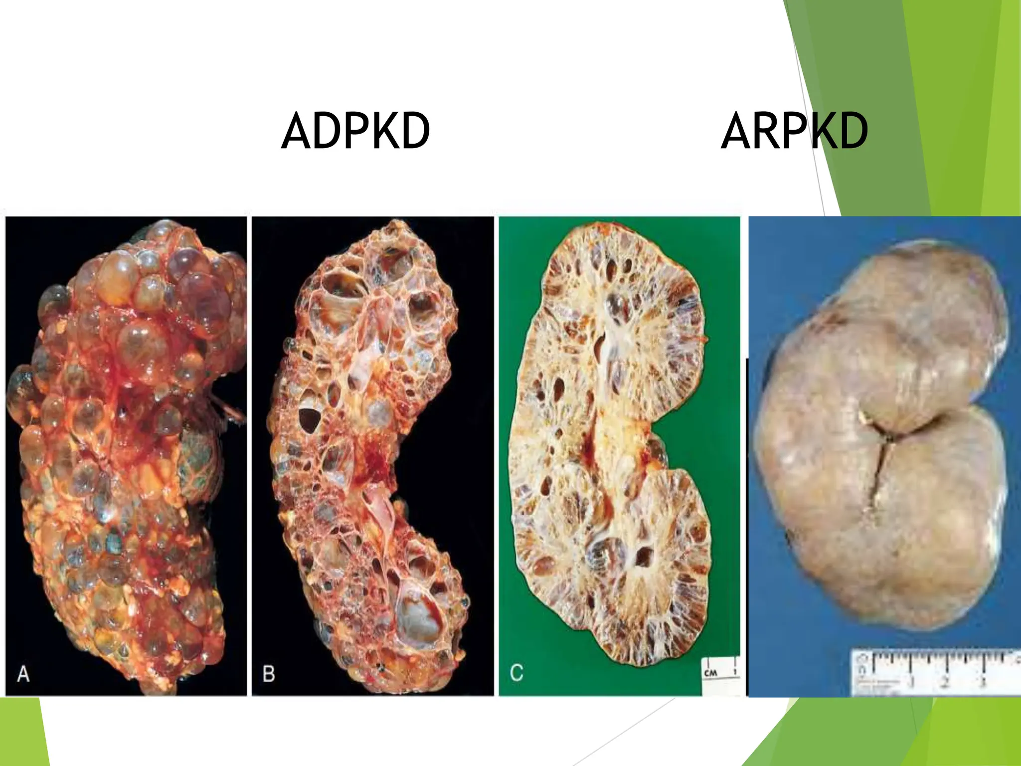
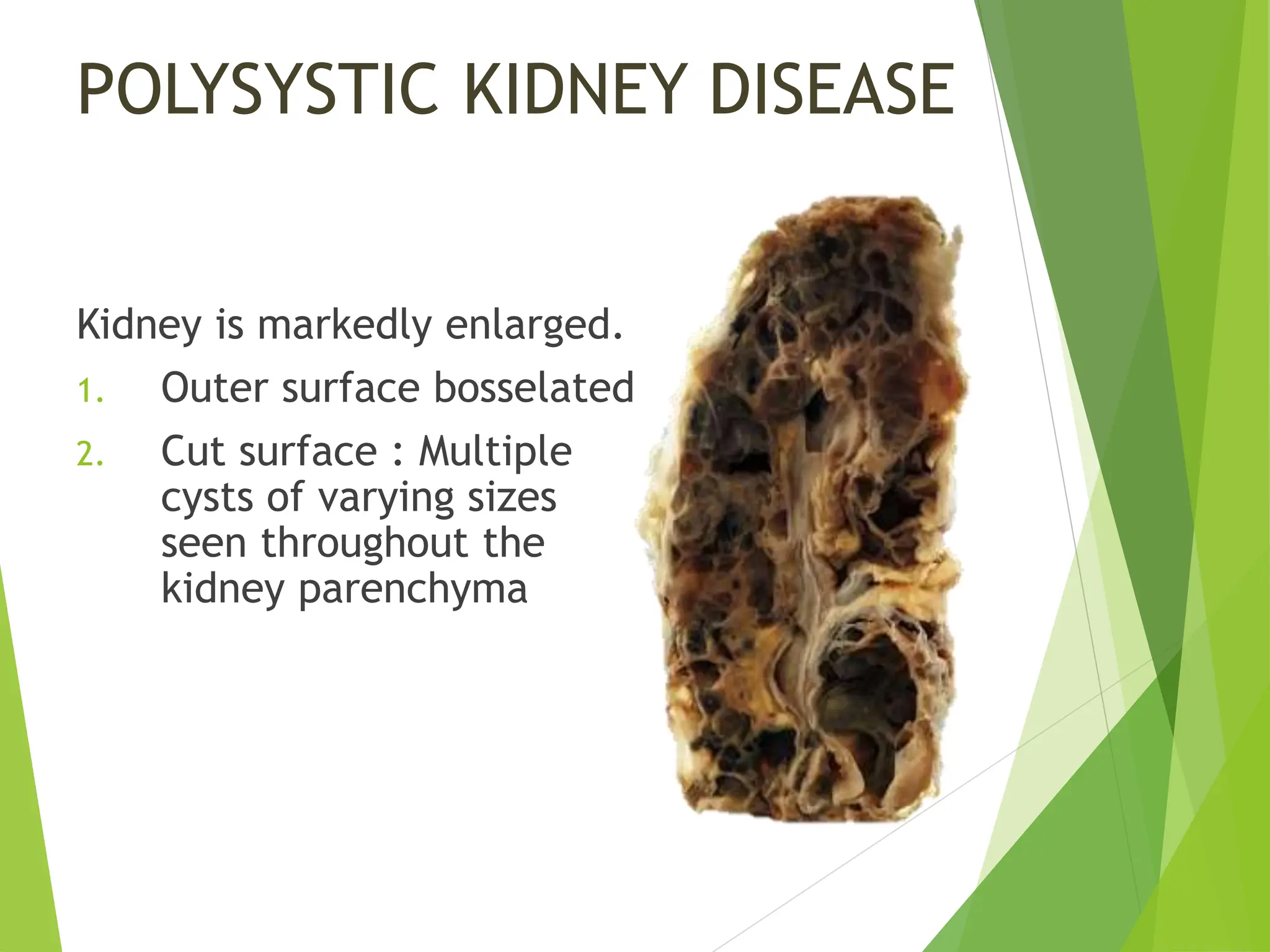
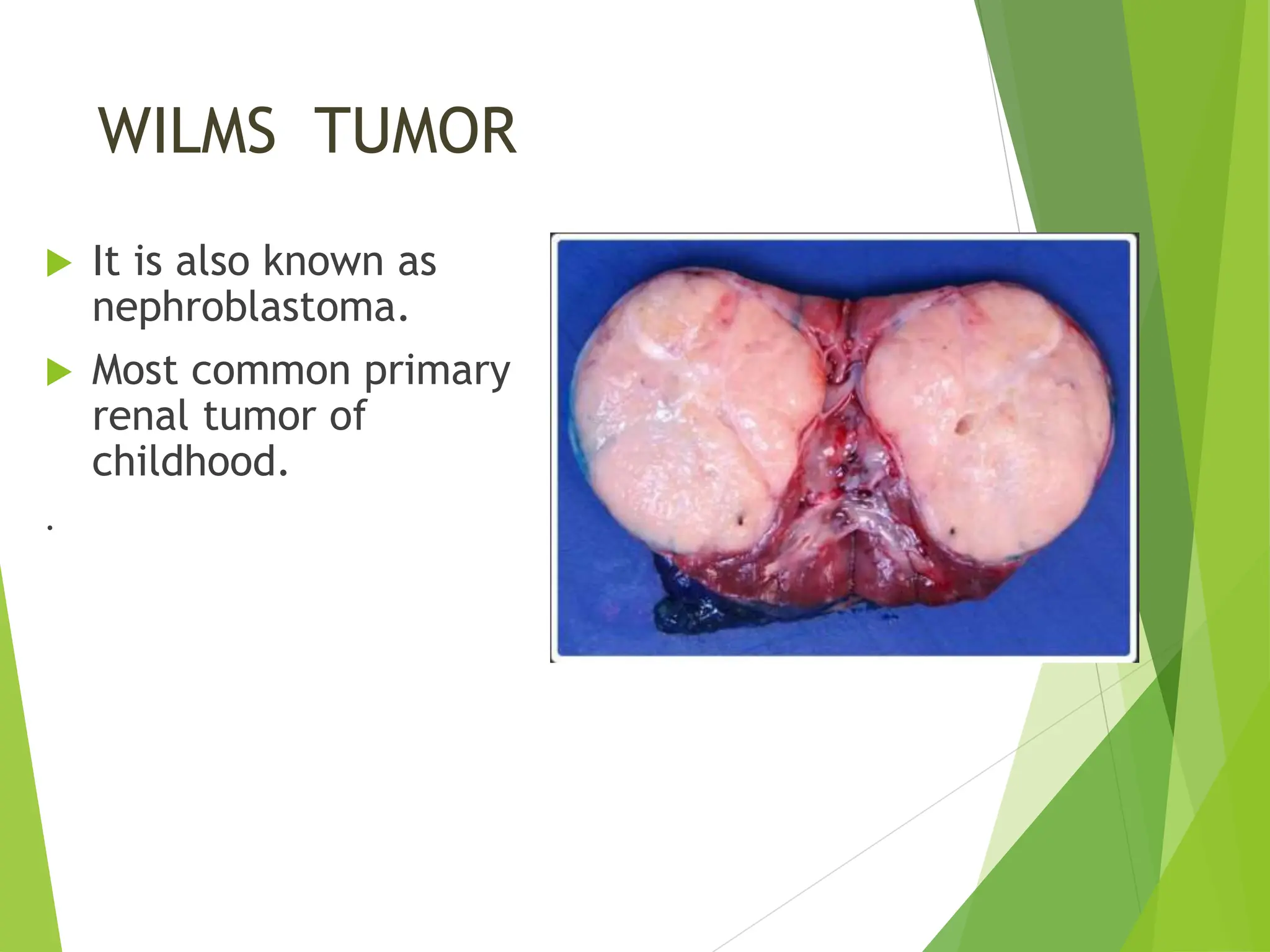
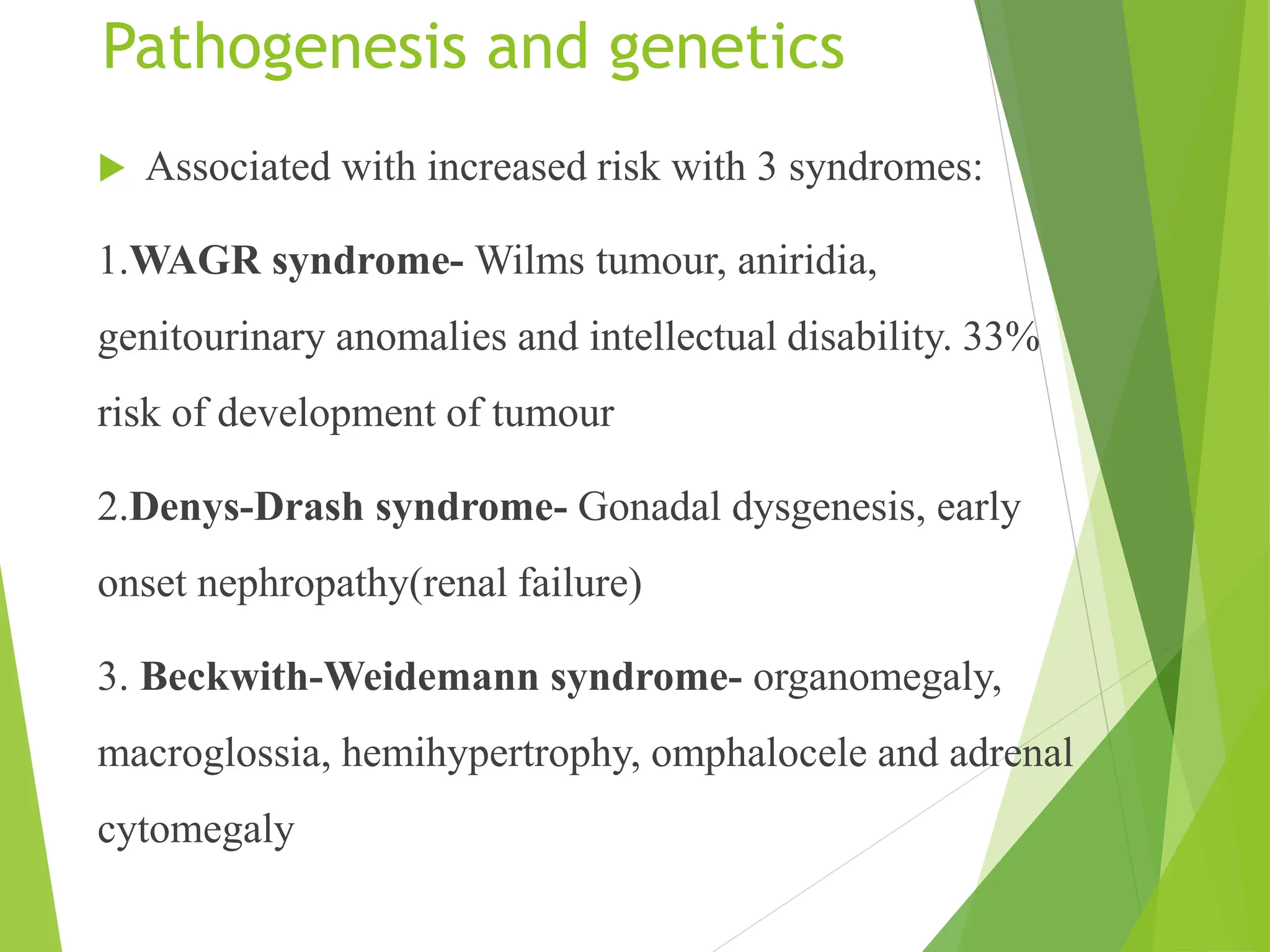
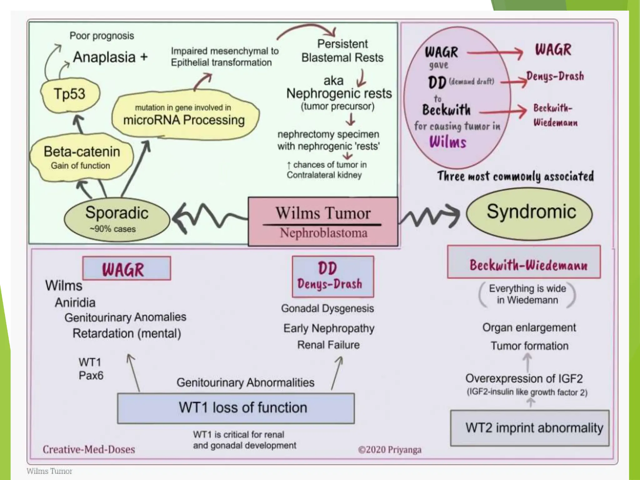
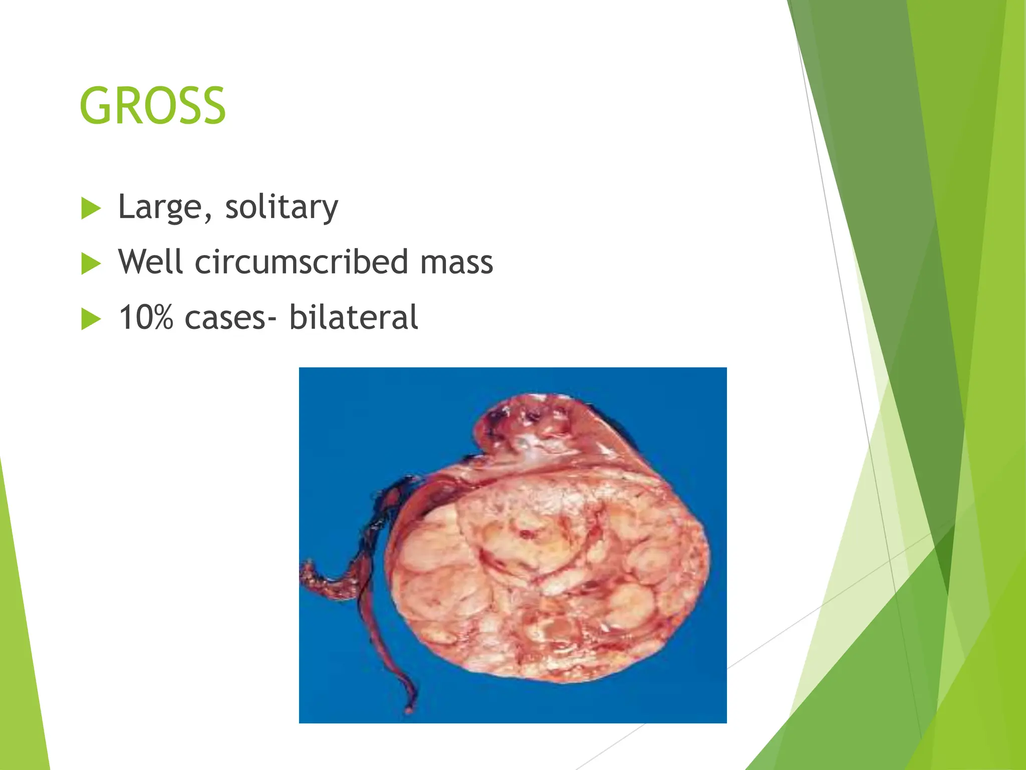
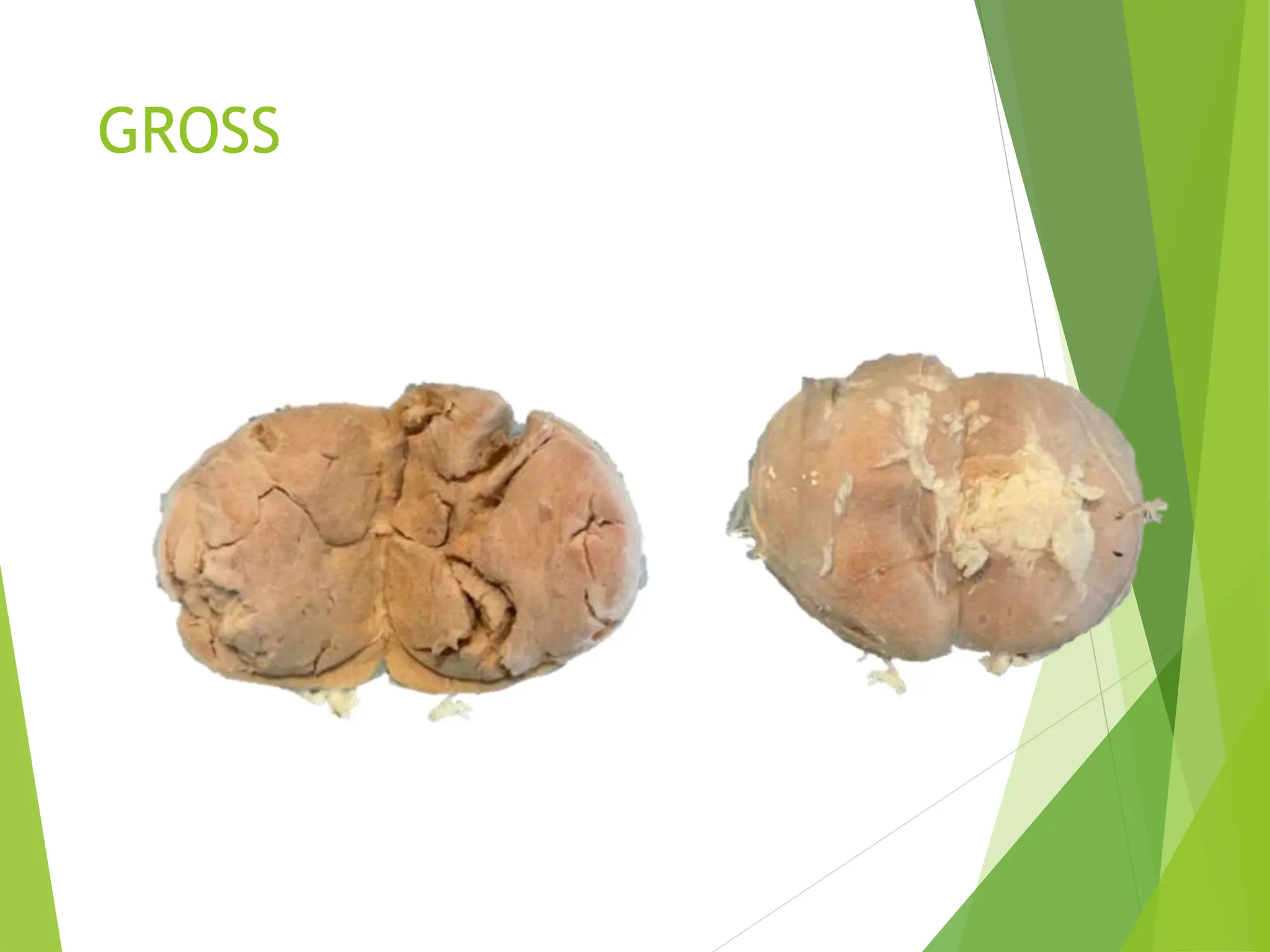
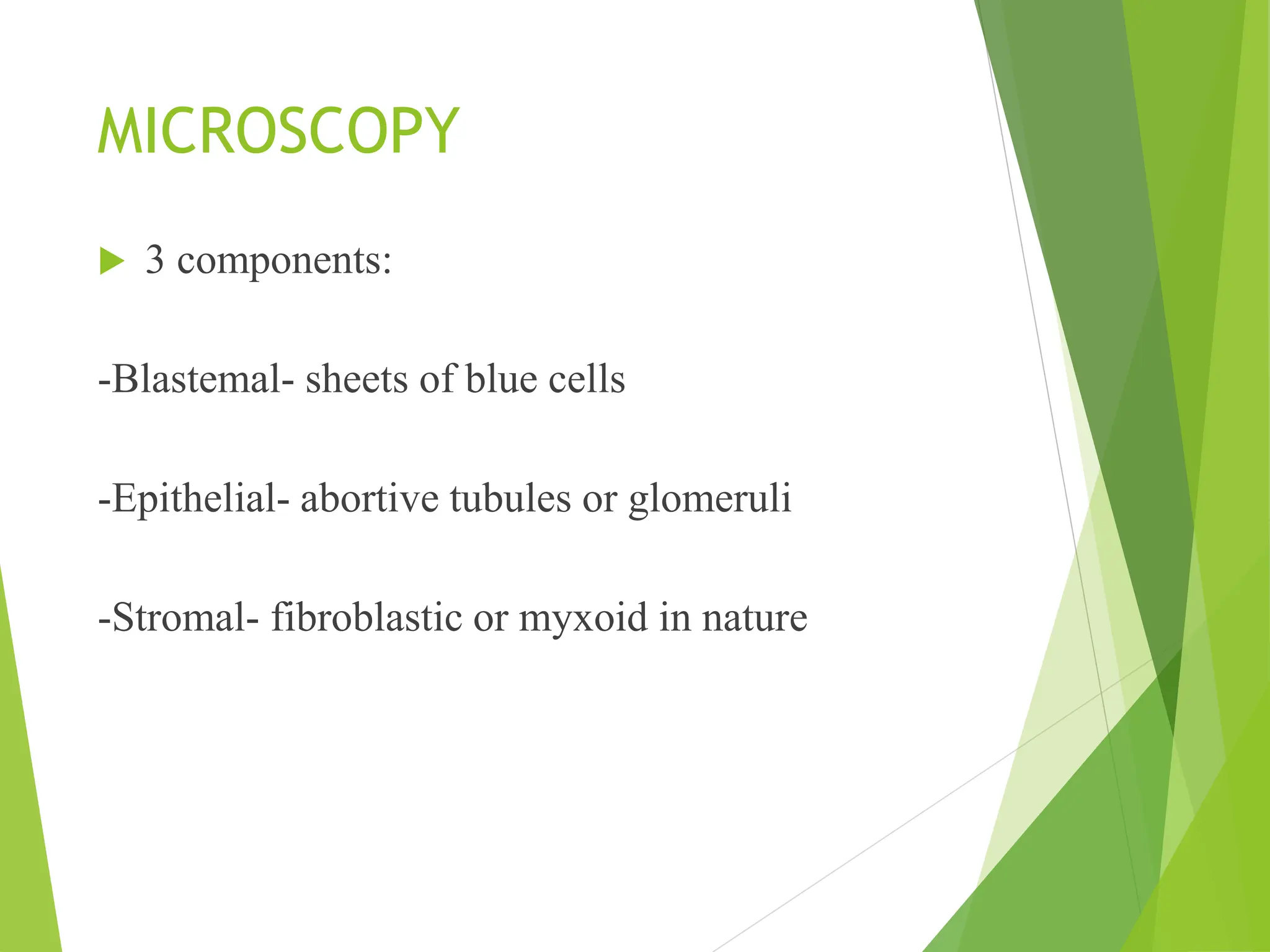
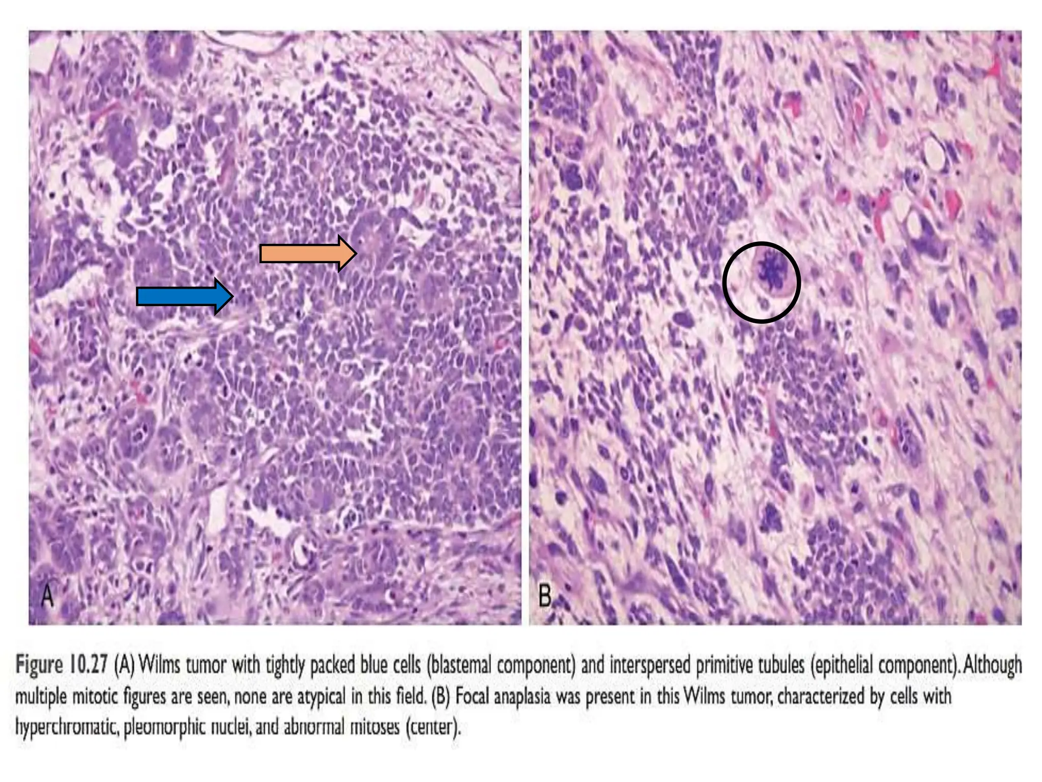
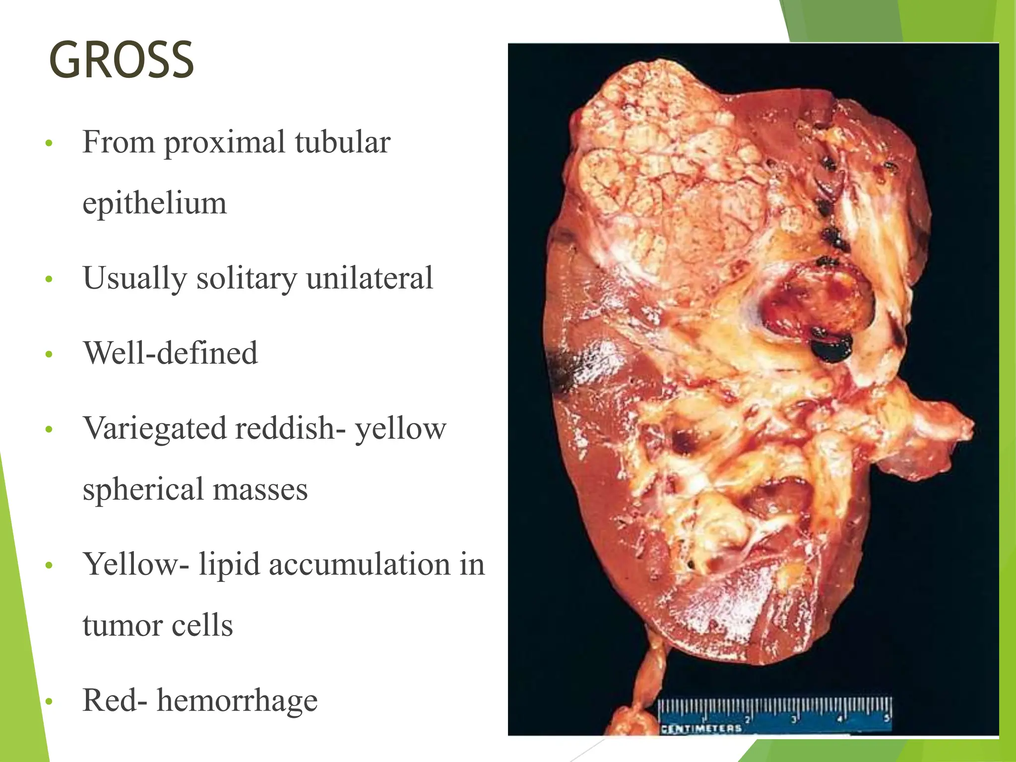
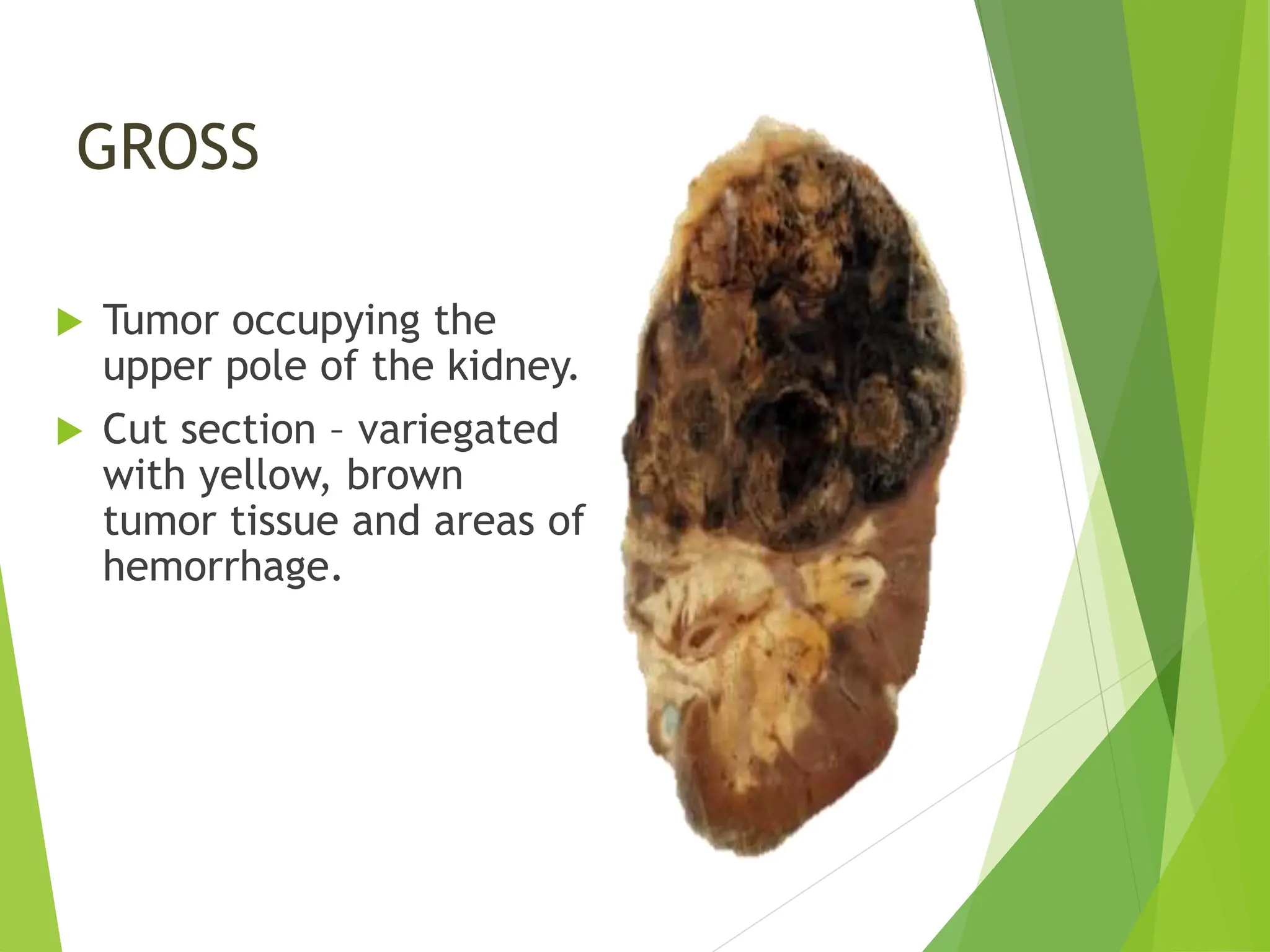
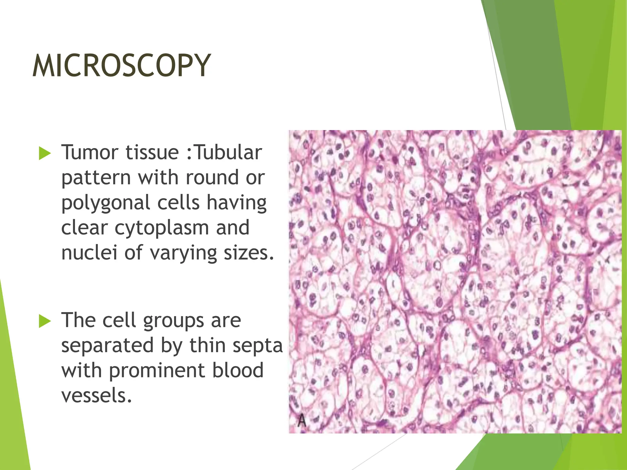
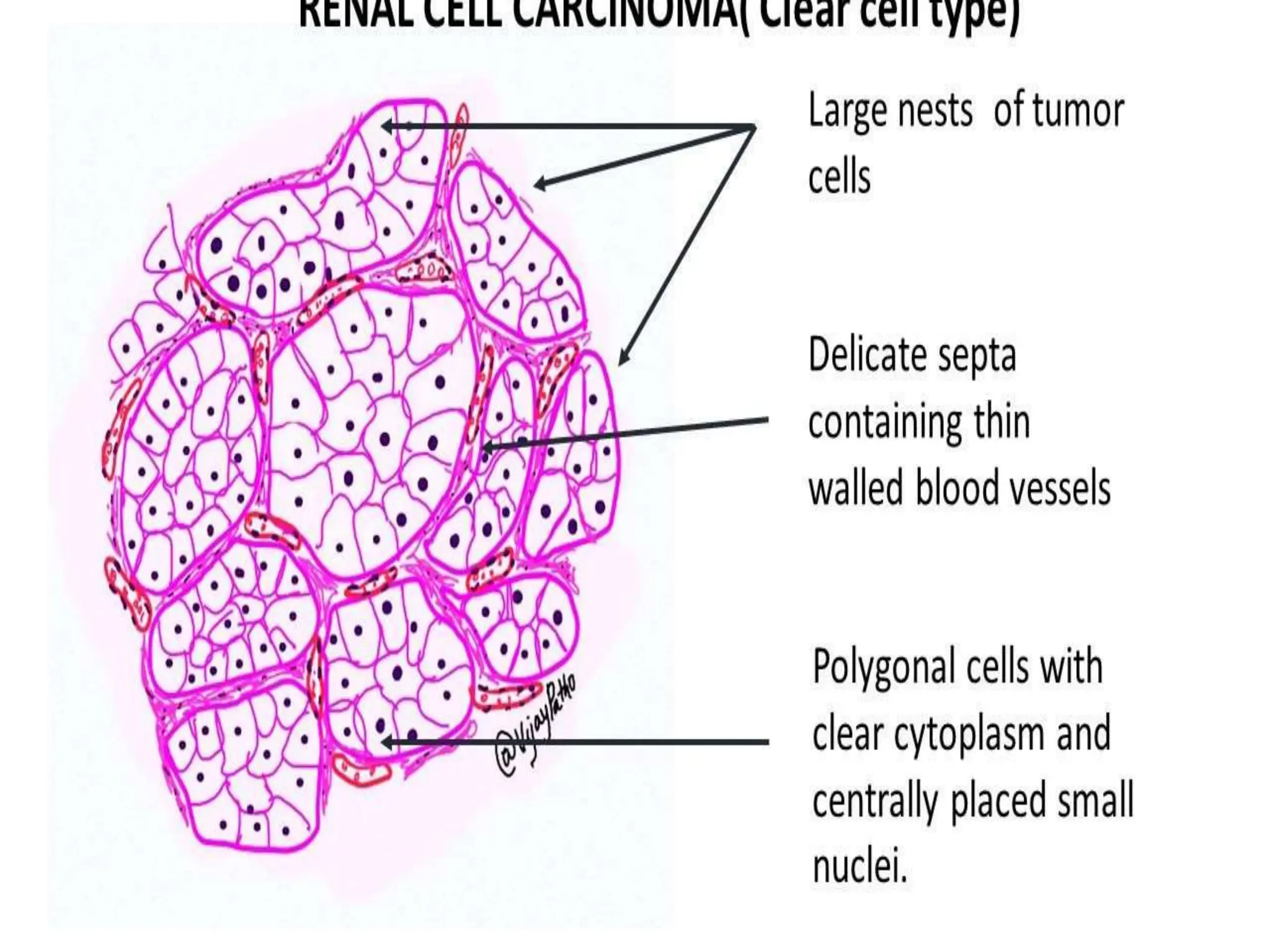
The document discusses cystic lesions and tumors of the kidney and urinary bladder. It covers cystic kidney diseases including polycystic kidney disease (ADPKD and ARPKD), tumors of the kidney including renal cell carcinoma (clear cell and papillary subtypes), Wilms tumor, and transitional cell carcinoma of the renal pelvis and urinary bladder. It provides details on pathogenesis, morphology, clinical features, and prognosis of these conditions.
This document summarizes the etiopathogenesis of renal tumors. It discusses benign renal tumors such as renal cysts, oncocytoma, angiomyolipoma and others. It describes genetic factors associated with various tumors. Risk factors for renal cell carcinoma include tobacco use, obesity, hypertension and familial syndromes like Von Hippel Lindau disease, hereditary papillary renal cell carcinoma, Birt-Hogg-Dubé syndrome and more. Various familial syndromes are outlined along with their associated genes and tumor types.
Cystic kidney diseases can be genetic or acquired. Autosomal dominant polycystic kidney disease (ADPKD) is a common genetic condition characterized by numerous fluid-filled cysts in the kidneys that worsen over time, potentially leading to kidney failure. Autosomal recessive polycystic kidney disease (ARPKD) presents in infancy with enlarged cystic kidneys and often liver disease, and can be fatal. Other cystic conditions include medullary sponge kidney cysts in the kidney papillae and nephronophthiasis-medullary cystic complex cysts at the corticomedullary junction. Simple cortical cysts are very common incidental findings. Ren
Cystic kidney diseases in children can be genetic or non-genetic in origin. Genetic causes include autosomal recessive polycystic kidney disease (ARPKD), autosomal dominant polycystic kidney disease (ADPKD), juvenile nephronophthisis, and glomerulocystic kidney disease. Non-genetic causes include simple cysts, multicystic dysplastic kidney, acquired cysts, and caliceal cysts. Ultrasound is the primary imaging modality used for diagnosis. ARPKD is the most severe genetic cause, occurring in 1 in 50,000, and is characterized by cystic dilatation of the collecting tubules affecting both kidneys
The document discusses various pediatric renal cystic diseases including their definitions, categories, genetics, presentations, associations, and management. Multicystic dysplastic kidney is defined as involving the entire kidney with primitive ducts and cysts present from early development. It is the renal cystic condition that arises prior to nephron formation.
This document discusses autosomal dominant polycystic kidney disease (ADPKD). It is characterized by multiple bilateral renal cysts and cysts in other organs caused by mutations in PKD1 and PKD2 genes. The proteins encoded by these genes, polycystin 1 and 2, are involved in maintaining renal tubule structure and calcium homeostasis. Disruption of their function leads to cyst formation through abnormal cell proliferation and fluid secretion. ADPKD causes kidney enlargement and failure but has a variable phenotype. Management involves blood pressure control, pain management, and treating complications like infections.
1. Benign renal tumors are the majority of renal masses found, often incidentally. They include renal cysts, angiomyolipomas, oncocytomas, and papillary adenomas.
2. Renal cell carcinoma (RCC) accounts for 2-3% of adult cancers. Risk factors include tobacco, obesity, hypertension, and family history. The most common subtypes are clear cell and papillary RCC.
3. Certain hereditary syndromes increase RCC risk, like von Hippel-Lindau disease, hereditary papillary RCC, and Birt-Hogg-Dubé syndrome. Genetic mutations contribute to tumor development in these conditions.
13. oldIntroduction to nephrology 1 & 2.pptYusuphShittu
This document provides an introduction to nephrology and outlines renal embryogenesis and common renal diseases in children. It discusses the significance of Congenital Anomalies of the Kidney and Urinary Tract (CAKUT) as a leading cause of chronic kidney failure in children. It describes tests of renal function and various renal malformations including obstructive uropathies, renal cystic diseases, renal agenesis, and horseshoe kidney. Surgical treatments for conditions like posterior urethral valves and bladder exstrophy are also mentioned.
This document provides a classification and overview of cystic diseases of the kidney. It discusses the main types of polycystic kidney diseases including autosomal recessive and dominant polycystic kidney disease. It also covers tubulointerstitial syndromes that can present with cysts, such as nephronophthisis and medullary cystic kidney disease. Cystic neoplasms and neoplastic cysts are described, including cystic nephroma and tubulocystic renal cell carcinoma. The pathogenesis, histopathology, and genetics of many of these conditions are summarized. Miscellaneous cysts like simple renal cysts and medullary sponge kidney are also briefly outlined.
Congenital anomalies of the kidney and urinary tractDhanya Raghu
CAKUT are a group of phenotypically diverse structural malformations characterized by defects in renal and urinary tract development.
Nearly half of children who develop end-stage renal disease (ESRD) have asymmetric, irregularly shaped kidneys, often referred to as bilateral renal scarring and frequently associated with lower urinary tract anomalies, including vesicoureteral reflux (VUR).
Congenitally abnormal kidneys may be large or small, cystic or irregular in outline, and absent or misplaced.
This document provides an overview of renal cell carcinoma (RCC), including its epidemiology, risk factors, pathology, classification, and staging. RCC accounts for 2-3% of adult cancers and its incidence is rising worldwide. The main subtypes are clear cell, papillary, and chromophobe RCC. RCC typically presents as a unilateral, solitary renal mass and can spread through direct extension or hematogenous routes. Staging involves evaluating the extent of disease to determine the appropriate treatment.
Adult polycystic kidney disease (ADPKD) is a genetic disorder characterized by the growth of numerous cysts in the kidneys and other organs. It is caused by mutations in one of two genes, PKD1 or PKD2, and affects around 1 in 1000 people. Symptoms include back or abdominal pain, hematuria, and hypertension. Extrarenal manifestations include cysts in the liver and pancreas as well as cerebral and coronary artery aneurysms. Diagnosis is made based on imaging and family history. While there is no cure, treatment focuses on slowing disease progression, managing complications like hypertension, and renal replacement therapy for end-stage renal disease.
1. Autosomal dominant polycystic kidney disease (ADPKD) is a genetic disorder characterized by multiple bilateral renal cysts and cysts in other organs caused by mutations in PKD1 and PKD2 genes.
2. The renal cysts enlarge over time, ultimately leading to renal failure in half of patients by age 60.
3. Treatment focuses on controlling blood pressure, treating infections, reducing pain, and delaying renal failure through medications such as ACE inhibitors or ARBs.
Polycystic kidney disease is caused by mutations in genes located on chromosomes 16p13.3 and 4q21. The kidneys become massively enlarged due to the growth of multiple cysts filled with fluid. Patients may be asymptomatic for years until renal insufficiency develops. Symptoms can include pain, bleeding, and high blood pressure as the cysts enlarge and damage the kidney tissue. Complications include liver and heart cysts as well as brain aneurysms. While the condition progresses slowly over decades, it ultimately leads to kidney failure and often death from heart or brain related issues.
This document discusses congenital malformations of the kidneys, focusing on cystic diseases. It describes three broad groups of kidney malformations: abnormalities in renal tissue amount, anomalies of position/form/orientation, and anomalies of differentiation. It then classifies and describes cystic kidney diseases in detail. The major cystic diseases discussed are multicystic renal dysplasia, adult polycystic kidney disease (ADPKD), and infantile polycystic kidney disease (ARPKD). ADPKD is inherited dominantly and manifests in adulthood, while ARPKD is inherited recessively and presents in infancy, often leading to death from renal failure.
This document discusses different types of kidney cysts. It identifies 5 main classifications: 1) Cystic Renal Dysplasia, which involves abnormal development leading to enlarged kidneys with varied sized cysts; 2) Polycystic Renal Disease, which can be autosomal dominant or recessive, causing multiple cysts through genetic defects affecting cell differentiation; 3) Medullary Cystic Disease, involving cysts in the kidney medulla due to defects in 3 genes leading to fibrosis; 4) Acquired/Dialysis Associated Cystic Disease, where numerous cortical and medullary cysts develop in patients on long-term dialysis due to tubular obstruction; and 5) Simple Cyst
This document discusses congenital malformations of the kidney. It is divided into three sections: abnormalities in renal tissue amount, anomalies in position and shape, and cystic diseases. Cystic diseases are further classified as multicystic renal dysplasia and polycystic kidney disease. Polycystic kidney disease occurs in two forms - an adult autosomal dominant form and a rare infantile autosomal recessive form. The adult form typically manifests between ages 30-50 with back pain, bleeding, infections and hypertension, while the infantile form causes renal failure in early childhood.
Pathology of intracranial tumors lectureEffiong Akang
This document provides an overview of intracranial tumors, including:
1. It classifies intracranial neoplasms according to histological group and WHO grade and discusses some of the major tumor types such as astrocytic tumors, meningiomas, pituitary adenomas and others.
2. It reviews the epidemiology and pathogenesis of intracranial tumors. Most are sporadic but some have familial causes.
3. The objectives are for students to understand the classification, clinical manifestations, gross and microscopic features of common intracranial neoplasms.
discuss the CLASSIFICATION OF KIDNEY TUMOURS.pptxHarunausman10
This document provides an overview of the classification of kidney tumours according to the 2016 WHO guidelines. It begins with an introduction and epidemiology section noting the rise in incidentally detected kidney masses. It then reviews clinical features and classification. The WHO categories include clear cell and papillary renal cell carcinomas, chromophobe RCC, and collecting duct carcinoma. Emerging entities like ALK-associated RCC are also discussed. Classification is based on morphology, architecture, genetic alterations, and immunochemistry. Precise diagnosis has improved with advances in imaging, pathology, and understanding of molecular underpinnings of lesions.
Seminar RKS kidney.pptxkidney slides- pathogenesis features of rccPriyankaPriyadarshin93
The document discusses various types of renal tumors, including clear cell renal cell carcinoma, papillary renal cell carcinoma, chromophobe renal cell carcinoma, collecting duct carcinoma, and others. It describes their epidemiology, histopathological features, genetic abnormalities, immunohistochemistry profiles, and associations with hereditary syndromes. In particular, it provides detailed information on clear cell renal cell carcinoma, succinate dehydrogenase-deficient renal cell carcinoma, and fumarate hydratase-deficient renal cell carcinoma, including their pathogenesis, clinical presentation, gross and microscopic appearance, IHC staining patterns, and genetic factors.
RCC typically presents in the 6th and 7th decade of life and accounts for 2-3% of adult malignancies. Clear cell RCC arises from the proximal convoluted tubules and is associated with VHL gene mutations. Established risk factors include tobacco, obesity, and hypertension. CT scan is the preferred imaging modality and can identify enhancing renal masses. Surgical resection is the main treatment, with partial nephrectomy preferred for smaller tumors when possible to preserve renal function. Follow up involves history, exam, bloodwork and imaging depending on pathologic stage.
This document discusses chronic kidney disease, including its etiology, pathogenesis, and complications. It begins by covering the normal anatomy and embryology of the kidney. It then describes the staging of CKD using the CGA classification system. The main causes of CKD are discussed, including congenital diseases, glomerular diseases, vascular diseases, tubulointerstitial diseases, and obstructive uropathies. The pathogenesis of glomerular injury is examined in depth. Finally, common complications of advanced CKD are outlined, such as anemia, cardiovascular abnormalities, and disorders of calcium and phosphate metabolism.
This document discusses cystic diseases of the kidney. It begins by classifying kidney cysts by cause, features, and location. It then categorizes cysts as developmental, genetic, associated with systemic diseases, acquired, or malignant. Several genetic cystic kidney diseases are described in detail, including their pathophysiology and clinical presentations. Bosniak and Bonsib classifications of renal cysts are presented. The document concludes with epidemiology, differential diagnosis of cyst types, and morbidity and mortality of cystic kidney diseases.
Congenital Development of Urinary System.pptchallenger0326
1. Congenital anomalies of the kidney and urinary tract are abnormalities that develop before birth and can affect the structure and function of the kidneys and urinary system.
2. Some of the main anomalies discussed include renal agenesis (absent kidney), hypoplasia (small kidney), dysplasia (abnormal structure of kidney), and abnormalities in kidney position. Other anomalies affect the collecting system like duplex kidneys, hydronephrosis, and posterior urethral valves.
3. These anomalies can have various clinical presentations ranging from asymptomatic to end stage renal disease. They often require long term follow up and management to monitor renal and bladder function and address any complications. Bilateral involvement or
Similar to Renal system -DOAP pathology ppt class.ppt (20)
NAVIGATING THE HORIZONS OF TIME LAPSE EMBRYO MONITORING.pdfRahul Sen
Time-lapse embryo monitoring is an advanced imaging technique used in IVF to continuously observe embryo development. It captures high-resolution images at regular intervals, allowing embryologists to select the most viable embryos for transfer based on detailed growth patterns. This technology enhances embryo selection, potentially increasing pregnancy success rates.
DECLARATION OF HELSINKI - History and principlesanaghabharat01
This SlideShare presentation provides a comprehensive overview of the Declaration of Helsinki, a foundational document outlining ethical guidelines for conducting medical research involving human subjects.
Co-Chairs, Val J. Lowe, MD, and Cyrus A. Raji, MD, PhD, prepared useful Practice Aids pertaining to Alzheimer’s disease for this CME/AAPA activity titled “Alzheimer’s Disease Case Conference: Gearing Up for the Expanding Role of Neuroradiology in Diagnosis and Treatment.” For the full presentation, downloadable Practice Aids, and complete CME/AAPA information, and to apply for credit, please visit us at https://bit.ly/3PvVY25. CME/AAPA credit will be available until June 28, 2025.
Are you looking for a long-lasting solution to your missing tooth?
Dental implants are the most common type of method for replacing the missing tooth. Unlike dentures or bridges, implants are surgically placed in the jawbone. In layman’s terms, a dental implant is similar to the natural root of the tooth. It offers a stable foundation for the artificial tooth giving it the look, feel, and function similar to the natural tooth.
These lecture slides, by Dr Sidra Arshad, offer a simplified look into the mechanisms involved in the regulation of respiration:
Learning objectives:
1. Describe the organisation of respiratory center
2. Describe the nervous control of inspiration and respiratory rhythm
3. Describe the functions of the dorsal and respiratory groups of neurons
4. Describe the influences of the Pneumotaxic and Apneustic centers
5. Explain the role of Hering-Breur inflation reflex in regulation of inspiration
6. Explain the role of central chemoreceptors in regulation of respiration
7. Explain the role of peripheral chemoreceptors in regulation of respiration
8. Explain the regulation of respiration during exercise
9. Integrate the respiratory regulatory mechanisms
10. Describe the Cheyne-Stokes breathing
Study Resources:
1. Chapter 42, Guyton and Hall Textbook of Medical Physiology, 14th edition
2. Chapter 36, Ganong’s Review of Medical Physiology, 26th edition
3. Chapter 13, Human Physiology by Lauralee Sherwood, 9th edition
Cell Therapy Expansion and Challenges in Autoimmune DiseaseHealth Advances
There is increasing confidence that cell therapies will soon play a role in the treatment of autoimmune disorders, but the extent of this impact remains to be seen. Early readouts on autologous CAR-Ts in lupus are encouraging, but manufacturing and cost limitations are likely to restrict access to highly refractory patients. Allogeneic CAR-Ts have the potential to broaden access to earlier lines of treatment due to their inherent cost benefits, however they will need to demonstrate comparable or improved efficacy to established modalities.
In addition to infrastructure and capacity constraints, CAR-Ts face a very different risk-benefit dynamic in autoimmune compared to oncology, highlighting the need for tolerable therapies with low adverse event risk. CAR-NK and Treg-based therapies are also being developed in certain autoimmune disorders and may demonstrate favorable safety profiles. Several novel non-cell therapies such as bispecific antibodies, nanobodies, and RNAi drugs, may also offer future alternative competitive solutions with variable value propositions.
Widespread adoption of cell therapies will not only require strong efficacy and safety data, but also adapted pricing and access strategies. At oncology-based price points, CAR-Ts are unlikely to achieve broad market access in autoimmune disorders, with eligible patient populations that are potentially orders of magnitude greater than the number of currently addressable cancer patients. Developers have made strides towards reducing cell therapy COGS while improving manufacturing efficiency, but payors will inevitably restrict access until more sustainable pricing is achieved.
Despite these headwinds, industry leaders and investors remain confident that cell therapies are poised to address significant unmet need in patients suffering from autoimmune disorders. However, the extent of this impact on the treatment landscape remains to be seen, as the industry rapidly approaches an inflection point.
Histololgy of Female Reproductive System.pptxAyeshaZaid1
Dive into an in-depth exploration of the histological structure of female reproductive system with this comprehensive lecture. Presented by Dr. Ayesha Irfan, Assistant Professor of Anatomy, this presentation covers the Gross anatomy and functional histology of the female reproductive organs. Ideal for students, educators, and anyone interested in medical science, this lecture provides clear explanations, detailed diagrams, and valuable insights into female reproductive system. Enhance your knowledge and understanding of this essential aspect of human biology.
5-hydroxytryptamine or 5-HT or Serotonin is a neurotransmitter that serves a range of roles in the human body. It is sometimes referred to as the happy chemical since it promotes overall well-being and happiness.
It is mostly found in the brain, intestines, and blood platelets.
5-HT is utilised to transport messages between nerve cells, is known to be involved in smooth muscle contraction, and adds to overall well-being and pleasure, among other benefits. 5-HT regulates the body's sleep-wake cycles and internal clock by acting as a precursor to melatonin.
It is hypothesised to regulate hunger, emotions, motor, cognitive, and autonomic processes.
- Video recording of this lecture in English language: https://youtu.be/Pt1nA32sdHQ
- Video recording of this lecture in Arabic language: https://youtu.be/uFdc9F0rlP0
- Link to download the book free: https://nephrotube.blogspot.com/p/nephrotube-nephrology-books.html
- Link to NephroTube website: www.NephroTube.com
- Link to NephroTube social media accounts: https://nephrotube.blogspot.com/p/join-nephrotube-on-social-media.html
10 Benefits an EPCR Software should Bring to EMS Organizations Traumasoft LLC
The benefits of an ePCR solution should extend to the whole EMS organization, not just certain groups of people or certain departments. It should provide more than just a form for entering and a database for storing information. It should also include a workflow of how information is communicated, used and stored across the entire organization.
Nano-gold for Cancer Therapy chemistry investigatory projectSIVAVINAYAKPK
chemistry investigatory project
The development of nanogold-based cancer therapy could revolutionize oncology by providing a more targeted, less invasive treatment option. This project contributes to the growing body of research aimed at harnessing nanotechnology for medical applications, paving the way for future clinical trials and potential commercial applications.
Cancer remains one of the leading causes of death worldwide, prompting the need for innovative treatment methods. Nanotechnology offers promising new approaches, including the use of gold nanoparticles (nanogold) for targeted cancer therapy. Nanogold particles possess unique physical and chemical properties that make them suitable for drug delivery, imaging, and photothermal therapy.
5. ADPKD ARPKD
INCIDENCE 1:400 to 1:1000 1:10000 to 1:40000
INVOLVED GENE(S) PKD 1 on chr 16
PKD 2 on chr 4
PKHD 1 on chr 6
PROTEIN DEFECTS Polycystin-1
Polycystin-2
Fibrocystin
HISTOLOGIC
APPEARANCE
Haphazardly arranged, cystic
dilation of all parts of the
involved nephron with normal
renal parenchyma
interspersed
Diffuse cystic dilation of the
collecting ducts with the
long axis of the cyst
perpendicular to the capsule
AGE AT SYMPTOM ONSET Middle-aged adulthood Infancy
COMPLICATIONS Renal failure
Rupture of berry aneurysms
In utero foetal demise
Neonatal respiratory distress
Renal failure
Liver failure
8. CLINICAL FEATURES
Asymptomatic until presence of renal
insufficiency
Haematuria
Flank pain
UTI
Renal stones
Hypertension
Hepatic fibrosis
9. POLYSYSTIC KIDNEY DISEASE
Kidney is markedly enlarged.
1. Outer surface bosselated
2. Cut surface : Multiple
cysts of varying sizes
seen throughout the
kidney parenchyma
10. POLYCYSTIC KIDNEY
Cysts maybe filled with clear serous fluid or turbid
haemorrhagic fluid
Enlarging cysts may encroach on the pelvis and
calyces to produce pressure defects
20. RCC account for 85% of renal cancers in adults.
Occurs most often in older individuals, usually in
6th and 7th decades of life.
Arises from tubular epithelium.
May arise in any portion of kidney, more commonly in poles.
RENAL CELL CARCINOMA
22. GROSS
• From proximal tubular
epithelium
• Usually solitary unilateral
• Well-defined
• Variegated reddish- yellow
spherical masses
• Yellow- lipid accumulation in
tumor cells
• Red- hemorrhage
23. GROSS
Tumor occupying the
upper pole of the kidney.
Cut section – variegated
with yellow, brown
tumor tissue and areas of
hemorrhage.
24. MICROSCOPY
Tumor tissue :Tubular
pattern with round or
polygonal cells having
clear cytoplasm and
nuclei of varying sizes.
The cell groups are
separated by thin septa
with prominent blood
vessels.