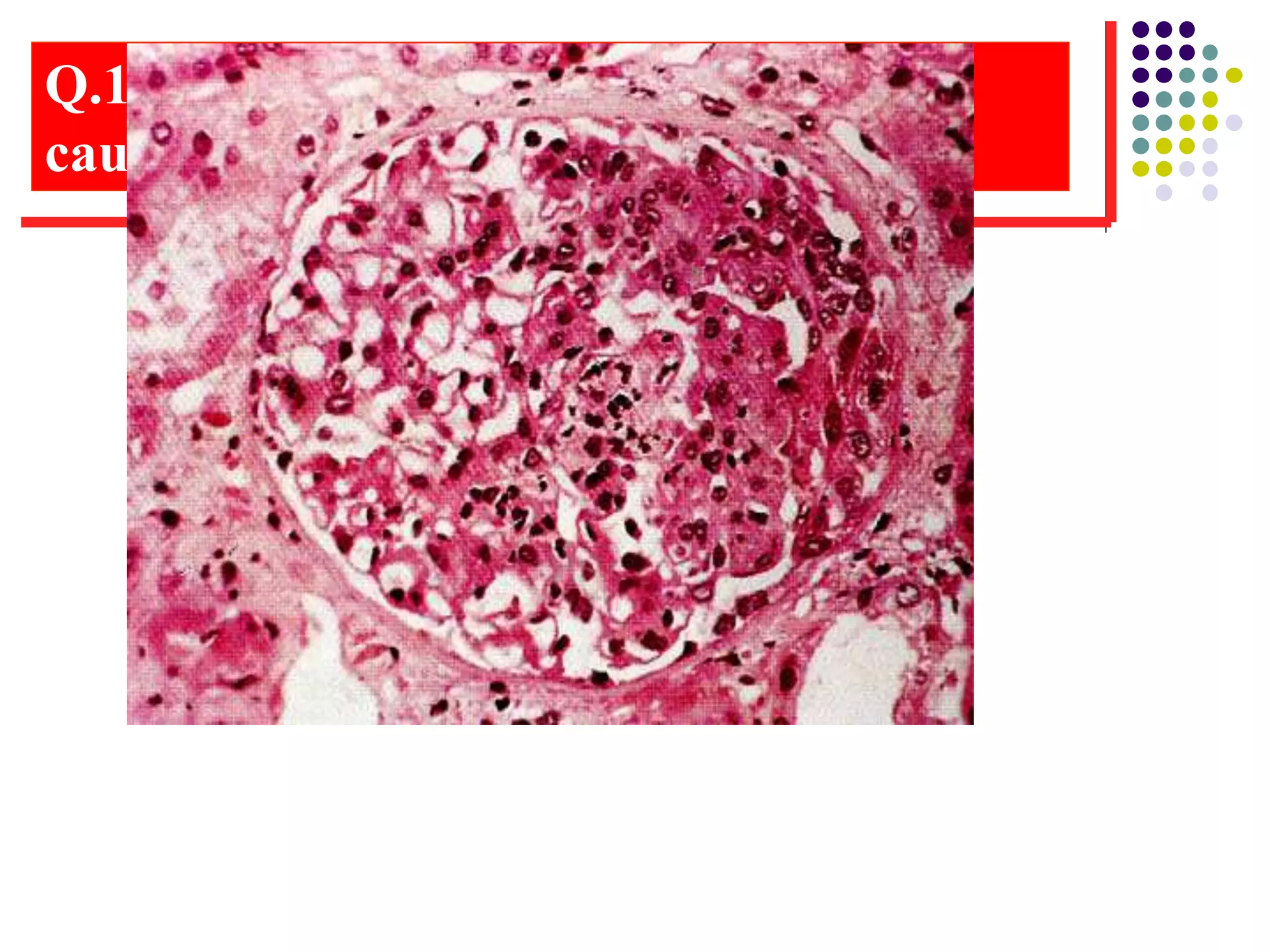
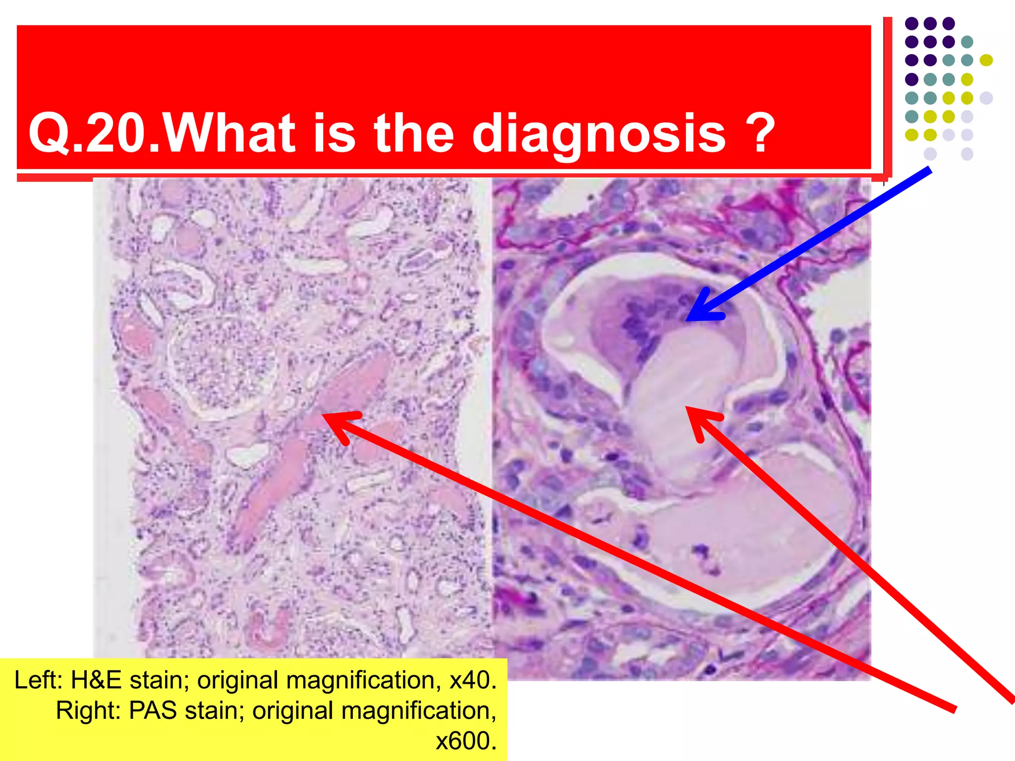
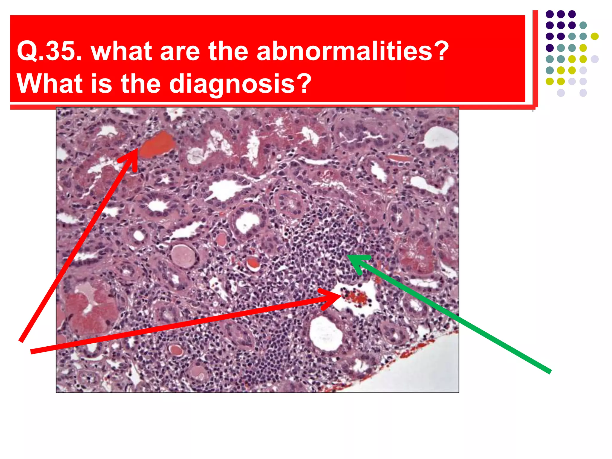
CLINICAL NEPHROLOGY
COURSE 2013
RENAL BIOPSY TESTING
PART 1 & 2
By
Dr. Abdel-Bassit Shaarawy
Professor of Internal Medicine
& Nephrology Ain Shams
University
2013
Bow
man’s
space
Dr.Abdelbassit Shaarway collection 2013
Q.1: What are the 4 structures you must
comment in renal biopsy? what is needed
for adequate biopsy report ?
.
1
3
The cortex contain glomeruli
and tubules
Tubule
Malpighian or renal corpuscles
The Malpighian corpuscle
(glomerulus+ Bowman’s capsule)
Glomerulus
– a ball of
capillaries
Bowman’s capsule
Medulla contains mainly
tubules
Tubules
Capillaries
Q.2.What are the 5 -normal components of
glomerulus? Could we identify these cells
on L/M? how could we identify them well?
Non-cellular components
normally present in glomerulus
Cells normally present in
glomerulus
1-
1-
2-
2-
3-
What are the structures that could be
abnormally present
Non-cellular components
abnormally present in
glomerulus
Cells abnormally present in
glomerulus
1-
1-
2- 3-
Q.3 Mention the structures of
this normal glomerulus Tufts of capillaries
6
4
1
3
5
Dr.Abdelbassit Shaarway collection 2013
Here is Bowman’s
space
Bowman’s space
accepts the filtrate
mesangium
Efferent A
Afferent A
Dr.Abdelbassit Shaarway collection 2013
Structure of glomerulus
Cells of glomerulus
cannot be
differantiated by LM
a = the afferent
e = the efferent
arteriole.
mesangial areas,
As a rule of thumb, no
more than 3
mesangial cells per
capillary loop are
usually seen in
normal glomerulus
Q.4 : Normal Glomerulus you must comment on :
name structures 1-how many cells in capillary area
(cellularity ) ,2-are capillaries empty ,collapsed
,filled ,3- what is their wall thickness, 4-what is in
area 3 and around them
Q.5. What is this structure? Is it
normal or abnormal ? Why?
Q.6. Comment on cellularity of these lesions
, which ones are increased ? What are the
cells increased ?What are the lesions ?
Q.7. A. How would you classify
Glomerular Diseases?
1 2 3
4 5 6
Q.8.B. what is the stain ,why used?classify this
lesion ? Is this lesion reversible?
Dr.Abdelbassit Shaarway collection 2013
Q.9. what are the stains used ? Mention 2
main abnormalities? Mention 4 diseases
produce this lesion?
Q.10. What is the lesion? Mention 2
causes of it
Q.11.What is the pattern of this renal
injury? Mention 5 secondary causes?
Q.12. This present with Nephrotic syndrome &
heavy proteinuria . Mention abnormalities
(comment in cellularity , capillary thickness,
mesangium
Q.13. what is the stain why used ? Mention
the investigations to do to exclude
secondary causes of this lesion
Q.14. Mention 3 abnormalities in this
biopsy (small ,large arrow ,astrixis ). What
is the most common cause of this lesion
Mention abnormalities
MPGN
 Light micrograph showing a
membranoproliferative pattern in lupus nephritis,
characterized by areas of cellular proliferation
(long arrows) and by thickening of the
glomerular capillary wall (due to immune
deposits) that may be prominent enough to form
a "wire-loop" (short arrows). Although
proliferative changes can be focal (affecting less
than 50 percent of glomeruli), disease of this
severity is usually diffuse.
Q.15. This patient present with acute renal failure,
what are the lesions, how to assess severity, Is it
reversible in this biopsy?
Q.16. These slides shows pathognomonic
features of a metabolic disease, give DD of
these lesions
Q.17.What are the DD of this
lesion?
Q.18.What is the diagnosis? What
further investigations should be
done?
Congo Red stain, right image under polarized
light (original magnification, x200).
Q.19.DM pt ,HTN,NS with microscopic
hemmaturia+no retinopathy + creat 3, a small IgG κ
monoclonal spike in serum & urine what is
diagnosis?
A. Nodular diabetic glomerulosclerosis
B. Monoclonal immunoglobulin (κ light chain)
deposition disease
C. Amyloidosis
Top: PAS stain; original
magnification, x400. Bottom:
EM; original magnification,
x20,000
Q.20.What is the diagnosis ?
Left: H&E stain; original magnification, x40.
Right: PAS stain; original magnification,
x600.
Q.21. What are the 5 normal components of
glomerulus that are well seen in E/M
1
2
5
3
4
3
2
1
4
6
.
Dr.Abdelbassit Shaarway collection 2013
Q.22.What are lesions diagnosed
mainly by electron microscope
• Alport’s syndrome
• Thin glomerular basement membrane
nephropathy
• Fibrillary glomerulopathy
• immunotactoid glomerulopathy
• Nail–patella syndrome
• Lipoprotein glomerulopathy
• Collagenofibrotic glomerulopathy
Q.23.What is important feature seen in
heavy proteinuria or any nephrotic
syndrome: compare to normal
Q.24.What is the examination? Where is the
immune deposit? what the lesion?
Q.25.What is the examination? Where is the
immune deposit? what the lesion?
Q.26.Where are the deposits?
Q.27. compared to normal glomerulus
, mention 3 abnormalities in this EM
biopsy
Q.28. What is this disease?
Q.29. A.What is 1,2,D,Ep, arrow indicates ?
What is the disease? Primary or secondary?
Q.29.B.These show 2 pathognomonic
features of Lupus Nephritis.
Electron microscope
characterizes fibrils
Q.30.A What is these histopathologic
examinations? Where are the deposits? what is the
pattern? What pathologic process ?mention a
disease with each
Mention 1 serologic test to support diagnosis?
Q.31.What is the pattern of IF?
IgG4 is diagnostic of ?
Q.32.What are the stains used to
identify deposits by
Immunofluorescence Microscopy ?
What are the diseases diagnosed only
by IF microscope ?
• Sections are stained b for immunoglobulins (IgG, IgM, IgA, Kappa and
Lambda light chains), Complements (C1q, C3 and C4), Fibrinogen,
Transferrin, Alpha-2-Macroglobulin.,
Light chain-associated diseases ,AL amyloid
IgA nephropathy, IgM nephropathy C1q nephropathy, C3
nephropathy
AGBM , Fibronectin glomerulopathy ,Humoral (C4d)
transplant rejection
• Dr.Abdelbassit Shaarway collection 2013
Q.33.What is this structure of
nephron?
Q.34. What are these 2 diseases
affecting tubules, what comments you
should report
1 2
Q.35. what are the abnormalities?
What is the diagnosis?
Q.36.what part of these renal biopsies is
affected? How do you report abnormalities
of this structure
1
4
3
2
Vascular affection in renal biopsy. Report
on Cellular changes ( Intima, Media,
Adventitia...)
 The afferent and efferent arterioles are examined for thickness, fibrin,
degeneration...
 The interstitial arteries and arterioles are also noted for thickness, duplication of
elastic lamina, inflammatory infiltrate of any abnormal findings
 Think of Cellular changes ( Intima, Media, Adventitia...)
 The afferent and efferent arterioles are examined for thickness, fibrin,
degeneration...
 The interstitial arteries and arterioles are also noted for thickness, duplication of
elastic lamina, inflammatory infiltrate of any abnormal findings
 1-Interlobular artery in a renal biopsy from a patient with microscopic polyangiitis
showing circumferential fibrinoid necrosis with associated leukocyte infiltration
and leukocytoclasia (H&E).2- Arterial and arteriolar medial thickening, intimal
and interstitial fibrosis, tubular atrophy and global sclerosis in
arterionephrosclerosis (PAS).
 3-Vascular fibrinoid necrosis and thrombosis in malignant hypertension
(Jones silver stain).
 4-glomeruli capillaries foull of fibrin thrombi in DIC
Q.37.what is the lesion,
DD?
 fibrin thrombi in glomerular and interstitial capillaries.
 All the capillaries are filled with foamy endothelial cells and inflammatory
cells.
 The acute HUS, the acute TTP, in other words, the acute thrombotic
angiopathy to use a generic term
 This is what HUS looks like otherwise or TTP
 TTP ,hemolytic-uremic syndrome (HUS).
 HUS can be a leading cause for acute renal failure in children.
 Ingestion of foods, such as poorly cooked ground beef, introduces a
verotoxin-producing E. coli infection into the GI tract.
 Such strains are often identified by serotyping, typically type O157:H7.
 A bloody diarrhea is followed by renal failure caused by endothelial
injury from the toxin,
 Most patients recover in a few weeks with supportive dialysis.
Q.38 A 22 year-old woman presents with the acute onset of renal failure and
hypertension one month after giving birth.
Findings on renal biopsy light micrographs showed.
A. Comment on histopathology
B. What is your diagnosis?
Q.39. What is this lesion that
follows cardiac catheterization?
Renal cholesterol emboli showing the
characteristic cholesterol biconvex clefts surrounding mononuclear
and early fibrotic reaction (PAS). induce an inflammatory reaction, &
adventitial fibrosis that obliterates the vessel lumen. The crystals of
cholesterol are dissolved during fixation, leaving biconvex "ghosts"
within the affected arterioles.

Renal biopsy workshop1,2.ppt

  • 1.
    CLINICAL NEPHROLOGY COURSE 2013 RENALBIOPSY TESTING PART 1 & 2 By Dr. Abdel-Bassit Shaarawy Professor of Internal Medicine & Nephrology Ain Shams University 2013 Bow man’s space Dr.Abdelbassit Shaarway collection 2013
  • 2.
    Q.1: What arethe 4 structures you must comment in renal biopsy? what is needed for adequate biopsy report ? . 1 3
  • 3.
    The cortex containglomeruli and tubules Tubule Malpighian or renal corpuscles
  • 4.
    The Malpighian corpuscle (glomerulus+Bowman’s capsule) Glomerulus – a ball of capillaries Bowman’s capsule
  • 5.
  • 6.
    Q.2.What are the5 -normal components of glomerulus? Could we identify these cells on L/M? how could we identify them well? Non-cellular components normally present in glomerulus Cells normally present in glomerulus 1- 1- 2- 2- 3- What are the structures that could be abnormally present Non-cellular components abnormally present in glomerulus Cells abnormally present in glomerulus 1- 1- 2- 3-
  • 7.
    Q.3 Mention thestructures of this normal glomerulus Tufts of capillaries 6 4 1 3 5
  • 8.
  • 9.
    Here is Bowman’s space Bowman’sspace accepts the filtrate mesangium Efferent A Afferent A Dr.Abdelbassit Shaarway collection 2013
  • 10.
    Structure of glomerulus Cellsof glomerulus cannot be differantiated by LM a = the afferent e = the efferent arteriole. mesangial areas, As a rule of thumb, no more than 3 mesangial cells per capillary loop are usually seen in normal glomerulus
  • 11.
    Q.4 : NormalGlomerulus you must comment on : name structures 1-how many cells in capillary area (cellularity ) ,2-are capillaries empty ,collapsed ,filled ,3- what is their wall thickness, 4-what is in area 3 and around them
  • 12.
    Q.5. What isthis structure? Is it normal or abnormal ? Why?
  • 13.
    Q.6. Comment oncellularity of these lesions , which ones are increased ? What are the cells increased ?What are the lesions ?
  • 14.
    Q.7. A. Howwould you classify Glomerular Diseases? 1 2 3 4 5 6
  • 15.
    Q.8.B. what isthe stain ,why used?classify this lesion ? Is this lesion reversible? Dr.Abdelbassit Shaarway collection 2013
  • 16.
    Q.9. what arethe stains used ? Mention 2 main abnormalities? Mention 4 diseases produce this lesion?
  • 17.
    Q.10. What isthe lesion? Mention 2 causes of it
  • 18.
    Q.11.What is thepattern of this renal injury? Mention 5 secondary causes?
  • 19.
    Q.12. This presentwith Nephrotic syndrome & heavy proteinuria . Mention abnormalities (comment in cellularity , capillary thickness, mesangium
  • 20.
    Q.13. what isthe stain why used ? Mention the investigations to do to exclude secondary causes of this lesion
  • 21.
    Q.14. Mention 3abnormalities in this biopsy (small ,large arrow ,astrixis ). What is the most common cause of this lesion
  • 22.
  • 23.
    MPGN  Light micrographshowing a membranoproliferative pattern in lupus nephritis, characterized by areas of cellular proliferation (long arrows) and by thickening of the glomerular capillary wall (due to immune deposits) that may be prominent enough to form a "wire-loop" (short arrows). Although proliferative changes can be focal (affecting less than 50 percent of glomeruli), disease of this severity is usually diffuse.
  • 24.
    Q.15. This patientpresent with acute renal failure, what are the lesions, how to assess severity, Is it reversible in this biopsy?
  • 25.
    Q.16. These slidesshows pathognomonic features of a metabolic disease, give DD of these lesions
  • 26.
    Q.17.What are theDD of this lesion?
  • 27.
    Q.18.What is thediagnosis? What further investigations should be done? Congo Red stain, right image under polarized light (original magnification, x200).
  • 28.
    Q.19.DM pt ,HTN,NSwith microscopic hemmaturia+no retinopathy + creat 3, a small IgG κ monoclonal spike in serum & urine what is diagnosis? A. Nodular diabetic glomerulosclerosis B. Monoclonal immunoglobulin (κ light chain) deposition disease C. Amyloidosis Top: PAS stain; original magnification, x400. Bottom: EM; original magnification, x20,000
  • 29.
    Q.20.What is thediagnosis ? Left: H&E stain; original magnification, x40. Right: PAS stain; original magnification, x600.
  • 30.
    Q.21. What arethe 5 normal components of glomerulus that are well seen in E/M 1 2 5 3 4 3 2 1 4 6
  • 31.
  • 32.
  • 33.
    Q.22.What are lesionsdiagnosed mainly by electron microscope • Alport’s syndrome • Thin glomerular basement membrane nephropathy • Fibrillary glomerulopathy • immunotactoid glomerulopathy • Nail–patella syndrome • Lipoprotein glomerulopathy • Collagenofibrotic glomerulopathy
  • 34.
    Q.23.What is importantfeature seen in heavy proteinuria or any nephrotic syndrome: compare to normal
  • 35.
    Q.24.What is theexamination? Where is the immune deposit? what the lesion?
  • 36.
    Q.25.What is theexamination? Where is the immune deposit? what the lesion?
  • 37.
  • 38.
    Q.27. compared tonormal glomerulus , mention 3 abnormalities in this EM biopsy
  • 39.
    Q.28. What isthis disease?
  • 40.
    Q.29. A.What is1,2,D,Ep, arrow indicates ? What is the disease? Primary or secondary?
  • 41.
    Q.29.B.These show 2pathognomonic features of Lupus Nephritis.
  • 42.
  • 46.
    Q.30.A What isthese histopathologic examinations? Where are the deposits? what is the pattern? What pathologic process ?mention a disease with each Mention 1 serologic test to support diagnosis?
  • 47.
    Q.31.What is thepattern of IF? IgG4 is diagnostic of ?
  • 48.
    Q.32.What are thestains used to identify deposits by Immunofluorescence Microscopy ? What are the diseases diagnosed only by IF microscope ? • Sections are stained b for immunoglobulins (IgG, IgM, IgA, Kappa and Lambda light chains), Complements (C1q, C3 and C4), Fibrinogen, Transferrin, Alpha-2-Macroglobulin., Light chain-associated diseases ,AL amyloid IgA nephropathy, IgM nephropathy C1q nephropathy, C3 nephropathy AGBM , Fibronectin glomerulopathy ,Humoral (C4d) transplant rejection • Dr.Abdelbassit Shaarway collection 2013
  • 49.
    Q.33.What is thisstructure of nephron?
  • 50.
    Q.34. What arethese 2 diseases affecting tubules, what comments you should report 1 2
  • 51.
    Q.35. what arethe abnormalities? What is the diagnosis?
  • 52.
    Q.36.what part ofthese renal biopsies is affected? How do you report abnormalities of this structure 1 4 3 2
  • 53.
    Vascular affection inrenal biopsy. Report on Cellular changes ( Intima, Media, Adventitia...)  The afferent and efferent arterioles are examined for thickness, fibrin, degeneration...  The interstitial arteries and arterioles are also noted for thickness, duplication of elastic lamina, inflammatory infiltrate of any abnormal findings  Think of Cellular changes ( Intima, Media, Adventitia...)  The afferent and efferent arterioles are examined for thickness, fibrin, degeneration...  The interstitial arteries and arterioles are also noted for thickness, duplication of elastic lamina, inflammatory infiltrate of any abnormal findings  1-Interlobular artery in a renal biopsy from a patient with microscopic polyangiitis showing circumferential fibrinoid necrosis with associated leukocyte infiltration and leukocytoclasia (H&E).2- Arterial and arteriolar medial thickening, intimal and interstitial fibrosis, tubular atrophy and global sclerosis in arterionephrosclerosis (PAS).  3-Vascular fibrinoid necrosis and thrombosis in malignant hypertension (Jones silver stain).  4-glomeruli capillaries foull of fibrin thrombi in DIC
  • 54.
    Q.37.what is thelesion, DD?
  • 55.
     fibrin thrombiin glomerular and interstitial capillaries.  All the capillaries are filled with foamy endothelial cells and inflammatory cells.  The acute HUS, the acute TTP, in other words, the acute thrombotic angiopathy to use a generic term  This is what HUS looks like otherwise or TTP  TTP ,hemolytic-uremic syndrome (HUS).  HUS can be a leading cause for acute renal failure in children.  Ingestion of foods, such as poorly cooked ground beef, introduces a verotoxin-producing E. coli infection into the GI tract.  Such strains are often identified by serotyping, typically type O157:H7.  A bloody diarrhea is followed by renal failure caused by endothelial injury from the toxin,  Most patients recover in a few weeks with supportive dialysis.
  • 56.
    Q.38 A 22year-old woman presents with the acute onset of renal failure and hypertension one month after giving birth. Findings on renal biopsy light micrographs showed. A. Comment on histopathology B. What is your diagnosis?
  • 57.
    Q.39. What isthis lesion that follows cardiac catheterization? Renal cholesterol emboli showing the characteristic cholesterol biconvex clefts surrounding mononuclear and early fibrotic reaction (PAS). induce an inflammatory reaction, & adventitial fibrosis that obliterates the vessel lumen. The crystals of cholesterol are dissolved during fixation, leaving biconvex "ghosts" within the affected arterioles.