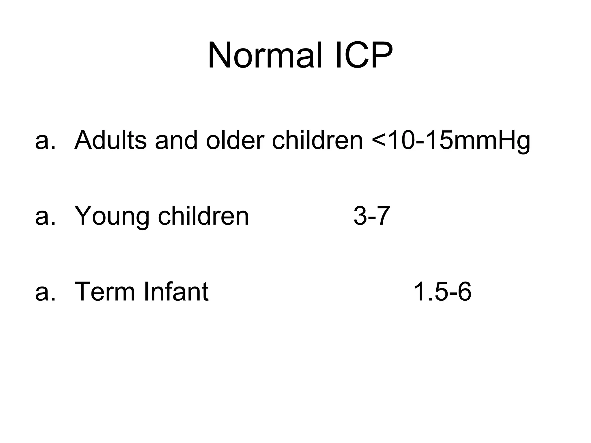
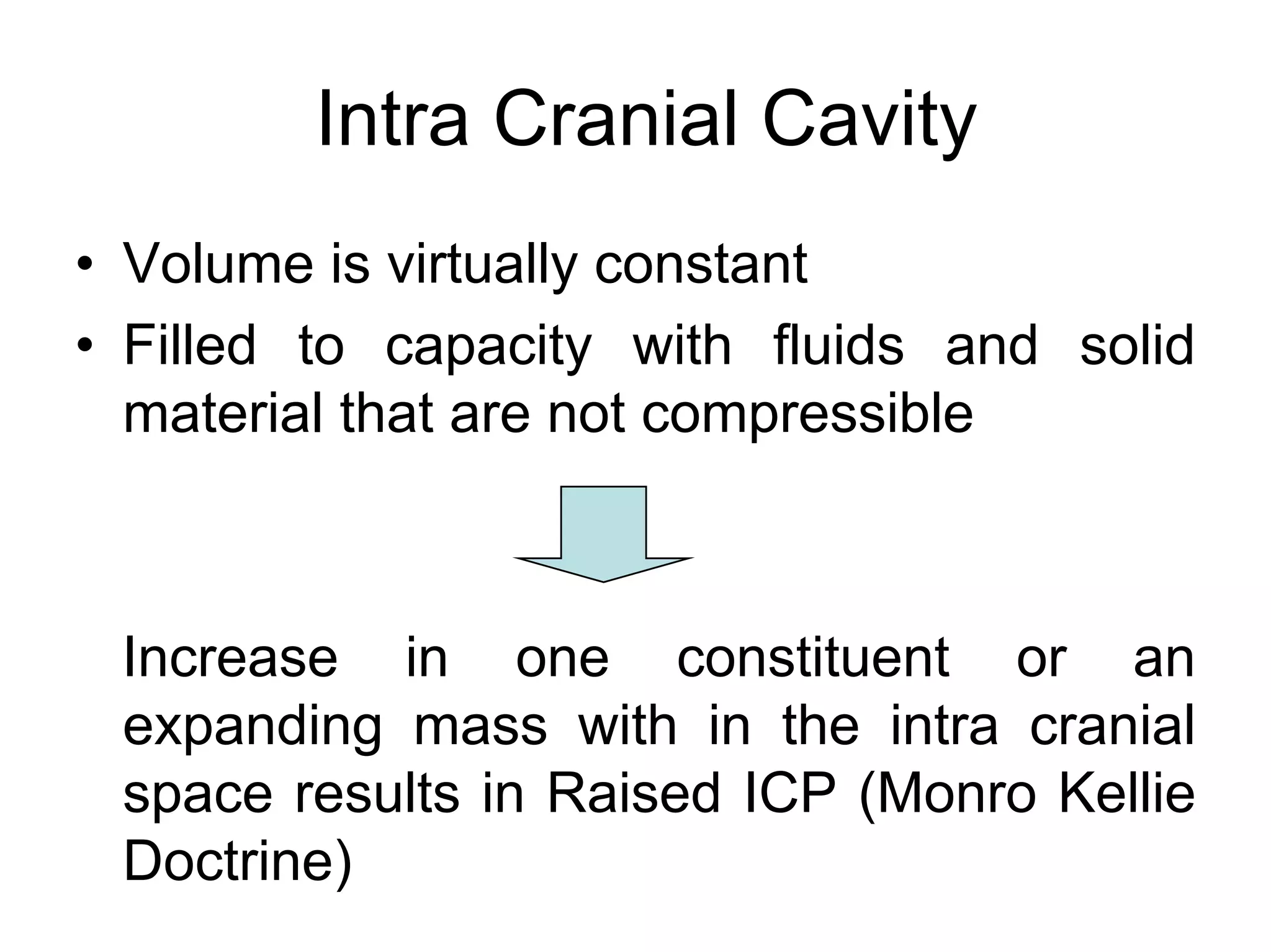
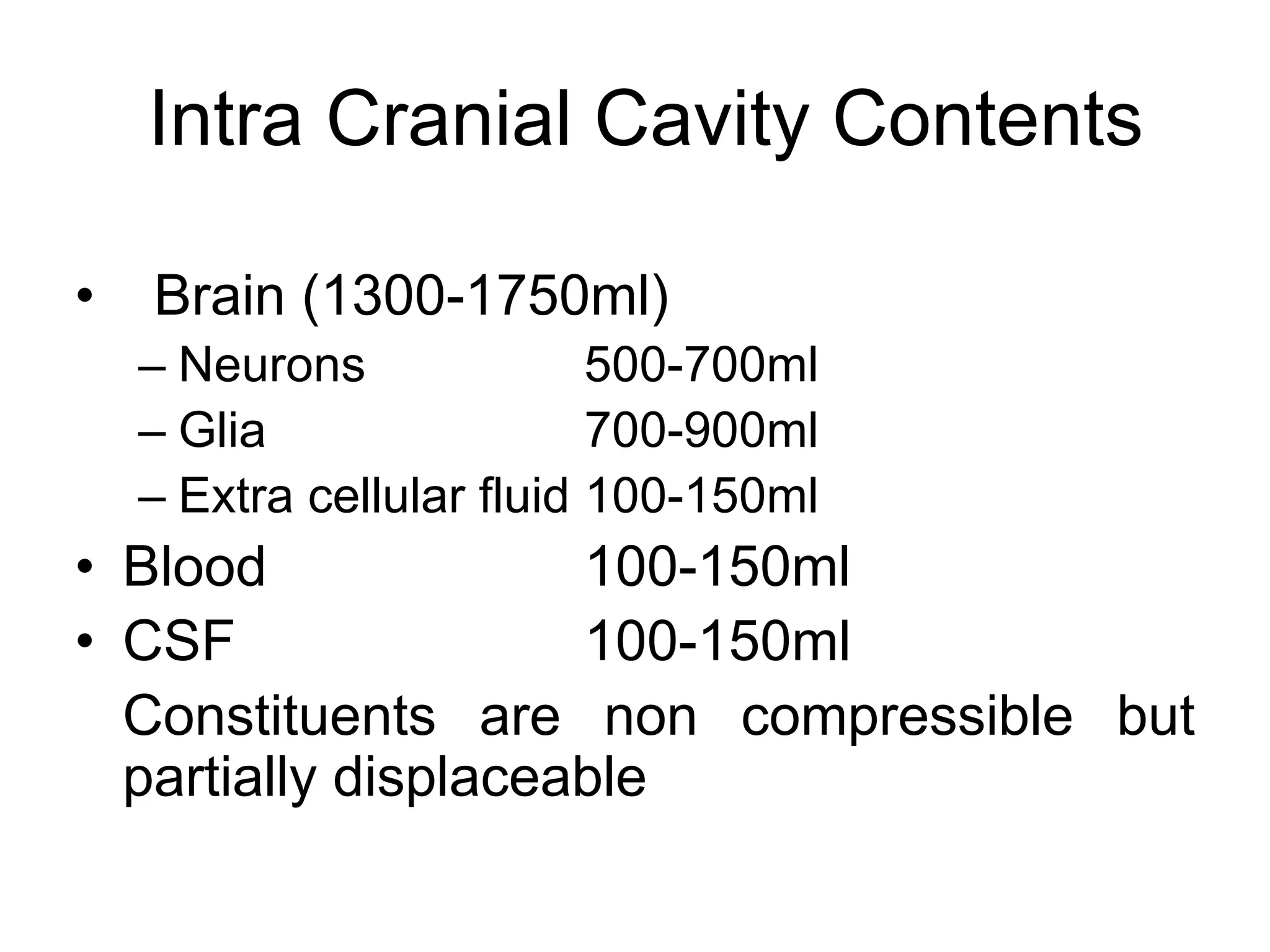
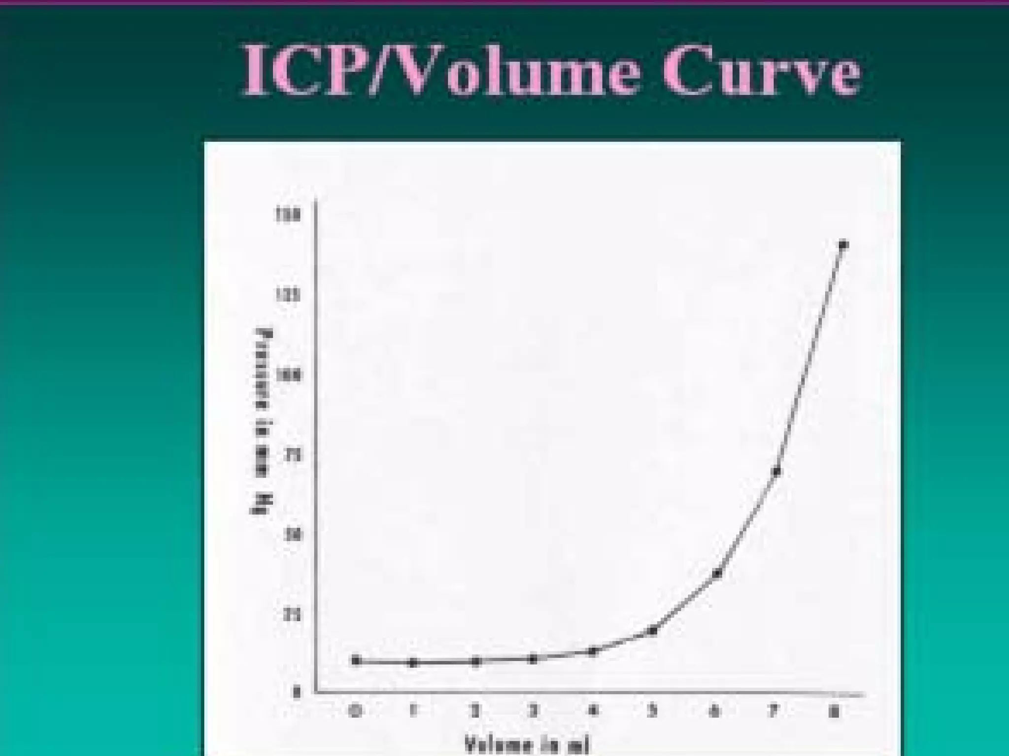
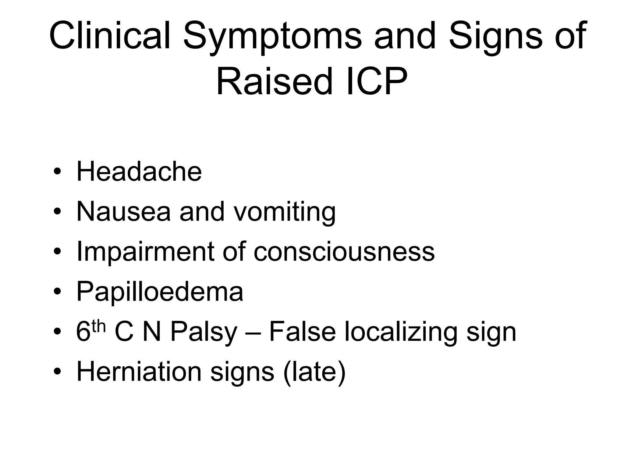
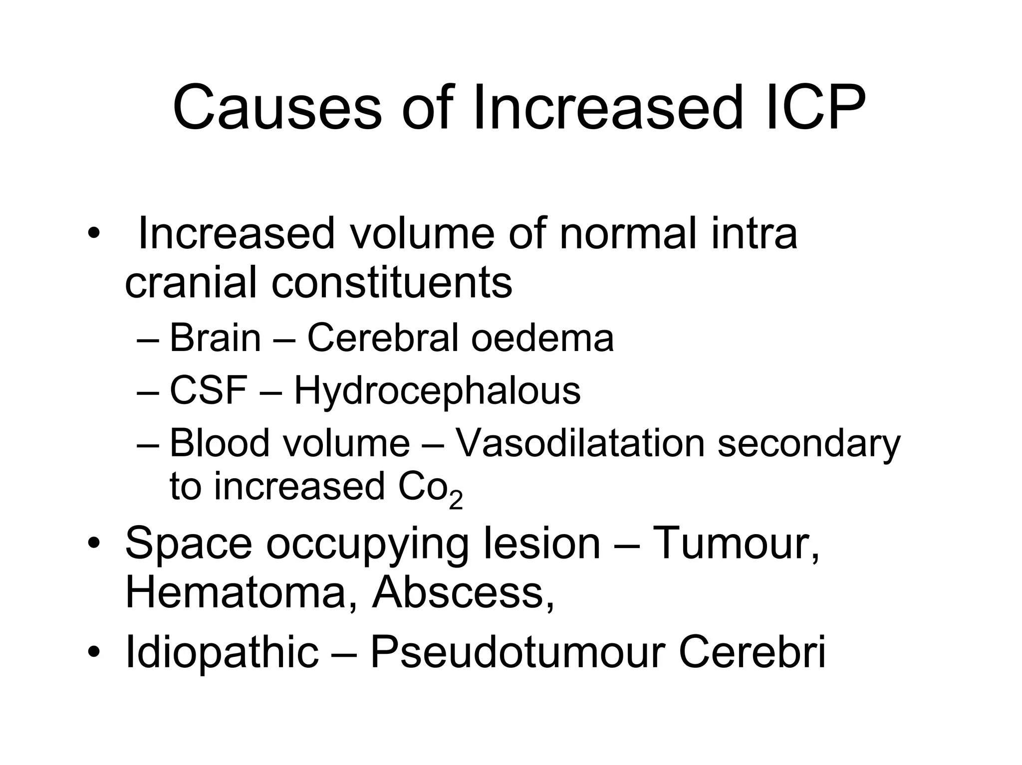
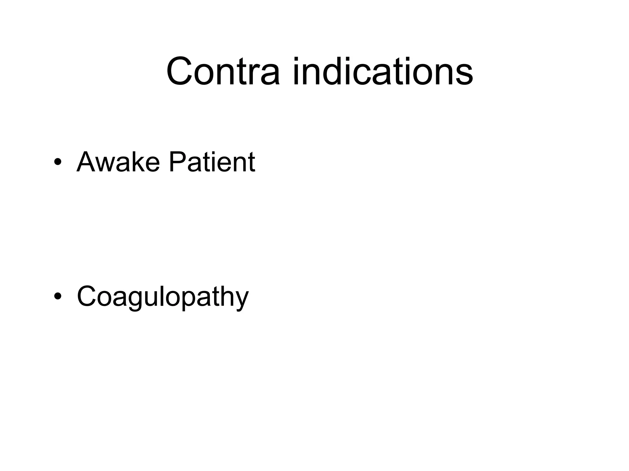
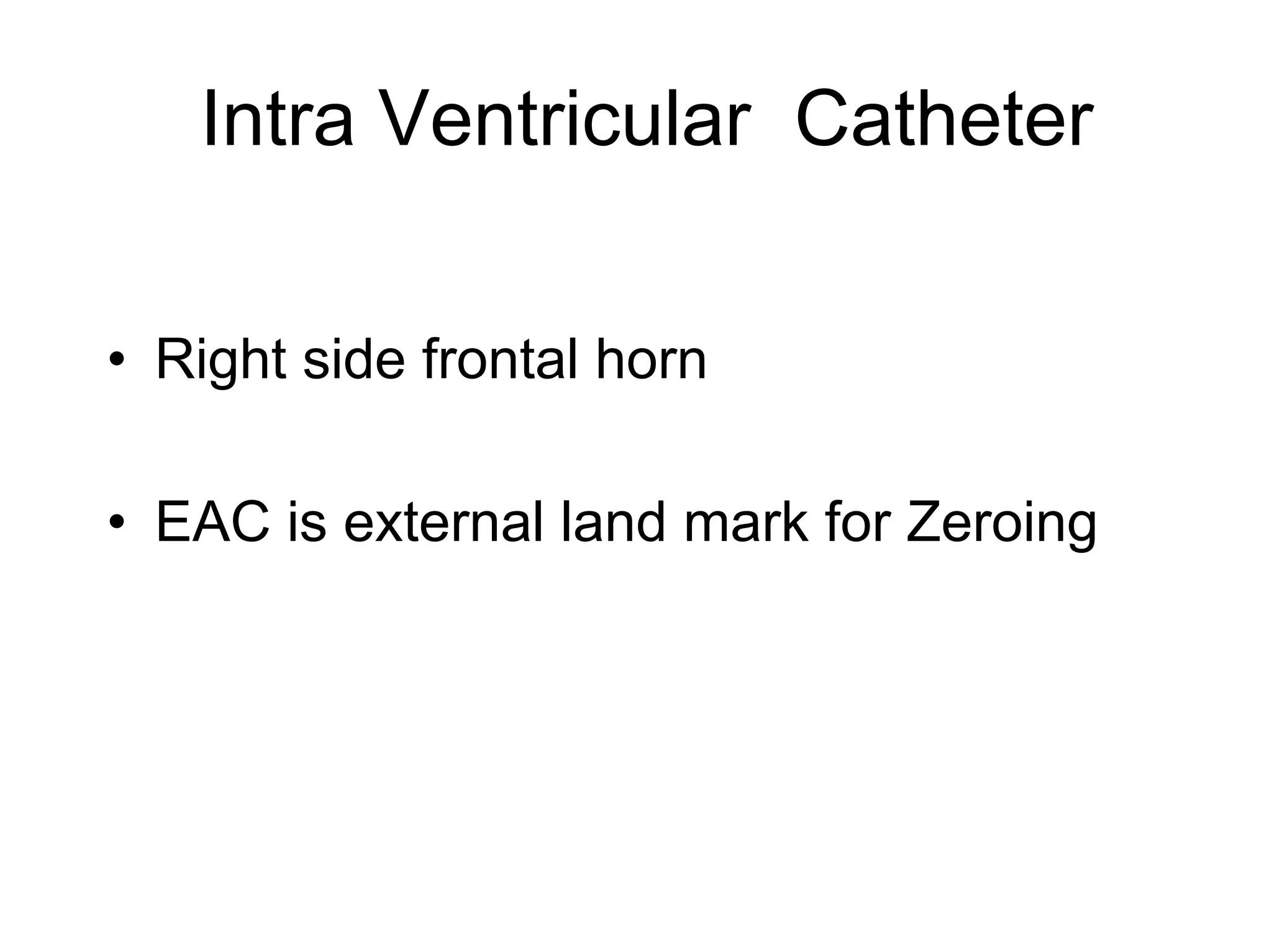
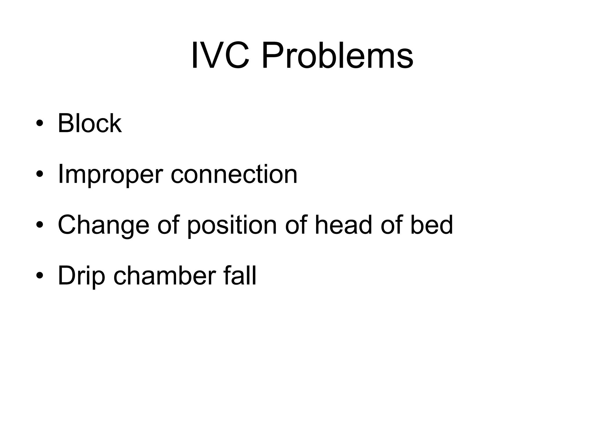
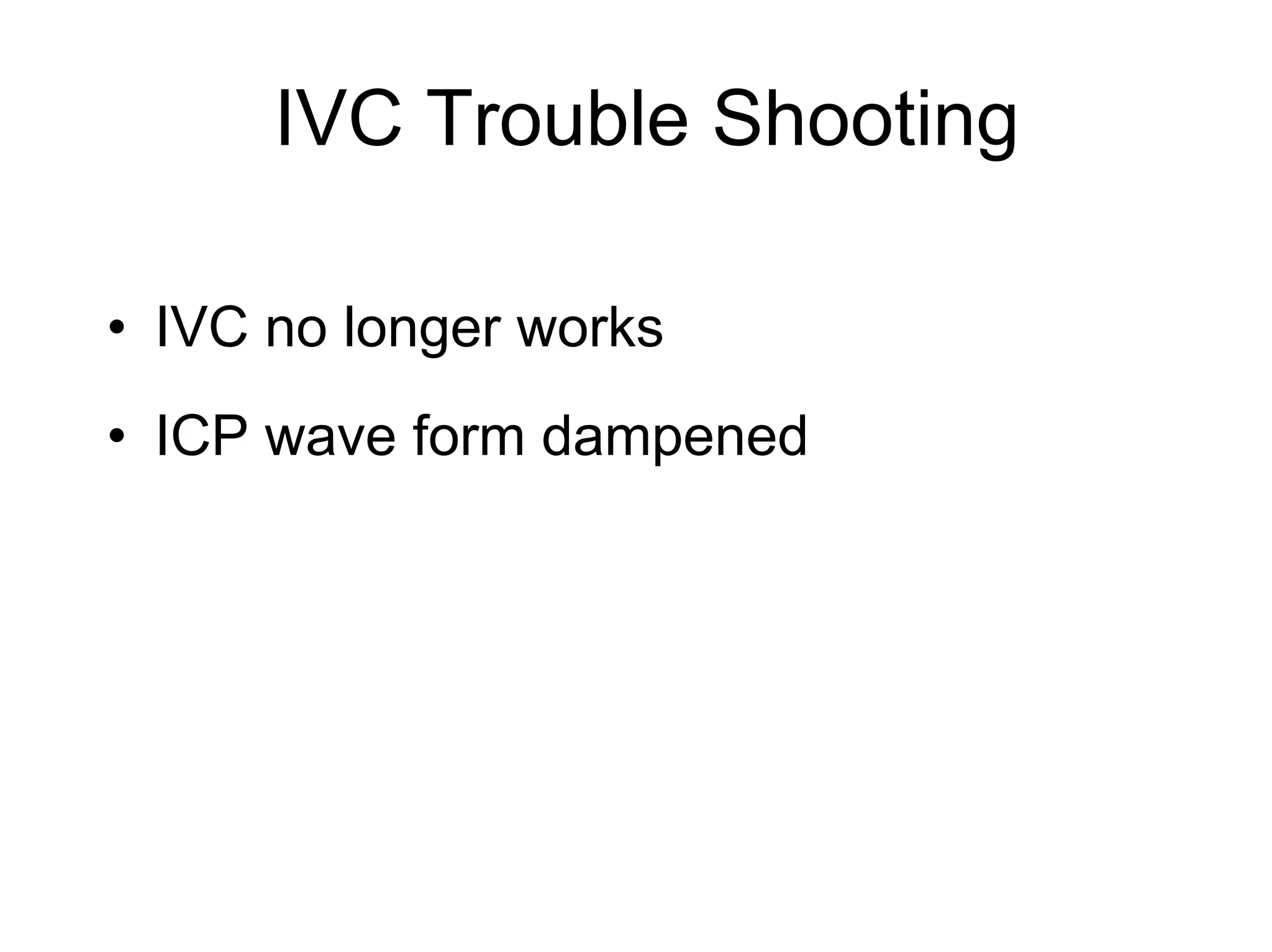
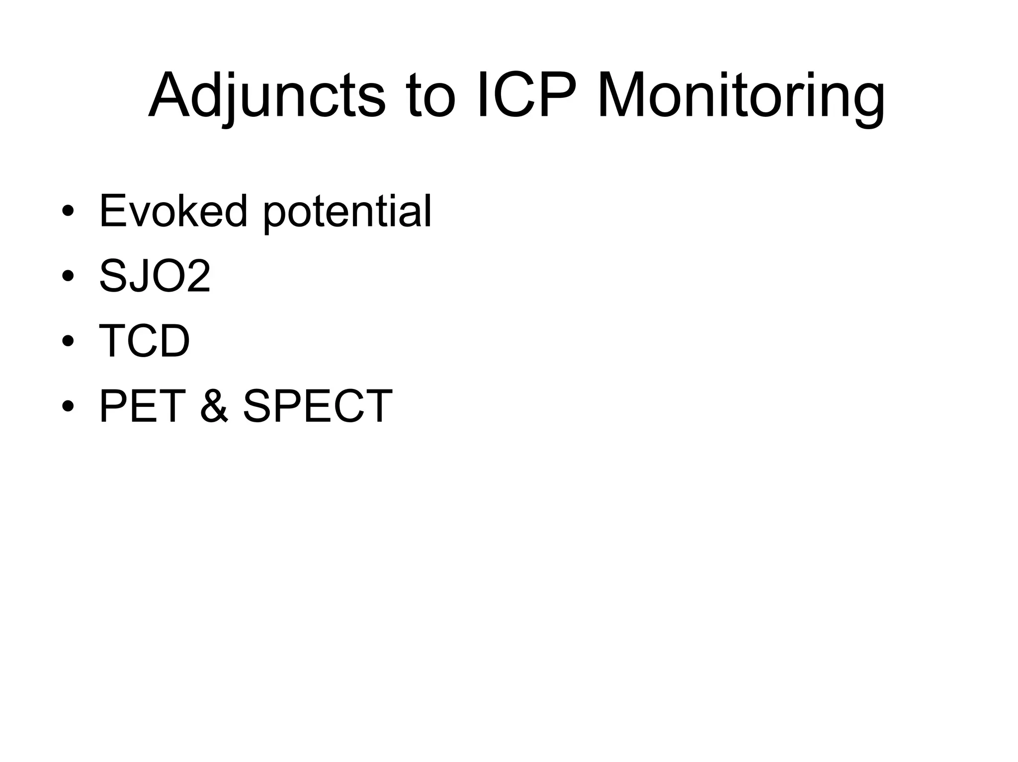
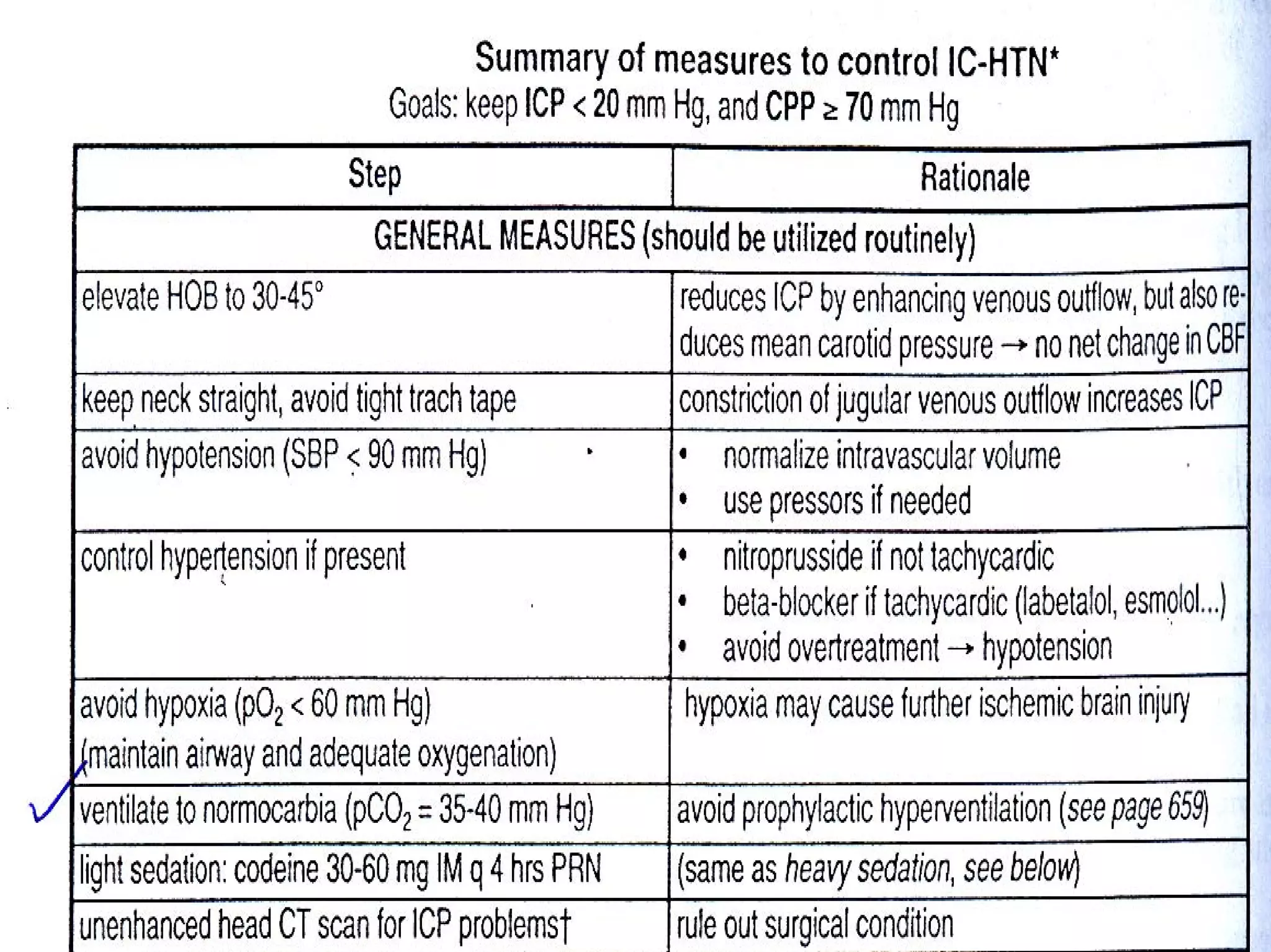
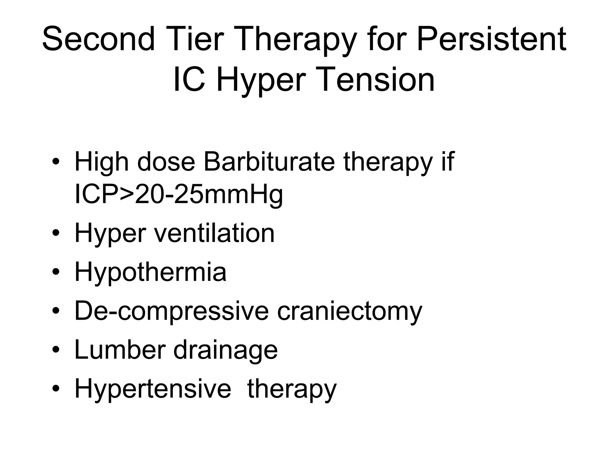
This document discusses monitoring and management of raised intracranial pressure. It begins by defining normal intracranial pressure ranges and describing the components of the intracranial cavity. Clinical signs of raised ICP are then outlined, followed by common causes and mechanisms. Monitoring of ICP is described including indications, contraindications and different monitoring device types. Treatment measures to lower ICP are then covered at both general and specific levels, including surgical intervention, ventilation management, barbiturate therapy, and decompressive craniectomy.
Vip Call Girls Anna Salai Chennai 👉 8250192130 ❣️💯 Top Class Girls Available
Raised ICP_.pdf
1. MONITORING OF RAISED INTRA
CRANIAL PRESSURE AND
MANAGEMENT OF RAISED INTRA
CRANIAL PRESSURE
Presented By : Pankaj Singh
2. Normal ICP
a. Adults and older children <10-15mmHg
a. Young children 3-7
a. Term Infant 1.5-6
3. Intra Cranial Cavity
• Volume is virtually constant
• Filled to capacity with fluids and solid
material that are not compressible
Increase in one constituent or an
expanding mass with in the intra cranial
space results in Raised ICP (Monro Kellie
Doctrine)
4. Intra Cranial Cavity Contents
• Brain (1300-1750ml)
– Neurons 500-700ml
– Glia 700-900ml
– Extra cellular fluid 100-150ml
• Blood 100-150ml
• CSF 100-150ml
Constituents are non compressible but
partially displaceable
8. Clinical Symptoms and Signs of
Raised ICP
• Headache
• Nausea and vomiting
• Impairment of consciousness
• Papilloedema
• 6th C N Palsy – False localizing sign
• Herniation signs (late)
9. Causes of Increased ICP
• Increased volume of normal intra
cranial constituents
– Brain – Cerebral oedema
– CSF – Hydrocephalous
– Blood volume – Vasodilatation secondary
to increased Co2
• Space occupying lesion – Tumour,
Hematoma, Abscess,
• Idiopathic – Pseudotumour Cerebri
10. Cerebral Perfusion Pressure
and Auto Regulation
CPP = MAP-ICP
Cerebral auto regulation is a
mechanism were by over a wide large
changes in systemic BP produces only
small changes in CBF
11. ICP
• Cerebral oedema
• Hyperemia
• Intra cranial Mass
• HCP
• Hypoventilation
• Systemic Hypertension
• Venous sinus thrombosis
• Increased Muscle tone and valsalva maneuver
• Sustained Post traumatic seizure
12. Secondary Increase in ICP (3-
10 Days Following trauma)
• Delayed hematoma formation
• Cerebral vasospasm
• ARDS with Hypoventilation
• Delayed Oedema formation
• Hyponatremia
14. ICP Monitoring
• Indication
– Severe Head injury (GCS< 8 after CPR) and
either
• Abnormal admitting CT
Or
• Normal CT with > 2 of risk factors (Age >40yr, SBP
<90mm Hg, Decerebrate or decorticate posturing
– Multiple System Injured with altered level of
consciousness
– Traumatic Intra cranial mass
• A physician may choose to monitor ICP in some the
patients
• Post op subsequent to removal
17. Types of Monitor
• Intra ventricular catheter (Standard) –
– Advantages –
• Low cost
• Therapeutic CSF Drainage
• May be recalibrated to minimize measurement drift
– Disadvantages –
• Difficult to insert
• Obstruction of fluid column
• Some effort is required to check any monitoring function
• Transducer must be consistently maintain at a fixed
reference point
18. – Intra parenchymal monitor – Similar to IVC
usually not subjected to measurement drift
– Less Accurate-
• Subarachnoid screw- At high ICP brain may
block Lumen
• Subdural
• Epidural
• Infants-
– Aplanation Principle
– Fontometry
21. Type of Wave Form
• Normal wave form
– Small pulsation transmitted from the systolic BP to
intra cranial cavity
• Large 1-2mm Hg Peak corresponding to altered systolic
Pressure wave with small dicrotic notch.
• The Peak is followed by smaller and less distinct peak
• Followed by peaks corresponding to central venous A
wave from right atrium.
– Blood pressure pulsation are superimposed on
slower respiratory variation
During expiration – SVC Pressure Increases so ICP
increase
22. Pathological Wave Form
As ICP rises cerebral compliance decreased
the venous component disappears and
arterial pulsation become more pronounced
a. Lundberg A- (Plateau wave)
b. Lundberg B- amplitude 10-20mmHg, last
30 second to 2 minutes. Respiratory
variation present.
c. Lundberg C- frequency 4-8 per Minutes.
Also called as Traube Hering Waves
23.
24. Normal Function of IVC System
• Wave form
• Pulsation
• Output
• Lowering of Head of Bed increases ICP.
Bilateral JV pressure increases ICP
25. IVC Problems
• Block
• Improper connection
• Change of position of head of bed
• Drip chamber fall
27. Adjuncts to ICP Monitoring
• Evoked potential
• SJO2
• TCD
• PET & SPECT
28. ICP Treatment Measures
• Guidelines
– ICP treatment should be initiated for ICP >20-
25mm Hg
• Options
– Interpretation and treatment of ICP based on
any threshold should be corroborated by
frequent clinical examination and CPP data
– CPP should be maintain ≥70mm Hg
29. ICP Management Protocol
• Goals of therapy
– Keep ICP <20mmHg (Prevents plateau waves
from compromising CBF and causing cerebral
ischemia and or brain death
– CPP >70mmHg (avoid hypotension)
30.
31. • Surgical treatment
– Any subdural or epidural hematoma larger
than 1cm maximal thickness should be
removed
– Patient with contused brain with
progressive deterioration should be
removed.
– De-compressive craniectomy should be
considered for ICP not controlled
medically.
• General care
– Avoid Hypoxia PaO2<60mmHg
– Avoid Hypo tension SBP ≤90mmHg
32. Specific Treatment
• Prophylaxis against stress ulcer
• Control fever
• Arterial line
• CVP line
• IV fluid –
– NS +20Meqkcl/let.
– Avoid hypotonic solution
– Normalize intra vascular volume
– Concept of running patient dry is obsolete
– If injury to other system present they may dictate
Fluid management
– Pressors or preferable to IV fluid bolus in head
injury
36. High Dose Barbiturate Therapy
• Guidelines
– May be considered in hemodynamically stable
salvageable severe TBI patient with Intra cranial Hyper
tension refractory to maximal Medical and surgical ICP
lowering therapy
– Theoretical benefits of barbiturates in head injury is
derived from
1. vasoconstriction in normal areas (shunting blood to
ischemic brain tissue)
2. Decreased metabolic demand for oxygen (CMRO2)
with accompanying reduction of CBF.
3. Free radical scavenging
4. Reduced intra cellular calcium
5. Lysosomal stabilization
37. • Pentobarbitol is choice
Load 10mg/kg/hr for 4 hrs.
1hr. 2.5mg/kg every 15 min.
four doses
Next 3hrs. 10mg/kg /hr infusion
Maintain 1.5mg/kg/hr infusion
BSAER early. Repeat if Pentobarbitol
level >6mg%
Goal ICP <24mmHg level 3-5mg%
If ICP <20 mmHg continue treatment for
48hrs. And then Taper dose