Download to read offline
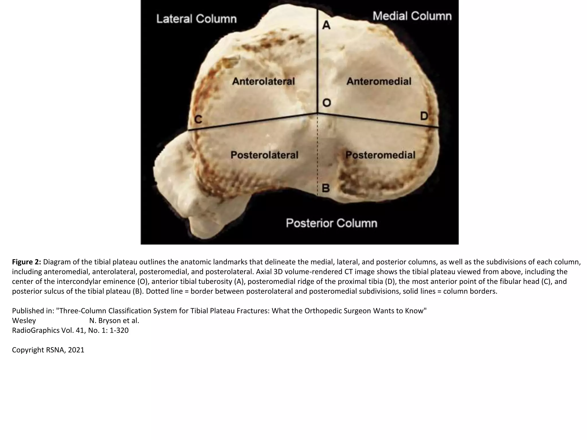
The diagram outlines the anatomical landmarks that delineate the medial, lateral, and posterior columns of the tibial plateau, including their subdivisions of anteromedial, anterolateral, posteromedial, and posterolateral. An axial CT image shows the top view of the tibial plateau, labeling landmarks like the intercondylar eminence, anterior tibial tuberosity, posteromedial ridge, fibular head, and posterior sulcus of the tibial plateau. The dotted line indicates the border between the posterolateral and posteromedial subdivisions and solid lines denote the column borders.


