Project Management The Managerial Process 5th Edition Larson Test Bank
Project Management The Managerial Process 5th Edition Larson Test Bank
Project Management The Managerial Process 5th Edition Larson Test Bank
Project Management The Managerial Process 5th Edition Larson Test Bank
Project Management The Managerial Process 5th Edition Larson Test Bank
1.
Project Management TheManagerial Process 5th
Edition Larson Test Bank download
https://testbankfan.com/product/project-management-the-
managerial-process-5th-edition-larson-test-bank/
Explore and download more test bank or solution manual
at testbankfan.com
2.
We have selectedsome products that you may be interested in
Click the link to download now or visit testbankfan.com
for more options!.
Project Management The Managerial Process 5th Edition
Larson Solutions Manual
https://testbankfan.com/product/project-management-the-managerial-
process-5th-edition-larson-solutions-manual/
Project Management The Managerial Process 7th Edition
Larson Test Bank
https://testbankfan.com/product/project-management-the-managerial-
process-7th-edition-larson-test-bank/
Project Management The Managerial Process 6th Edition
Larson Test Bank
https://testbankfan.com/product/project-management-the-managerial-
process-6th-edition-larson-test-bank/
South Western Federal Taxation 2013 Individual Income
Taxes 36th Edition Hoffman Solutions Manual
https://testbankfan.com/product/south-western-federal-
taxation-2013-individual-income-taxes-36th-edition-hoffman-solutions-
manual/
3.
Organizational Behaviour Understandingand Managing Life
at Work Canadian 10th Edition Johns Solutions Manual
https://testbankfan.com/product/organizational-behaviour-
understanding-and-managing-life-at-work-canadian-10th-edition-johns-
solutions-manual/
Operations Management Canadian 6th Edition Stevenson Test
Bank
https://testbankfan.com/product/operations-management-canadian-6th-
edition-stevenson-test-bank/
Principles of Financial Accounting 12th Edition Needles
Test Bank
https://testbankfan.com/product/principles-of-financial-
accounting-12th-edition-needles-test-bank/
Macroeconomics 2nd Edition Hubbard Test Bank
https://testbankfan.com/product/macroeconomics-2nd-edition-hubbard-
test-bank/
Taxation for Decision Makers 2019 9th Edition Escoffier
Test Bank
https://testbankfan.com/product/taxation-for-decision-makers-2019-9th-
edition-escoffier-test-bank/
Chapter 07 -Managing Risk
7-1
Chapter 07
Managing Risk
Multiple Choice Questions
1. An uncertain event or condition that, if it occurs, has a positive or negative effect on a
project objectives is termed.
A. Random chance
B. A disaster
C. Risk
D. Hazard
E. Bad luck
2. The chances of a risk event occurring as a project proceeds through its life cycle tends to
A. Slowly rise
B. Drop sharply and then level out
C. Rise sharply and then level out
D. Remain about the same
E. Slowly drop
3. The cost impact of a risk event occurring as a project proceeds through its life cycle tends
to
A. Slowly rise
B. Drop sharply and then level out
C. Rise sharply and then level out
D. Remain about the same
E. Slowly drop
4. The attempt to recognize and manage potential and unforeseen trouble spots that may occur
when a project is implemented is known as
A. Risk forecasting
B. Risk management
C. Contingency planning
D. Scenario analysis
E. Disaster protection
6.
Chapter 07 -Managing Risk
7-2
5. Which of the following is not one of the steps in the risk management process?
A. Risk response development
B. Risk assessment
C. Risk identification
D. Risk tracking
E. Risk response control
6. The initial step in the risk management process is to
A. Determine the level of acceptable risk
B. Assess the risk potential
C. Identify the risks
D. Set aside budget funds for managing the risks
E. Appoint a risk manager
7. One common mistake made early in the risk identification process is to
A. Not all possibilities are considered
B. Participants are over-optimistic
C. Participants are over-pessimistic
D. Focus on objectives and not on the events that could produce consequences.
E. Too much attention is given to past events
8. In the beginning the focus of risk management should be on risks that
A. Impact the whole project
B. Impact the critical path
C. Are known
D. Have the greatest cost impact
E. Have the greatest schedule impact
9. The 1999 NASA Mars Climate Orbiter is an example of
A. Disaster avoidance through proactive risk management
B. Murphy's Law
C. Proper use of critical thinking
D. Mismanaged risk control
E. Using historical records to assess risk
7.
Chapter 07 -Managing Risk
7-3
10. Which of the following would not be considered a threat?
A. Inflation
B. Meeting the project schedule
C. International disruptions
D. Economic conditions
E. Competition
11. A list of questions that address traditional areas of uncertainty on a project is termed a
risk
A. Risk profile
B. Questionnaire
C. Research
D. Query
E. Checklist
12. Which of the following is typically included in risk profiles?
A. Management aspects
B. Market aspects
C. Technical aspects
D. Both A and C are included
E. A, B, and C are all included
13. All of the following are included in the risk identification process except
A. Customers
B. Subcontractors
C. Competitors
D. Vendors
E. None of these are included
8.
Chapter 07 -Managing Risk
7-4
14. One of the keys to success in risk identification is
A. Critical thinking
B. Optimism
C. Pessimism
D. A "can do" attitude
E. All of these are correct
15. The easiest and most commonly used technique for analyzing risks is _____ analysis.
A. Probability
B. Scenario
C. Payback
D. Risk/reward
E. Impact
16. A risk profile is a list of questions that address traditional areas of uncertainty on a project
that answers developed from:
A. When the event might occur in the project
B. Chances of the event occurring
C. Interaction with other parts of the project or with other projects
D. From previous, similar projects
E. Magnitude or severity of the event's impact
17. The risk management tool that is divided into three color-coded zones representing major,
moderate, and minor risks is the risk
A. Assessment form
B. Responsibility matrix
C. Scenario assessment
D. Impact assessment
E. Risk severity matrix
9.
Chapter 07 -Managing Risk
7-5
18. The risk assessment form contains all of the following except
A. Likelihood of the risk event occurring
B. Potential impact of the risk event
C. Who will detect the occurrence of the risk event.
D. Difficulty of detecting the occurrence of the risk event
E. When the risk event may occur
19. The two scales of a risk severity matrix measure
A. Time, cost
B. Cost, schedule
C. Impact, cost
D. Time, impact
E. Likelihood, impact
20. Which of the following is not one of the probability analysis tools?
A. Ratio/range analysis
B. Decision tree
C. PERT simulation
D. PERT
E. All of these are probability analysis tools
21. This risk assessment tool is a variation of the risk severity matrix that includes the ease of
detection for each of the identified risks.
A. PERT simulation
B. FMEA analysis
C. Ratio/range analysis
D. Probability analysis
E. Semi-quantitative analysis
10.
Chapter 07 -Managing Risk
7-6
22. Which of the following is not included in a Failure Mode and Effects Analysis?
A. Impact
B. Probability
C. Detection
D. Risk value
E. All of these are included
23. Which of the following is used to review activity and project risk?
A. NPV
B. S-curves
C. PERT
D. Decision trees
E. All of these can be used
24. Which of the following is not one of the potential responses to a specific risk event?
A. Mitigating
B. Retaining
C. Ignoring
D. Transferring
E. Sharing
25. A Risk Response Matrix contains all of the following except
A. Contingency plan
B. Trigger
C. Who is responsible?
D. Response
E. All of these are included in the matrix
11.
Chapter 07 -Managing Risk
7-7
26. The demolition of the Seattle Kingdome (Snapshot from Practice) is an example of which
of the following?
A. Mitigating
B. Retaining
C. Ignoring
D. Transferring
E. Sharing
27. The risk associated with one of the key members being stuck by lightning would most
likely be handled by which of the following?
A. Mitigating
B. Retaining
C. Ignoring
D. Transferring
E. Sharing
28. Funds that are for identified risks that have a low probability of occurring and that
decrease as the project progresses are called ______ reserves.
A. Management
B. Budget
C. Contingency
D. Padded
E. Just in case
29. Technical risks are:
A. Can often be the kind that cause the project to be shut down.
B. Problematic
C. Imposed duration dates
D. Both A and B are correct
E. A, B, and C are all correct
12.
Chapter 07 -Managing Risk
7-8
30. Detailing all identified risks, including descriptions, category, and probability of
occurring, impact, responses, contingency plans, owners and current status is called:
A. Management reserves
B. Change control
C. Contingency reserves
D. Risk register
E. Risk profiles
31. Which of the following is identified to cover major unforeseen risks and, hence, are
applied to the total project?
A. Budget reserves
B. Management reserves
C. Time buffers
D. Both B and C are correct
E. A, B, and C are all correct
32. Change management systems are designed to accomplish all of the following except:
A. Track all changes that are to be implemented
B. Review, evaluates, and approve/disapprove proposed changes formally
C. Identify expected effects of proposed changes on schedule and budget
D. Reflect scope changes in baseline and performance measures
E. All of the above are correct
Fill in the Blank Questions
33. The ________ impact of a risk event in a project is less if the event occurs earlier rather
than later.
________________________________________
34. The likelihood of a risk event occurring ________ as a project goes through its life cycle.
________________________________________
13.
Chapter 07 -Managing Risk
7-9
35. The first step in the Risk Management process is ________.
________________________________________
36. Risk events such as inflation, market acceptance, and government regulations are referred
to as ________.
________________________________________
37. A ________ is a list of questions that address traditional areas of uncertainty on a project.
________________________________________
38. The easiest and most commonly used technique for analyzing risks is ________.
________________________________________
39. The ________ form identifies each risk event, the likelihood of it occurring, the potential
impact, when it may occur, and the degree of difficulty in detecting it.
________________________________________
40. The ________ matrix is divided into red, yellow, and green zones representing major,
moderate, and minor risks.
________________________________________
41. The vertical scale on the Risk Severity Matrix measures the _________ of a potential risk
event.
________________________________________
42. The horizontal scale on the Risk Severity Matrix measures the _________ of a potential
risk event.
________________________________________
14.
Chapter 07 -Managing Risk
7-10
43. In __________ the Risk Severity Matrix is extended by including the ease of detecting a
risk event occurring.
________________________________________
44. In a ________, three different estimates of activity times are used to statistically predict
the time an activity will take to complete.
________________________________________
45. The "Snapshot from Practice" case where Ellipsus Systems developed parallel prototype
systems (WAP and JAVA) is an example of _________ a risk.
________________________________________
46. Testing a new project on a smaller isolated area prior to installing it for the entire
organization is an example of ________ a risk.
________________________________________
47. Performance bonds, warranties, and insurance are examples of ________ a risk.
________________________________________
48. When the entertainment industry formed a consortium to define a common operating
format for DVD it was ________ the risk.
________________________________________
49. If a risk event is very unlikely to occur the project owner would probably ________ the
risk.
________________________________________
Chapter 07 -Managing Risk
7-11
50. A ________ identifies what to do if a potential risk event actually occurs.
________________________________________
51. ________ reserves are identified for specific work packages and cover risks that have a
low probability of occurring.
________________________________________
52. ________ reserves are controlled by the project manager and used to cover major
unforeseen risks to the entire project.
________________________________________
53. A ____________ is an alternative that will be used if a possible foreseen risk event
becomes a reality.
________________________________________
54. A ____________ is useful for summarizing how the project team plans to manage risks
that have been identified.
________________________________________
True / False Questions
55. The probability that a risk event will occur is higher during the initial stages of a project.
True False
56. Due to the impact over a long period of time, risk events that occur in the early stages of a
project will have a greater cost impact than those that occur in later stages.
True False
17.
Chapter 07 -Managing Risk
7-12
57. During risk identification the smaller risks should be identified first because they will
naturally lead to identifying the larger risks.
True False
58. One common mistake that is made early on in the risk identification process is to focus on
consequences and not on the events that could produce consequences.
True False
59. Risks such as inflation and monetary exchange rates are not usually included in a project's
risk assessment.
True False
60. The first step in the risk management process is Risk Assessment.
Refer to Figure 7.2
True False
61. A risk profile is a list of questions that have been developed and refined from previous,
similar projects.
True False
62. The risk identification process should be limited to just the core project team.
True False
63. Since the goal is to find problems before they happen, the project manager should
encourage critical thinking when it comes to risk identification.
True False
18.
Chapter 07 -Managing Risk
7-13
64. The Risk Severity Matrix rates risk events based upon schedule and cost.
Refer to Figure 7.7
True False
65. In a Risk Severity Matrix a green zone risk is considered inconsequential and ignored
unless their status changes.
True False
66. The quality and credibility of the risk analysis process requires that different levels of risk
probabilities and impacts be defined.
True False
67. Adopting proven technology instead of experimental technology is an example of
mitigating a risk.
True False
68. The FMEA method calculates a risk value by assigning ease of detection ratings to the key
risk elements.
True False
69. Performance bonds, warranties, and guarantees are financial instruments used to share
risk.
True False
70. Fixed price contracts are an example of transferring risk from an owner to a contractor.
True False
19.
Chapter 07 -Managing Risk
7-14
71. Scheduling outdoor work in the summer, investing in up front safety training, and
choosing high quality materials are examples of retaining a risk.
True False
72. Budget reserves are setup to cover identified risks associated with specific segments of a
project while management reserves are set up to cover unidentified risks associated with the
total project.
True False
73. Change management systems involve reporting, controlling, and recording changes to the
project baseline.
True False
74. Project managers need to establish an environment in which participants feel comfortable
raising concerns and admitting mistakes.
True False
75. Contingency funding is made up of budget reserves and management reserves.
True False
Short Answer Questions
76. Describe the relationship between the likelihood of a risk event occurring and the cost of
fixing the risk event as a project proceeds through its life cycle.
20.
Chapter 07 -Managing Risk
7-15
77. Identify and briefly describe the four steps in risk management.
78. Describe the process for identifying project risks.
79. What is a risk profile and what benefits does it provide to risk management?
80. Identify at least six items that may be included on a Risk Profile.
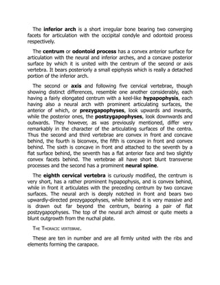
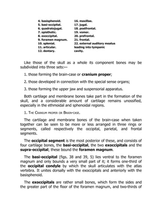
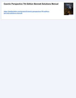
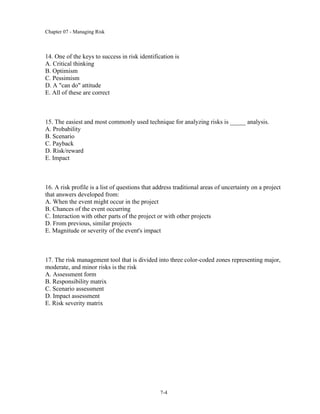
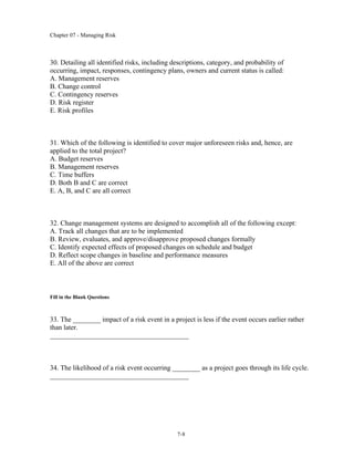
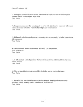
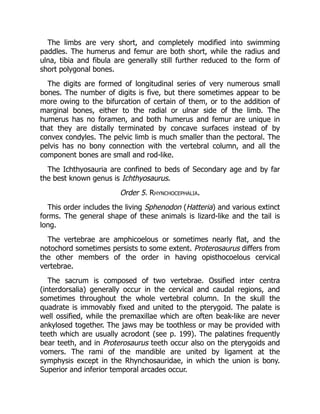
Fig. 32. Lateral(below) and dorsal (above) views of the skull of an Ichthyosaurus.
(Modified from Deslongchamps.)
1. premaxillae. 12. squamosal.
2. maxillae. 13. supratemporal.
3. nasal. 14. quadratojugal.
4. prefrontal[74]. 15. sclerotic ring.
5. frontal. 16. postorbital.
6. postfrontal[74]. 17. jugal.
7. anterior nares. 18. lachrymal.
8. orbit. 19. dentary.
9. supratemporal fossa. 20. articular.
10. interparietal foramen. 21. angular.
11. parietal.
The ribs are long, and the anterior ones have capitula and tubercula.
There is no sternum, but the ventral body wall is strengthened by a
complex system of abdominal splint ribs.
The pectoral girdle is strongly developed, the scapulae are narrow, the
coracoids broad, and meet ventrally without overlapping. There are
probably no precoracoids, but clavicles and a T-shaped interclavicle are
well developed.
23.
The limbs arevery short, and completely modified into swimming
paddles. The humerus and femur are both short, while the radius and
ulna, tibia and fibula are generally still further reduced to the form of
short polygonal bones.
The digits are formed of longitudinal series of very numerous small
bones. The number of digits is five, but there sometimes appear to be
more owing to the bifurcation of certain of them, or to the addition of
marginal bones, either to the radial or ulnar side of the limb. The
humerus has no foramen, and both humerus and femur are unique in
that they are distally terminated by concave surfaces instead of by
convex condyles. The pelvic limb is much smaller than the pectoral. The
pelvis has no bony connection with the vertebral column, and all the
component bones are small and rod-like.
The Ichthyosauria are confined to beds of Secondary age and by far
the best known genus is Ichthyosaurus.
Order 5. Rhynchocephalia.
This order includes the living Sphenodon (Hatteria) and various extinct
forms. The general shape of these animals is lizard-like and the tail is
long.
The vertebrae are amphicoelous or sometimes nearly flat, and the
notochord sometimes persists to some extent. Proterosaurus differs from
the other members of the order in having opisthocoelous cervical
vertebrae.
The sacrum is composed of two vertebrae. Ossified inter centra
(interdorsalia) generally occur in the cervical and caudal regions, and
sometimes throughout the whole vertebral column. In the skull the
quadrate is immovably fixed and united to the pterygoid. The palate is
well ossified, while the premaxillae which are often beak-like are never
ankylosed together. The jaws may be toothless or may be provided with
teeth which are usually acrodont (see p. 199). The palatines frequently
bear teeth, and in Proterosaurus teeth occur also on the pterygoids and
vomers. The rami of the mandible are united by ligament at the
symphysis except in the Rhynchosauridae, in which the union is bony.
Superior and inferior temporal arcades occur.
24.
The ribs havecapitula and tubercula, and often uncinate processes
(see p. 190) as in birds. A pectoral girdle and sternum, with clavicles and
a T-shaped interclavicle are developed, and abdominal ribs are always
found. The precoracoid is however absent. The limbs are pentedactylate.
Sphenodon[75] (Hatteria) now living in some of the islands of the New
Zealand group, is certainly the most generalised of all living reptiles.
Though lizard-like in form it differs from all living lizards in the
possession of two temporal arcades, abdominal ribs and a fixed
quadrate; and is often considered to be nearly allied in many respects to
the type of reptile from which all the others took their origin.
Among the better known extinct forms are Proterosaurus of Permian
and Hyperodapedon of Triassic age.
Order 6. Squamata.
This order includes the extinct Mosasaurians, and the lizards and
snakes which form the vast majority of living reptiles. The trunk may be
moderately elongated and provided with four short limbs as in lizards, or
it may be limbless, extremely elongated, and passing imperceptibly into
the tail. The surface is generally completely covered with overlapping
horny epidermal scales, below which bony dermal scutes may be
developed.
The vertebrae are procoelous, rarely amphicoelous. There are no inter
centra, and the neural arches are firmly united to the centra. Additional
articulating surfaces, the zygosphenes and zygantra, are often
developed[76]. The sacrum is formed of two or rarely three vertebrae, or
may be wanting as in Ophidia. In the skull an infratemporal arcade
forming the lower boundary of the infratemporal fossa is absent, and the
quadrate, except in the Chamaeleons, is movably articulated to the
squamosal. The palatal vacuities are large and the nares are separate.
There is often a distinct parasphenoid. The teeth are either acrodont (i.e.
ankylosed to the summit of the jaw), or pleurodont, i.e. ankylosed to the
inner side of the jaw. The thoracic ribs each have a single head which
articulates with the centrum of the vertebra; while uncinate processes
and abdominal ribs never occur.
25.
A pectoral girdleand sternum may be present, or may be completely
absent as in snakes. Except in snakes there are generally four
pentedactylate limbs which may either form paddles or be adapted for
walking.
Suborder (1). Lacertilia
[77].
The body is elongated, and as a rule four short pentedactylate limbs
are present, but sometimes limbs are vestigial or absent. The
exoskeleton generally has the form of horny plates, spines, or scales;
while sometimes as in the Chamaeleons and Amphisbaenians it is
absent. In other forms such as Tiliqua and Scincus, the body has a
complete armour of bony scutes, whose shape corresponds with that of
the overlying horny scales.
The vertebrae are procoelous, rarely as in the Geckos amphicoelous;
they are usually without zygosphenes and zygantra, but these structures
occur in the Iguanidae. The sacral vertebrae of living forms are not
ankylosed together, and the caudal vertebrae usually have well-
developed chevron bones.
In the skull[78] the orbits are separated from one another, only by an
imperfectly developed interorbital septum, the cranial cavity not
extending forwards between them, while the alisphenoidal region is
unossified. The premaxillae may be paired or united (Amphisbaenidae),
and there is usually an interparietal foramen. There may be a complete
supratemporal[79] arcade bounding the lower margin of the
supratemporal fossa, or the supratemporal fossa may be open below.
The quadratojugal is not ossified, and the quadrate articulates with the
exoccipital. There is no infratemporal arcade. There is commonly a rod-
like epipterygoid[80] (fig. 33, 14) connecting the pterygoid and parietal.
Teeth are always present, and may be confined to the jaws or may be
developed also on the pterygoids and rarely on the palatines; they are
either acrodont or pleurodont. The rami of the mandible are suturally
united.
A pectoral girdle is always present, and generally also a sternum.
Clavicles and a T-shaped interclavicle are commonly present, but are
26.
absent in theChamaeleons.
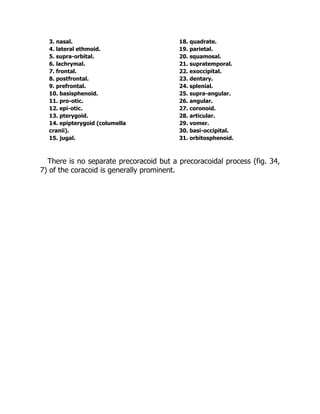
Fig. 33. A, Lateral view, and B, longitudinal section of the skull of a Lizard
(Varanus varius). × 3/5. (Brit. Mus.)
1. premaxillae. 16. transpalatine.
2. maxillae. 17. parasphenoid.
27.
3. nasal. 18.quadrate.
4. lateral ethmoid. 19. parietal.
5. supra-orbital. 20. squamosal.
6. lachrymal. 21. supratemporal.
7. frontal. 22. exoccipital.
8. postfrontal. 23. dentary.
9. prefrontal. 24. splenial.
10. basisphenoid. 25. supra-angular.
11. pro-otic. 26. angular.
12. epi-otic. 27. coronoid.
13. pterygoid. 28. articular.
14. epipterygoid (columella 29. vomer.
cranii). 30. basi-occipital.
15. jugal. 31. orbitosphenoid.
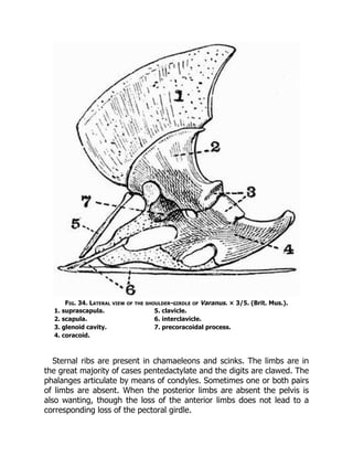
There is no separate precoracoid but a precoracoidal process (fig. 34,
7) of the coracoid is generally prominent.
28.
Fig. 34. Lateralview of the shoulder-girdle of Varanus. × 3/5. (Brit. Mus.).
1. suprascapula. 5. clavicle.
2. scapula. 6. interclavicle.
3. glenoid cavity. 7. precoracoidal process.
4. coracoid.
Sternal ribs are present in chamaeleons and scinks. The limbs are in
the great majority of cases pentedactylate and the digits are clawed. The
phalanges articulate by means of condyles. Sometimes one or both pairs
of limbs are absent. When the posterior limbs are absent the pelvis is
also wanting, though the loss of the anterior limbs does not lead to a
corresponding loss of the pectoral girdle.
29.
The pubis correspondsto the pre-pubis of Dinosaurs, and both pubes
and ischia meet in ventral symphyses.
The suborder includes the Lizards, Chamaeleons and Amphisbaenians.
Suborder (2). Ophidia
[81].
The Ophidia or snakes are characterised by their greatly elongated
body and want of limbs. The body is covered with overlapping horny
scales and bony dermal scutes are never present. The vertebrae are
procoelous, and are distinguishable into two groups only, precaudal or
rib-bearing, and caudal or ribless. The atlas vertebra is also ribless. The
neural arches are always provided with zygosphenes and zygantra. Many
of the vertebrae have strong hypapophyses, and the caudal vertebrae
are without chevron bones.
In the skull the cranial cavity extends forwards between the orbits,
and is closed in front by downgrowths from the frontals and parietals
which meet the well-ossified alisphenoids and orbitosphenoids[82]. The
cranium is strongly ossified, and there are no parotic processes or
interparietal foramen. There are no temporal arcades and no
epipterygoid. The premaxillae if present are very small (fig. 51, 1) and
usually toothless. The quadrates articulate with the squamosals, and do
not as in Lacertilia meet the exoccipitals. The palatines do not unite
directly with the vomers or with the base of the cranium, and the whole
palato-maxillary apparatus is more loosely connected with the cranium
than it is in Lacertilia. The pterygoids, and in most cases also the
palatines, bear teeth. The dentition is acrodont, and the rami of the
mandible are united only by an elastic ligament—an important point
serving to distinguish the Ophidia from the Lacertilia. There is an
imperfectly developed interorbital septum, the ventral part of which is
formed by the parasphenoid. The postfrontal is generally well developed,
while the jugals and quadratojugals are absent. There are never any
traces of the anterior limbs or pectoral girdle, but occasionally there are
vestiges of a pelvis and posterior limbs.
Suborder (3). Pythonomorpha
[83].
30.
This suborder includesMosasaurus and its allies, a group of enormous
extinct marine reptiles found in beds of Cretaceous age.
The skin is in most forms at any rate unprovided with dermal scutes.
The vertebrae may be with or without zygosphenes and zygantra. The
skull resembles that of lizards, having an interparietal foramen, and a
cranial cavity open in front. The squamosal takes part in the formation of
the cranial wall, and the quadrate articulates with the squamosal, not as
in Lacertilia with the exoccipital. There are large supratemporal fossae,
bounded below by supratemporal arcades. The teeth are large and
acrodont, and occur on the pterygoids as well as on the jaws. The two
rami of the mandible are united by ligament only. Pectoral and pelvic
girdles are present, but clavicles are wanting, and the pelvis is not as a
rule united to any sacrum.
The limbs are pentedactylate, and are adapted for swimming, while all
the limb bones except the phalanges are relatively very short. The
number of phalanges is not increased beyond the normal, and they
articulate with one another by flat surfaces. The terminal phalanges are
without claws.
Order 7. Dinosauria
[84].
The extinct reptiles comprising this order were all terrestrial, and
include the largest terrestrial animals known. They vary greatly in size
and in the structure of the limbs, some approach close to the type of
structure met with in birds, others are allied to crocodiles.
Passing to the more detailed characters:—there is sometimes a well-
developed exoskeleton having the form of bony plates or spines. The
vertebrae may be solid or their centra may be hollowed internally; their
surfaces may be flat, biconcave or opisthocoelous. The sacrum is
composed of from two to six vertebrae.
As regards the skull, the quadrate is large and fixed, and
supratemporal and infratemporal fossae bounded by bone occur. The
teeth are more or less laterally compressed, and often have serrated
edges; they may be placed in distinct sockets or in a continuous groove.
The ribs have capitula and tubercula, and sternal ribs often occur. The
scapula is very large, the coracoid small, and there is no precoracoid, or
31.
T-shaped interclavicle. Claviclesare only known in a few cases. In the
pelvis the ilium is elongated both in front of, and behind, the
acetabulum, sometimes the pre-pubis, sometimes the post-pubis is the
better developed. The anterior limbs are shorter than the posterior, and
the long bones are sometimes solid, sometimes hollow.
There are three well-marked suborders of the Dinosauria.
Suborder (1). Sauropoda
[85].
The reptiles belonging to this group were probably quadrupedal and
herbivorous.
They have the cervical and anterior trunk vertebrae opisthocoelous,
while the posterior vertebrae are biconcave; all the presacral, and
sometimes the sacral vertebrae are hollowed internally. The teeth are
spatulate and without serrated edges, they are always planted in distinct
sockets, and some of them are borne by the premaxillae.
Fig. 35. Restored skeleton of Ceratosaurus nasicornis. × 1/30. (After Marsh.)
32.
1. anterior nares.5. scapula.
2. prominence on the nasal 6. coracoid.
bones which probably 7. ilium.
carried a horn. 8. pubis (pre-pubis).
3. pre-orbital vacuity. 9. ischium.
4. orbit.
The nares have the form of long slits and there are large pre-orbital
vacuities.
The limb bones are solid, and the anterior limbs are not much shorter
than the posterior ones. All the limbs are plantigrade and pentedactylate,
and the digits of the pes are clawed. There is a large pre-pubis directed
downwards and forwards, meeting its fellow in a ventral symphysis, but
there is no post-pubis.
The Sauropoda are found in the secondary rocks of Europe and N.
America and include the largest land animals that are known to have
existed. Many of the best known forms such as Brontosaurus and
Morosaurus are North American.
Suborder (2). Theropoda.
The members of this suborder were all carnivorous, and from the
small comparative size of the anterior limbs many of them were probably
bipedal.
The vertebrae are opisthocoelous or amphicoelous, their neural arches
are provided with zygosphenes and zygantra, and their centra are
frequently hollowed internally; the limb bones are also hollow, and in
fact the whole skeleton is extremely light. The tail is of great length. The
teeth are pointed and recurved, and have one or both borders serrated;
they are always planted in distinct sockets, and some of them are borne
by the premaxillae. There are large pre-orbital vacuities. The digits of
both manus and pes are terminated by pointed ungual phalanges which
must have borne claws. In the pelvis the pre-pubes and ischia are
slender bones, the former meeting in a ventral symphysis. The ilia are
very deep vertically and there are no post-pubes. The astragalus is
closely applied to the tibia, in front of which it sends an ascending
process, sometimes the two bones appear to have been ankylosed
33.
together, as inbirds. The metatarsals are elongated and the feet
digitigrade.
The Theropoda vary greatly in size, one of the best known genera
Compsognathus was about as large as a cat, another, Megalosaurus,
perhaps as large as an elephant. Ceratosaurus is the name of a well-
known North American form regarded by many authorities as identical
with Megalosaurus.
Suborder (3). Orthopoda.
This suborder includes the most specialised of the Dinosaurs, certain
of which resemble the Theropoda in being bipedal. In some of them
such as Stegosaurus the exoskeleton is strongly developed, in others
such as Iguanodon it is absent.
The vertebrae are solid and may be opisthocoelous, biconcave, or flat.
The teeth are compressed and serrated, often irregularly, and are
frequently not set in distinct sockets. The anterior part of the premaxillae
is without teeth, and a toothless predentary or mento-meckelian bone is
present. The pre-orbital vacuities are small or absent, and the nares are
large and placed far forwards.
The most characteristic features of the group are found in the pelvis
which, except in the Ceratopsia, bears a striking resemblance to that of
birds. The ischium and post-pubis are long slender bones directed
backwards parallel to one another, and the pre-pubis is also well
developed. The ischium has an obturator process. The limb bones are
sometimes hollow, sometimes solid. The anterior limbs are much shorter
than the posterior, pointing to a bipedal method of progression. The pes
is digitigrade or plantigrade, and has three, rarely four, digits.
The suborder Orthopoda may be further subdivided into three
sections:—
A. Stegosauria.
A dermal exoskeleton is strongly developed. The vertebral centra are
flat or biconcave, and neither they nor the limb bones are hollowed out
by internal cavities. The limbs are plantigrade, the anterior ones short,
34.
the posterior onesvery large and strong. The post-pubis is well
developed;
e.g. Stegosaurus from the Upper Jurassic of Colorado.
B. Ceratopsia.
There is sometimes a well-developed dermal exoskeleton formed of
small granules and plates of bone. The bones are solid, and the vertebral
centra flat. The cranium bears a pair of enormous pointed frontal horns,
and the parietal is greatly expanded and elevated behind, forming with
the squamosals a shield which overhangs the anterior cervical vertebrae.
The premaxillae are united, and in front of them is a pointed beak-like
bone which bites upon a toothless predentary ossification of the
mandible. The teeth have two roots. The anterior limbs are but little
shorter than the posterior ones. There is no post-pubis;
e.g. Polyonax from the uppermost Cretaceous of Montana.
C. Ornithopoda
[86].
There is no dermal exoskeleton. The cervical vertebrae are
opisthocoelous, and so are sometimes the thoracic. The limb bones are
hollow and the anterior limbs are much shorter than the posterior ones.
The feet are digitigrade and provided with long pointed claws. The post-
pubis is long and slender and directed back parallel to the ischium;
e.g. Iguanodon from the European Cretaceous.
Order 8. Crocodilia
[87].
This order includes the Crocodiles, Alligators and Garials and various
extinct forms, some of which are closely allied to the early Dinosaurs.
There is always a more or less complete exoskeleton formed of bony
scutes overlain by epidermal scales; these bony scutes are specially well
developed on the dorsal surface but may occur also on the ventral. The
vertebral column is divisible into the five regions commonly
distinguishable. In all living forms the vertebrae, with the exception of
the atlas and axis, the two sacrals, and first caudal, are procoelous, but
35.
in many extinctforms they are amphicoelous. The atlas (fig. 71) is
remarkable, consisting of four pieces, and the first caudal is biconvex.
The teeth are, in the adult, planted in separate deep sockets. The skull
is very dense and solid, and all the component bones including the
quadrate are firmly united. The dorsal surface of the skull is generally
characteristically sculptured. There is an interorbital septum, and the
orbitosphenoidal and presphenoidal regions are imperfectly ossified.
Supratemporal, infratemporal, and post-temporal fossae occur, but no
interparietal foramen. In living genera there is a long secondary palate
formed by the meeting in the middle line of the palatines, pterygoids and
maxillae (fig. 43, A).
Cervical ribs (fig. 41, 8 and 9) are well developed, and articulate with
rather prominent surfaces borne on the neural arches and centra
respectively. The thoracic ribs articulate with the long transverse
processes, and sternal ribs and abdominal splint ribs (fig. 46, 4) occur.
The sternum is cartilaginous, and both it and the shoulder-girdle are
very simple. The precoracoid is represented by merely a small process
on the coracoid, while the clavicles are absent, except in the Parasuchia.
In the pelvis (fig. 49) there is a large ilium, and an ischium meeting its
fellow in a ventral symphysis; these two bones form almost the whole of
the acetabulum. In front of the acetabulum, in the Eusuchia, projects a
bone which is generally called the pubis, but is in reality rather an
epipubis (fig. 49, 4), the true pubis being probably represented by a
fourth element which remains cartilaginous for some time, and later on
ossifies and attaches itself to the ischium. The limbs are small in
proportion to the size of the body, and are adapted for swimming or for
shuffling along the ground; they are plantigrade and the bones are all
solid. In living forms the anterior limbs have five digits and the posterior
four, the fifth being represented only by a short metatarsal. The first
three digits in each case are clawed. The calcaneum has a large
backwardly-projecting process.
The order Crocodilia may be subdivided into two suborders.
Suborder (1). Parasuchia.
36.
The vertebral centraare flat or biconcave. The premaxillae are very
large, and the nares are separated, and placed far back. The posterior
narial openings lie comparatively far forward between the anterior
extremities of the palatines.
The palatines and pterygoids do not form a secondary palate. The
supratemporal fossae are small, and open posteriorly, the lateral
temporal fossae are very large. The parietals and frontals are paired.
Clavicles are present. The best known and most important genus of
these extinct crocodiles is Belodon.
Suborder (2). Eusuchia.
The vertebrae are either biconcave or procoelous. The premaxillae are
small, and the anterior nares are united and placed far forwards. The
posterior nares lie far back, the palatines and in living genera the
pterygoids, meeting in the middle line, and giving rise to a closed palate.
The supratemporal fossae are surrounded by bone on all sides, and the
parietals, and often also the frontals are united. There are no clavicles.
The suborder includes the genera Crocodilus, Alligator, Garialis and
others living and extinct.
Order 9. Pterosauria
[88].
These animals, called also the pterodactyles or Ornithosauria, are a
group of extinct reptiles, whose structure has been greatly modified from
the ordinary reptilian type for the purpose of flight.
The skin was naked and they vary greatly in size and in the length of
the tail. The vertebrae and limb bones are pneumatic just as in birds.
The presacral vertebrae are procoelous and have their neural arches
firmly united to the centra. The neck is long, the caudal vertebrae are
amphicoelous, and from three to five vertebrae are fused together in the
sacral region. The skull is large and somewhat bird-like, the facial portion
being much drawn out anteriorly, and the sutures being obliterated. It
resembles that of other reptiles in having large supratemporal fossae;
large pre-orbital vacuities also occur. The jaws may be toothed or
toothless, and the teeth, when present, are imbedded in separate
sockets. The premaxillae are large, and the quadrate is firmly attached
to the skull. The rami of the mandible are united at the symphysis, and
37.
there is anossified ring in the sclerotic. The occurrence of a postfrontal
and its union with the jugal behind the orbit, are characteristic reptilian
features.
The ribs have capitula and tubercula, and sternal and abdominal ribs
occur. The sternum has a well-developed keel, and the scapula and
coracoid are large and bird-like. There are no clavicles or interclavicle.
The anterior limbs are modified to form wings by the great elongation
of the fifth digit, to which a membrane was attached. The second, third
and fourth digits are clawed and are not elongated in the way that they
are in bats. The pollex, if present at all, is quite vestigial.
The pelvis is weak and small, and though the ilia are produced both in
front of and behind the acetabula, in other features the pelvis is not
bird-like. The ischia are short and wide, and the pubes are represented
only by the pre-pubes. The posterior limbs are small and the fibula is
much reduced. The pes is quite reptilian in type, and has five separate
slender metatarsals. The two best known genera are Pterodactylus, in
which the tail is short, and Rhamphorhynchus, in which it is long. The
Pterosauria are found throughout the Jurassic and Cretaceous formations
in both Europe and North America.
38.
CHAPTER XIV.
THE SKELETONOF THE GREEN TURTLE. (Chelone
midas.)
The most striking feature as regards the skeleton of the Turtle is that
the trunk is enveloped in a bony box, the dorsal portion of which is
called the carapace, while the ventral portion is the plastron.
I. EXOSKELETON.
a. The epidermal exoskeleton in the Green Turtle as in all other
Chelonia except Dermochelys, Trionyx and their allies is strongly
developed, its most important part consisting of a series of horny
shields which cover over the bony plates of the carapace and plastron
but do not at all correspond to them in size and arrangement.
The shields covering over the carapace consist of three rows of larger
central shields,—five (vertebral) shields being included in the middle
row and four (costal) in each lateral row,—and of a number of smaller
marginal shields.
Of the marginal shields, that lying immediately in front of the first
vertebral is termed the nuchal, while the two succeeding the last
vertebral are called sometimes pygal, sometimes supracaudal; the
remainder are the marginal shields proper.
The epidermal covering of the plastron consists principally of six pairs
of symmetrically arranged shields, called respectively the gular,
humeral, pectoral, abdominal, femoral, and anal, the gular being
the most anterior. In front of the gular shields is an unpaired
intergular, and the shields of the plastron are connected laterally with
those of the carapace, by five or six pairs of rather irregular infra-
39.
marginal shields. Smallerhorny plates occur on other parts of the body,
especially on the limbs and head.
Two other sets of structures belong also to the epidermal exoskeleton,
viz. (a) horny beaks with denticulated edges which ensheath both upper
and lower jaws, (b) claws, which as a rule are borne only by the first
digit of each limb. Sometimes in young individuals the second digit is
also clawed.
b. The dermal exoskeleton is strongly developed, and is combined
with endoskeletal structures derived from the ribs and vertebrae to form
the carapace.
The Carapace (fig. 36) consists of a number of plates firmly united to
one another by sutures. They have a very definite arrangement and
include:
(a) the nuchal plate (fig. 36, 1), a wide plate forming the whole of
the anterior margin of the carapace. It is succeeded by three series of
plates, eight in each series, which together make up the main part of the
carapace. Of these the small
(b) neural plates[1] (fig. 36, A, 2) form the middle series. They are
closely united with the neural arches of the underlying vertebrae;
(c) the costal plates[89] (fig. 36, A, 3) are broad arched plates united
to one another by long straight sutures. They are united at their inner
extremities with the neural plates, but the boundaries of the two sets of
plates do not regularly correspond. Each is united ventrally with a rib
which projects beyond it laterally for some distance; (d) the marginal
plates (fig. 36, 4) are twenty-three in number, eleven lying on each
side, while an unpaired one lies in the middle line posteriorly. Many of
them are marked by slight depressions into which the ends of the ribs
fit; (e) the pygal plates (fig. 36, 5) are two unpaired plates lying
immediately posterior to the last neural.
40.
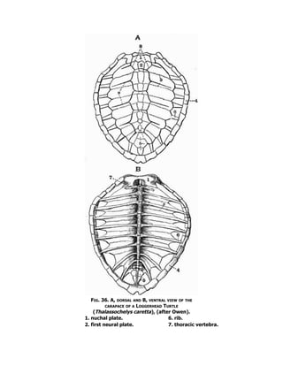
Fig. 36. A,dorsal and B, ventral view of the
carapace of a Loggerhead Turtle
(Thalassochelys caretta), (after Owen).
1. nuchal plate. 6. rib.
2. first neural plate. 7. thoracic vertebra.
41.
3. second costalplate. 8. first vertebral shield.
4. marginal plate. 9. costal shield.
5. pygal plate.
The sculpturing due to the epidermal shields is very obvious on the
carapace.
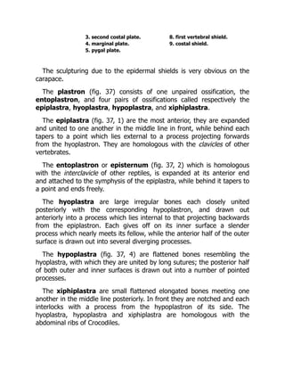
The plastron (fig. 37) consists of one unpaired ossification, the
entoplastron, and four pairs of ossifications called respectively the
epiplastra, hyoplastra, hypoplastra, and xiphiplastra.
The epiplastra (fig. 37, 1) are the most anterior, they are expanded
and united to one another in the middle line in front, while behind each
tapers to a point which lies external to a process projecting forwards
from the hyoplastron. They are homologous with the clavicles of other
vertebrates.
The entoplastron or episternum (fig. 37, 2) which is homologous
with the interclavicle of other reptiles, is expanded at its anterior end
and attached to the symphysis of the epiplastra, while behind it tapers to
a point and ends freely.
The hyoplastra are large irregular bones each closely united
posteriorly with the corresponding hypoplastron, and drawn out
anteriorly into a process which lies internal to that projecting backwards
from the epiplastron. Each gives off on its inner surface a slender
process which nearly meets its fellow, while the anterior half of the outer
surface is drawn out into several diverging processes.
The hypoplastra (fig. 37, 4) are flattened bones resembling the
hyoplastra, with which they are united by long sutures; the posterior half
of both outer and inner surfaces is drawn out into a number of pointed
processes.
The xiphiplastra are small flattened elongated bones meeting one
another in the middle line posteriorly. In front they are notched and each
interlocks with a process from the hypoplastron of its side. The
hyoplastra, hypoplastra and xiphiplastra are homologous with the
abdominal ribs of Crocodiles.
42.
Fig. 37. ThePlastron of a Green Turtle (Chelone midas). × 1/7. (Camb. Mus.)
1. epiplastron (clavicle). 4. hypoplastron.
2. entoplastron (interclavicle). 5. xiphiplastron.
3. hyoplastron.
II. ENDOSKELETON.
1. The Axial Skeleton.
The axial skeleton includes the vertebral column, the ribs, and the
skull.
43.
A. The Vertebralcolumn and Ribs.
The number of vertebrae in the Green Turtle is thirty-eight, not a great
number as compared with that in many reptiles, and of these eighteen
are caudal.
The vertebral column is divisible into four regions only—cervical,
thoracic, sacral, and caudal.
The Cervical vertebrae.
These are eight in number, and are chiefly remarkable for the great
variety of articulating surfaces which their centra present, and for their
mobility upon one another.
The first or atlas vertebra differs much from all the others and
consists of the following parts:—
a. the neural arch, formed of two separate ossifications united in the
mid-dorsal line;
b. the inferior arch;
c. the centrum, which is detached from the rest and forms the
odontoid process of the second vertebra.
Each half of the neural arch consists of a ventral portion, the
pedicel, which lies more or less vertically and is united ventrally to the
inferior arch, and of a dorsal portion, the lamina, which lies more or
less horizontally and meets its fellow in the middle line in front, partially
roofing over the neural canal. Each pedicel bears a facet on its anterior
surface, which, with a corresponding one on the inferior arch, articulates
with the occipital condyle of the skull. Three similar facets occur also on
the posterior surface of the pedicel and inferior arch, and articulate with
the odontoid process. The laminae meet one another in front, but do not
fuse, while behind they are separated by a wide triangular space. They
bear a pair of small downwardly-directed facets, the
postzygapophyses, for articulation with the prezygapophyses of the
second vertebra.
44.
The inferior archis a short irregular bone bearing two converging
facets for articulation with the occipital condyle and odontoid process
respectively.
The centrum or odontoid process has a convex anterior surface for
articulation with the neural and inferior arches, and a concave posterior
surface by which it is united with the centrum of the second or axis
vertebra. It bears posteriorly a small epiphysis which is really a detached
portion of the inferior arch.
The second or axis and following five cervical vertebrae, though
showing distinct differences, resemble one another considerably, each
having a fairly elongated centrum with a keel-like hypapophysis, each
having also a neural arch with prominent articulating surfaces, the
anterior of which, or prezygapophyses, look upwards and inwards,
while the posterior ones, the postzygapophyses, look downwards and
outwards. They however, as was previously mentioned, differ very
remarkably in the character of the articulating surfaces of the centra.
Thus the second and third vertebrae are convex in front and concave
behind, the fourth is biconvex, the fifth is concave in front and convex
behind. The sixth is concave in front and attached to the seventh by a
flat surface behind, the seventh has a flat anterior face and two slightly
convex facets behind. The vertebrae all have short blunt transverse
processes and the second has a prominent neural spine.
The eighth cervical vertebra is curiously modified, the centrum is
very short, has a rather prominent hypapophysis, and is convex behind,
while in front it articulates with the preceding centrum by two concave
surfaces. The neural arch is deeply notched in front and bears two
upwardly-directed prezygapophyses, while behind it is very massive and
is drawn out far beyond the centrum, bearing a pair of flat
postzygapophyses. The top of the neural arch almost or quite meets a
blunt outgrowth from the nuchal plate.
The Thoracic vertebrae.
These are ten in number and are all firmly united with the ribs and
elements forming the carapace.
45.
The first thoracicvertebra differs from the others, the centrum is short
and has a concave anterior surface articulating with the centrum of the
last cervical vertebra, and a pair of prezygapophyses borne on long
outgrowths. The neural spine arises only from the anterior half of the
centrum, and is not fused to the carapace. Arising laterally from the
anterior part of the centrum are a small pair of ribs each of which is
connected with a process arising from the rib of the succeeding vertebra.
The next seven thoracic vertebrae are all very similar, each has a long
cylindrical centrum, expanded at the ends, and firmly united to the
preceding and succeeding vertebrae. The neural arches are flattened and
expanded dorsally, and are united to one another and to the overlying
neural plates; each arises only from the anterior half of its respective
centrum, and overlaps the centrum of the vertebra in front of it.
Between the base of the neural arch and its successor is a small foramen
for the exit of the spinal nerve. There are no transverse processes or
zygapophyses.
To each thoracic vertebra from the second to ninth inclusive, there
corresponds a pair of ribs (fig. 36, 6) of a rather special character. Each
is suturally united with the anterior half of the edge of its own vertebra,
and overlaps on to the posterior half of the edge of the next preceding
vertebra. The ribs are much flattened, and each is fused with the
corresponding costal plate, beyond which it projects to fit into a pit in
one of the marginal plates.
The tenth thoracic vertebra is smaller than the others, and its neural
arch does not overlap the preceding vertebra, it bears a pair of small ribs
which are without costal plates, but meet those of the ninth vertebra.
There are no lumbar vertebrae.
The Sacral vertebrae.
The sacral vertebrae are two in number, they are short and wide,
their centra are ankylosed together, and their neural arches are not
united to the carapace.
The first has the anterior face of the centrum concave and the
posterior flat, while both faces of the second are flat. Each bears a pair
46.
of short ribswhich meet the ilia, but are not completely ankylosed either
with them or the centra.
The Caudal vertebrae.
The caudal vertebrae are eighteen in number. The centrum of the
first is flat in front and is ankylosed to the second sacral; behind it is
convex. The others are all very similar to one another, and decrease
gradually in size when followed back. Each has a moderately long
centrum, concave in front and convex behind, both terminations being
formed by epiphyses. The neural arch arises only from the anterior half
of the vertebra; it bears a blunt truncated neural spine and prominent
pre- and post-zygapophyses. The first seven caudal vertebrae bear short
ribs attached to their lateral margins, the similar outgrowths on the
succeeding vertebrae do not ossify from distinct centres, and are
transverse processes rather than ribs.
B. The Skull.
The skull of the Turtle is divisible into the following three parts:—
(1) the cranium;
(2) the lower jaw or mandible;
(3) the hyoid.
(1) The Cranium.
The cranium is a very compact bony box, containing a cavity in which
the brain lies, and which is a direct continuation of the neural canal of
the vertebrae.
47.
Fig. 38. Theskull of the Green Turtle (Chelone midas). × ½. A,
posterior half, B, anterior half. (Brit. Mus.)
1. parietal. 13. angular.
2. squamosal. 14. supra-angular.
3. quadrate. 15. premaxillae.
48.
4. basisphenoid. 16.maxillae.
5. basi-occipital. 17. jugal.
6. quadratojugal. 18. postfrontal.
7. opisthotic. 19. vomer.
8. exoccipital. 20. prefrontal.
9. foramen magnum. 21. frontal.
10. splenial. 22. external auditory meatus
11. articular. leading into tympanic
12. dentary. cavity.
Like those of the skull as a whole its component bones may be
subdivided into three sets:—
1. those forming the brain-case or cranium proper;
2. those developed in connection with the special sense organs;
3. those forming the upper jaw and suspensorial apparatus.
Both cartilage and membrane bones take part in the formation of the
skull, and a considerable amount of cartilage remains unossified,
especially in the ethmoidal and sphenoidal regions.
1. The Cranium proper or Brain-case.
The cartilage and membrane bones of the brain-case when taken
together can be seen to be more or less arranged in three rings or
segments, called respectively the occipital, parietal, and frontal
segments.
The occipital segment is the most posterior of these, and consists of
four cartilage bones, the basi-occipital, the two exoccipitals and the
supra-occipital; these bound the foramen magnum.
The basi-occipital (figs. 38 and 39, 5) lies ventral to the foramen
magnum and only bounds a very small part of it; it forms one-third of
the occipital condyle by which the skull articulates with the atlas
vertebra. It unites dorsally with the exoccipitals and anteriorly with the
basisphenoid.
The exoccipitals are rather small bones, which form the sides and
the greater part of the floor of the foramen magnum, and two-thirds of
49.
the occipital condyle.Laterally each is united with the pterygoid and
opisthotic of its side. At the sides of the occipital condyle each exoccipital
is pierced by a pair of foramina, the more dorsal and posterior of which
transmits the hypoglossal nerve.
The supra-occipital (fig. 39, 14) is a larger bone than the others of
the occipital segment. It forms the upper border of the foramen
magnum and is drawn out dorsally into a large crest which extends back
far beyond the occipital condyle. In the adult the supra-occipital is
completely ankylosed with the epi-otics.
The Parietal segment.
The ventral portion of the parietal segment is formed by the
basisphenoid (figs. 38 and 39, 4) which lies immediately in front of the
basi-occipital. A triangular portion of it is seen in a ventral view of the
skull, but it is quickly overlapped by the pterygoids. It gives off dorsally a
pair of short processes which meet the pro-otics.
The alisphenoidal region is unossified and the only other constituents
of the parietal segment are the parietals (fig. 39, 1). These are large
bones which, after roofing over the cranial cavity, extend upwards and
become expanded into a pair of broad plates which unite with the
squamosal and bones of the frontal segment to form a wide, solid, false
roof to the skull. Each also sends ventralwards a plate which meets an
upgrowth from the pterygoid and acts as an alisphenoid.
The Frontal segment.
Of the frontal segment the basal or presphenoidal and lateral or
orbitosphenoidal portions do not become ossified, the dorsal portion
however includes three pairs of membrane bones, the frontals,
prefrontals and postfrontals.
The frontals are a pair of small bones lying immediately in front of the
parietals, and in front of them are the prefrontals (figs. 38 and 39, 20), a
pair of similar but still smaller bones, which are produced ventrally to
meet the vomer and palatines. They form also the dorsal boundary of
the anterior nares. The postfrontals (figs. 38 and 39, 18) are larger
bones, united dorsally to the frontals and parietals, posteriorly to the
50.
squamosals, and ventrallyto the jugals and quadratojugals. All three
pairs of frontal bones, especially the postfrontals, take part in the
bounding of the orbits.
Fig. 39. Longitudinal vertical section through the Cranium of a Green Turtle (Chelone
midas). × 2/3. (Camb. Mus.)
1. parietal. 10. palatine. 17. jugal.
2. squamosal. 11. rod passed into narial 18. postfrontal.
3. quadrate. passage. 19. vomer.
4. basisphenoid. 12. exoccipital. 20. prefrontal.
5. basi-occipital. 13. epi-otic fused to 21. frontal.
6. quadratojugal. supra-occipital. V, VII, VIII, IX, X, XI, XII,
7. pro-otic. 14. supra-occipital. foramina for the exit of
8. opisthotic. 15. premaxillae. cranial nerves.
9. pterygoid. 16. maxillae.
2. The Sense capsules.
Skeletal structures occur in connection with each of the three special
sense organs of hearing, sight, and smell.
The Auditory capsules.
The auditory or periotic capsule of the turtle is rather large and its
walls are well ossified, epi-otic, pro-otic and opisthotic bones being
51.
present.
The epi-otic (fig.39, 13) is the more dorsal of the three bones, and
in the adult is completely ankylosed with the supra-occipital.
The opisthotic (fig. 39, 8) is the ventral posterior element. On its
inner side it is united to the supra-occipital above, and to the exoccipital
below; it sometimes becomes completely fused with the exoccipital. In
front it meets the pro-otic, and on its outer side the squamosal and
quadrate. Its anterior portion is hollowed out by the cavity in which the
auditory organ lies, it gives off also a process which is separated from
the exoccipital by an oval foramen through which the glossopharyngeal,
pneumogastric, and spinal accessory nerves leave the cranial cavity.
The pro-otic is the anterior element; it meets the supra-occipital and
opisthotic posteriorly, while anteriorly it is separated from the
alisphenoidal plate of the parietal and pterygoid by a large oval foramen
through which the maxillary and mandibular branches of the trigeminal
nerve pass out (fig. 39, V 1 & 2). It is hollowed out posteriorly by the
cavity in which the auditory organ lies, and its inner wall as seen in
longitudinal section is pierced by a foramen through which the external
carotid artery and facial nerve leave the cranial cavity,—the nerve finally
leaving the skull through a small oval foramen on the anterior face of the
pro-otic near its junction with the quadrate.
Between the pro-otic and opisthotic as seen in a longitudinal section of
the skull is a large opening constricted in the middle. This is the
internal auditory meatus (fig. 39, VIII.). Through it the auditory
nerve leaves the cranial cavity and enters the ear. The ramus vestibularis
leaves through the dorsal part of the hole, the ramus cochlearis through
the ventral.
The cavity of the auditory or periotic capsule communicates with the
exterior by a fairly large hole, the fenestra ovalis, which lies between
the opisthotic and pro-otic, and opens into a deep depression, the
tympanic cavity, which is seen in a posterior view of the skull lying
just external to the exoccipital. The cavity communicates with the
exterior by a large opening, the external auditory meatus (fig. 38,
22).
52.
Welcome to ourwebsite – the perfect destination for book lovers and
knowledge seekers. We believe that every book holds a new world,
offering opportunities for learning, discovery, and personal growth.
That’s why we are dedicated to bringing you a diverse collection of
books, ranging from classic literature and specialized publications to
self-development guides and children's books.
More than just a book-buying platform, we strive to be a bridge
connecting you with timeless cultural and intellectual values. With an
elegant, user-friendly interface and a smart search system, you can
quickly find the books that best suit your interests. Additionally,
our special promotions and home delivery services help you save time
and fully enjoy the joy of reading.
Join us on a journey of knowledge exploration, passion nurturing, and
personal growth every day!
testbankfan.com


![Fig. 32. Lateral (below) and dorsal (above) views of the skull of an Ichthyosaurus.
(Modified from Deslongchamps.)
1. premaxillae. 12. squamosal.
2. maxillae. 13. supratemporal.
3. nasal. 14. quadratojugal.
4. prefrontal[74]. 15. sclerotic ring.
5. frontal. 16. postorbital.
6. postfrontal[74]. 17. jugal.
7. anterior nares. 18. lachrymal.
8. orbit. 19. dentary.
9. supratemporal fossa. 20. articular.
10. interparietal foramen. 21. angular.
11. parietal.
The ribs are long, and the anterior ones have capitula and tubercula.
There is no sternum, but the ventral body wall is strengthened by a
complex system of abdominal splint ribs.
The pectoral girdle is strongly developed, the scapulae are narrow, the
coracoids broad, and meet ventrally without overlapping. There are
probably no precoracoids, but clavicles and a T-shaped interclavicle are
well developed.](https://image.slidesharecdn.com/4868-250411035259-be59b2b0/85/Project-Management-The-Managerial-Process-5th-Edition-Larson-Test-Bank-22-320.jpg)
![The ribs have capitula and tubercula, and often uncinate processes
(see p. 190) as in birds. A pectoral girdle and sternum, with clavicles and
a T-shaped interclavicle are developed, and abdominal ribs are always
found. The precoracoid is however absent. The limbs are pentedactylate.
Sphenodon[75] (Hatteria) now living in some of the islands of the New
Zealand group, is certainly the most generalised of all living reptiles.
Though lizard-like in form it differs from all living lizards in the
possession of two temporal arcades, abdominal ribs and a fixed
quadrate; and is often considered to be nearly allied in many respects to
the type of reptile from which all the others took their origin.
Among the better known extinct forms are Proterosaurus of Permian
and Hyperodapedon of Triassic age.
Order 6. Squamata.
This order includes the extinct Mosasaurians, and the lizards and
snakes which form the vast majority of living reptiles. The trunk may be
moderately elongated and provided with four short limbs as in lizards, or
it may be limbless, extremely elongated, and passing imperceptibly into
the tail. The surface is generally completely covered with overlapping
horny epidermal scales, below which bony dermal scutes may be
developed.
The vertebrae are procoelous, rarely amphicoelous. There are no inter
centra, and the neural arches are firmly united to the centra. Additional
articulating surfaces, the zygosphenes and zygantra, are often
developed[76]. The sacrum is formed of two or rarely three vertebrae, or
may be wanting as in Ophidia. In the skull an infratemporal arcade
forming the lower boundary of the infratemporal fossa is absent, and the
quadrate, except in the Chamaeleons, is movably articulated to the
squamosal. The palatal vacuities are large and the nares are separate.
There is often a distinct parasphenoid. The teeth are either acrodont (i.e.
ankylosed to the summit of the jaw), or pleurodont, i.e. ankylosed to the
inner side of the jaw. The thoracic ribs each have a single head which
articulates with the centrum of the vertebra; while uncinate processes
and abdominal ribs never occur.](https://image.slidesharecdn.com/4868-250411035259-be59b2b0/85/Project-Management-The-Managerial-Process-5th-Edition-Larson-Test-Bank-24-320.jpg)
![A pectoral girdle and sternum may be present, or may be completely
absent as in snakes. Except in snakes there are generally four
pentedactylate limbs which may either form paddles or be adapted for
walking.
Suborder (1). Lacertilia
[77].
The body is elongated, and as a rule four short pentedactylate limbs
are present, but sometimes limbs are vestigial or absent. The
exoskeleton generally has the form of horny plates, spines, or scales;
while sometimes as in the Chamaeleons and Amphisbaenians it is
absent. In other forms such as Tiliqua and Scincus, the body has a
complete armour of bony scutes, whose shape corresponds with that of
the overlying horny scales.
The vertebrae are procoelous, rarely as in the Geckos amphicoelous;
they are usually without zygosphenes and zygantra, but these structures
occur in the Iguanidae. The sacral vertebrae of living forms are not
ankylosed together, and the caudal vertebrae usually have well-
developed chevron bones.
In the skull[78] the orbits are separated from one another, only by an
imperfectly developed interorbital septum, the cranial cavity not
extending forwards between them, while the alisphenoidal region is
unossified. The premaxillae may be paired or united (Amphisbaenidae),
and there is usually an interparietal foramen. There may be a complete
supratemporal[79] arcade bounding the lower margin of the
supratemporal fossa, or the supratemporal fossa may be open below.
The quadratojugal is not ossified, and the quadrate articulates with the
exoccipital. There is no infratemporal arcade. There is commonly a rod-
like epipterygoid[80] (fig. 33, 14) connecting the pterygoid and parietal.
Teeth are always present, and may be confined to the jaws or may be
developed also on the pterygoids and rarely on the palatines; they are
either acrodont or pleurodont. The rami of the mandible are suturally
united.
A pectoral girdle is always present, and generally also a sternum.
Clavicles and a T-shaped interclavicle are commonly present, but are](https://image.slidesharecdn.com/4868-250411035259-be59b2b0/85/Project-Management-The-Managerial-Process-5th-Edition-Larson-Test-Bank-25-320.jpg)
![The pubis corresponds to the pre-pubis of Dinosaurs, and both pubes
and ischia meet in ventral symphyses.
The suborder includes the Lizards, Chamaeleons and Amphisbaenians.
Suborder (2). Ophidia
[81].
The Ophidia or snakes are characterised by their greatly elongated
body and want of limbs. The body is covered with overlapping horny
scales and bony dermal scutes are never present. The vertebrae are
procoelous, and are distinguishable into two groups only, precaudal or
rib-bearing, and caudal or ribless. The atlas vertebra is also ribless. The
neural arches are always provided with zygosphenes and zygantra. Many
of the vertebrae have strong hypapophyses, and the caudal vertebrae
are without chevron bones.
In the skull the cranial cavity extends forwards between the orbits,
and is closed in front by downgrowths from the frontals and parietals
which meet the well-ossified alisphenoids and orbitosphenoids[82]. The
cranium is strongly ossified, and there are no parotic processes or
interparietal foramen. There are no temporal arcades and no
epipterygoid. The premaxillae if present are very small (fig. 51, 1) and
usually toothless. The quadrates articulate with the squamosals, and do
not as in Lacertilia meet the exoccipitals. The palatines do not unite
directly with the vomers or with the base of the cranium, and the whole
palato-maxillary apparatus is more loosely connected with the cranium
than it is in Lacertilia. The pterygoids, and in most cases also the
palatines, bear teeth. The dentition is acrodont, and the rami of the
mandible are united only by an elastic ligament—an important point
serving to distinguish the Ophidia from the Lacertilia. There is an
imperfectly developed interorbital septum, the ventral part of which is
formed by the parasphenoid. The postfrontal is generally well developed,
while the jugals and quadratojugals are absent. There are never any
traces of the anterior limbs or pectoral girdle, but occasionally there are
vestiges of a pelvis and posterior limbs.
Suborder (3). Pythonomorpha
[83].](https://image.slidesharecdn.com/4868-250411035259-be59b2b0/85/Project-Management-The-Managerial-Process-5th-Edition-Larson-Test-Bank-29-320.jpg)
![This suborder includes Mosasaurus and its allies, a group of enormous
extinct marine reptiles found in beds of Cretaceous age.
The skin is in most forms at any rate unprovided with dermal scutes.
The vertebrae may be with or without zygosphenes and zygantra. The
skull resembles that of lizards, having an interparietal foramen, and a
cranial cavity open in front. The squamosal takes part in the formation of
the cranial wall, and the quadrate articulates with the squamosal, not as
in Lacertilia with the exoccipital. There are large supratemporal fossae,
bounded below by supratemporal arcades. The teeth are large and
acrodont, and occur on the pterygoids as well as on the jaws. The two
rami of the mandible are united by ligament only. Pectoral and pelvic
girdles are present, but clavicles are wanting, and the pelvis is not as a
rule united to any sacrum.
The limbs are pentedactylate, and are adapted for swimming, while all
the limb bones except the phalanges are relatively very short. The
number of phalanges is not increased beyond the normal, and they
articulate with one another by flat surfaces. The terminal phalanges are
without claws.
Order 7. Dinosauria
[84].
The extinct reptiles comprising this order were all terrestrial, and
include the largest terrestrial animals known. They vary greatly in size
and in the structure of the limbs, some approach close to the type of
structure met with in birds, others are allied to crocodiles.
Passing to the more detailed characters:—there is sometimes a well-
developed exoskeleton having the form of bony plates or spines. The
vertebrae may be solid or their centra may be hollowed internally; their
surfaces may be flat, biconcave or opisthocoelous. The sacrum is
composed of from two to six vertebrae.
As regards the skull, the quadrate is large and fixed, and
supratemporal and infratemporal fossae bounded by bone occur. The
teeth are more or less laterally compressed, and often have serrated
edges; they may be placed in distinct sockets or in a continuous groove.
The ribs have capitula and tubercula, and sternal ribs often occur. The
scapula is very large, the coracoid small, and there is no precoracoid, or](https://image.slidesharecdn.com/4868-250411035259-be59b2b0/85/Project-Management-The-Managerial-Process-5th-Edition-Larson-Test-Bank-30-320.jpg)
![T-shaped interclavicle. Clavicles are only known in a few cases. In the
pelvis the ilium is elongated both in front of, and behind, the
acetabulum, sometimes the pre-pubis, sometimes the post-pubis is the
better developed. The anterior limbs are shorter than the posterior, and
the long bones are sometimes solid, sometimes hollow.
There are three well-marked suborders of the Dinosauria.
Suborder (1). Sauropoda
[85].
The reptiles belonging to this group were probably quadrupedal and
herbivorous.
They have the cervical and anterior trunk vertebrae opisthocoelous,
while the posterior vertebrae are biconcave; all the presacral, and
sometimes the sacral vertebrae are hollowed internally. The teeth are
spatulate and without serrated edges, they are always planted in distinct
sockets, and some of them are borne by the premaxillae.
Fig. 35. Restored skeleton of Ceratosaurus nasicornis. × 1/30. (After Marsh.)](https://image.slidesharecdn.com/4868-250411035259-be59b2b0/85/Project-Management-The-Managerial-Process-5th-Edition-Larson-Test-Bank-31-320.jpg)
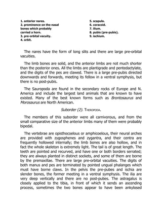
![the posterior ones very large and strong. The post-pubis is well
developed;
e.g. Stegosaurus from the Upper Jurassic of Colorado.
B. Ceratopsia.
There is sometimes a well-developed dermal exoskeleton formed of
small granules and plates of bone. The bones are solid, and the vertebral
centra flat. The cranium bears a pair of enormous pointed frontal horns,
and the parietal is greatly expanded and elevated behind, forming with
the squamosals a shield which overhangs the anterior cervical vertebrae.
The premaxillae are united, and in front of them is a pointed beak-like
bone which bites upon a toothless predentary ossification of the
mandible. The teeth have two roots. The anterior limbs are but little
shorter than the posterior ones. There is no post-pubis;
e.g. Polyonax from the uppermost Cretaceous of Montana.
C. Ornithopoda
[86].
There is no dermal exoskeleton. The cervical vertebrae are
opisthocoelous, and so are sometimes the thoracic. The limb bones are
hollow and the anterior limbs are much shorter than the posterior ones.
The feet are digitigrade and provided with long pointed claws. The post-
pubis is long and slender and directed back parallel to the ischium;
e.g. Iguanodon from the European Cretaceous.
Order 8. Crocodilia
[87].
This order includes the Crocodiles, Alligators and Garials and various
extinct forms, some of which are closely allied to the early Dinosaurs.
There is always a more or less complete exoskeleton formed of bony
scutes overlain by epidermal scales; these bony scutes are specially well
developed on the dorsal surface but may occur also on the ventral. The
vertebral column is divisible into the five regions commonly
distinguishable. In all living forms the vertebrae, with the exception of
the atlas and axis, the two sacrals, and first caudal, are procoelous, but](https://image.slidesharecdn.com/4868-250411035259-be59b2b0/85/Project-Management-The-Managerial-Process-5th-Edition-Larson-Test-Bank-34-320.jpg)
![The vertebral centra are flat or biconcave. The premaxillae are very
large, and the nares are separated, and placed far back. The posterior
narial openings lie comparatively far forward between the anterior
extremities of the palatines.
The palatines and pterygoids do not form a secondary palate. The
supratemporal fossae are small, and open posteriorly, the lateral
temporal fossae are very large. The parietals and frontals are paired.
Clavicles are present. The best known and most important genus of
these extinct crocodiles is Belodon.
Suborder (2). Eusuchia.
The vertebrae are either biconcave or procoelous. The premaxillae are
small, and the anterior nares are united and placed far forwards. The
posterior nares lie far back, the palatines and in living genera the
pterygoids, meeting in the middle line, and giving rise to a closed palate.
The supratemporal fossae are surrounded by bone on all sides, and the
parietals, and often also the frontals are united. There are no clavicles.
The suborder includes the genera Crocodilus, Alligator, Garialis and
others living and extinct.
Order 9. Pterosauria
[88].
These animals, called also the pterodactyles or Ornithosauria, are a
group of extinct reptiles, whose structure has been greatly modified from
the ordinary reptilian type for the purpose of flight.
The skin was naked and they vary greatly in size and in the length of
the tail. The vertebrae and limb bones are pneumatic just as in birds.
The presacral vertebrae are procoelous and have their neural arches
firmly united to the centra. The neck is long, the caudal vertebrae are
amphicoelous, and from three to five vertebrae are fused together in the
sacral region. The skull is large and somewhat bird-like, the facial portion
being much drawn out anteriorly, and the sutures being obliterated. It
resembles that of other reptiles in having large supratemporal fossae;
large pre-orbital vacuities also occur. The jaws may be toothed or
toothless, and the teeth, when present, are imbedded in separate
sockets. The premaxillae are large, and the quadrate is firmly attached
to the skull. The rami of the mandible are united at the symphysis, and](https://image.slidesharecdn.com/4868-250411035259-be59b2b0/85/Project-Management-The-Managerial-Process-5th-Edition-Larson-Test-Bank-36-320.jpg)
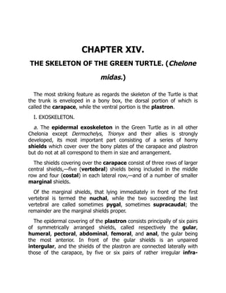
![marginal shields. Smaller horny plates occur on other parts of the body,
especially on the limbs and head.
Two other sets of structures belong also to the epidermal exoskeleton,
viz. (a) horny beaks with denticulated edges which ensheath both upper
and lower jaws, (b) claws, which as a rule are borne only by the first
digit of each limb. Sometimes in young individuals the second digit is
also clawed.
b. The dermal exoskeleton is strongly developed, and is combined
with endoskeletal structures derived from the ribs and vertebrae to form
the carapace.
The Carapace (fig. 36) consists of a number of plates firmly united to
one another by sutures. They have a very definite arrangement and
include:
(a) the nuchal plate (fig. 36, 1), a wide plate forming the whole of
the anterior margin of the carapace. It is succeeded by three series of
plates, eight in each series, which together make up the main part of the
carapace. Of these the small
(b) neural plates[1] (fig. 36, A, 2) form the middle series. They are
closely united with the neural arches of the underlying vertebrae;
(c) the costal plates[89] (fig. 36, A, 3) are broad arched plates united
to one another by long straight sutures. They are united at their inner
extremities with the neural plates, but the boundaries of the two sets of
plates do not regularly correspond. Each is united ventrally with a rib
which projects beyond it laterally for some distance; (d) the marginal
plates (fig. 36, 4) are twenty-three in number, eleven lying on each
side, while an unpaired one lies in the middle line posteriorly. Many of
them are marked by slight depressions into which the ends of the ribs
fit; (e) the pygal plates (fig. 36, 5) are two unpaired plates lying
immediately posterior to the last neural.](https://image.slidesharecdn.com/4868-250411035259-be59b2b0/85/Project-Management-The-Managerial-Process-5th-Edition-Larson-Test-Bank-39-320.jpg)