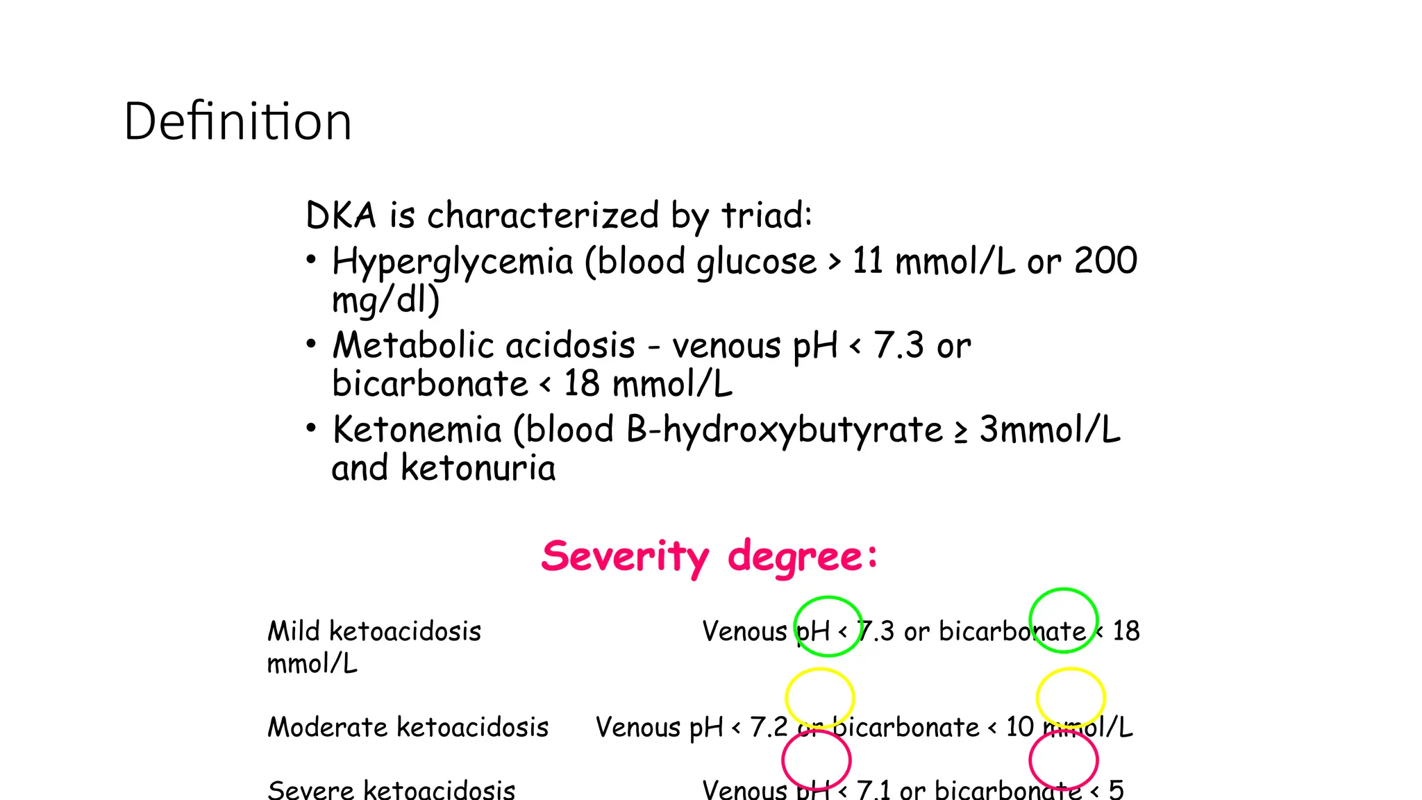
Diabetic Keto Acidosis
MBBS Lecture Series
(Paediatric Endocrinology)
INTRODUCTION
• Diabetes remain the commonest endocrine abnormality in children
• DKA is the commonest mode of presentation in our environment due
to poor health seeking behaviour.
• Failure to diagnose DKA at presentation invariably means mortality
• Even when diagnosed appropriate management is necessary for
survival
outline
• Definition
• Precipitants
• Pathophysiology
• Frequency
• Diagnosis
• Laboratory work up
• Treatment
• Complication of treatment and their management
OBJECTIVE
• To define DKA and know the diagnostic criteria
• To recognize clinical features of DKA
• To evaluate a child with DKA
• To plan treatment for DKA
DKA is characterized by triad:
• Hyperglycemia (blood glucose > 11 mmol/L or 200
mg/dl)
• Metabolic acidosis - venous pH < 7.3 or
bicarbonate < 18 mmol/L
• Ketonemia (blood B-hydroxybutyrate ≥ 3mmol/L
and ketonuria
Definition
Severity degree:
Mild ketoacidosis Venous pH < 7.3 or bicarbonate < 18
mmol/L
Moderate ketoacidosis Venous pH < 7.2 or bicarbonate < 10 mmol/L
Severe ketoacidosis Venous pH < 7.1 or bicarbonate < 5
Pathophysiology
DKA results from deficiency of circulating insulin and increased level of
the counterregulatory hormones.
In most case DKA is precipitated by
• New onset of diabetes
• Omission of insulin injections
• Interruption of insulin delivery in children using insulin pump
• Inadequate management of infection.
Counter-regulatory hormones:
Catecholamines
Glucagon
Cortisol
Growth hormone
Insulin
Absolute deficiency:
- previously undiagnosed T1DM
- patients on treatment that deliberately or
inadvertently do not take insulin
Relative insulin deficiency:
- increase concentrations of counter-regulatory
hormones in response to stress
- gastrointestinal illness
pathophysiology
•accelerated catabolic state
•increased glucose production
(via glycogenolysis and gluconeogenesis)
•impaired peripheral glucose utilization
(hyperglycemia and hyperosmolality)
•increased lipolysis and ketogenesis
(ketonemia and metabolic acidosis)
Absolute insulin deficiency OR
Stress, infection or insufficient insulin intake
Counter-regulatory hormones:  Glucagon, Cortisol,
Catecholamines, GH
Lipolysis
 FFA to liver
 Ketogenesis
 Alkali reserve
Acidosis
 Lactate
Glucose
utilization
 Proteolysis
 Protein synthesis
 Glycogenolysis
Gluconeogenic substrates
 Gluconeogenesis
Hyperglycemia
Glucosuria (osmotic diuresis)
Loss of water and electrolytes
Dehydration
Impaired renal function
Hyperosmolarity
Frequency of DKA
At disease onset
15-70% at onset of diabetes
>80% in series from Nig
Inversely correlate with the
regional incidence of T1DM
<5 yr of age
Families which do not have
ready access to medical care
for social or economic reasons
Poor metabolic control or previous
episodes of DKA
Peripubertal and adolescent girls
Psychiatric disorders
Difficult or unstable family
circumstances
Children who omit insulin
Children with limited access to
medical services
Insulin pump therapy
Children with
established diabetes
1-10% per patient per year
Diagnosis
Clinical History
• Polyuria, polydipsia, nausea, vomiting, rapid
breathing, abdominal pain, weakness, weight
loss, confusion, decreased level of
consciousness
Diagnosis
Clinical Signs
• Dehydration
• Deep sighing respiration (Kussmaul)
• Smell of ketones
• Drowsiness
Diagnosis
Biochemical
• Hyperglycaemia
• Ketonuria
• Ketonaemia
• Acidemia - Plasma bicarbonate < 18 mmol/l
Laboratory measurement:
 Glucose
 Blood urea nitrogen
 Creatinine
 Hemoglobin and complete blood count
 Venous pH, pCO2
 Electrolytes
 Osmolality
 Calcium
 Phosphorus
 Magnesium
 HbA1c
 Urinalysis for ketones
 Blood β-hydroxybutyrate concentration
Specimens for culture (blood, urine, throat)
Electrocardiogram (ECG)
Serum free insulin concentration
Secure the airway and
Empty the stomach by continuous nasogastric
suction of to prevent pulmonary aspiration in the
unconscious or severely obtunded patient.
Peripheral intravenous (IV) catheter
Arterial catheter (critically ill patients
managed in an intensive care unit)
Treatment-Supportive measures
Cardiac monitor
Oxygen to patients with severe circulatory
impairment or shock
Antibiotics
Catheterization of the bladder
Goals of therapy
• Correct dehydration
• Correct acidosis and reverse ketosis
• Restore blood glucose to near normal
• Monitor for complications of DKA and its therapy
• Identify and treat any precipitating event
Fluids and salt
• Patients with DKA have a deficit in body fluid that
usually is in the range 5–10% of body weight.
• Clinical estimates of degree of dehydration based on
physical examination and vital sign are inaccurate.
Therefore
Mild DKA – 5%
Moderate DKA – 7%
Severe DKA – 10%
• Increase Urea and anion gap at presentation
correlates well with volume deficit
• Begin with fluid replacement before insulin therapy.
Fluids and salt
Resuscitation fluid
• Expand intravascular volume with isotonic 0.9%
saline: 10–20 ml/kg body weight over 20 – 30
min
• For those in shock 20ml/kg of 0.9% saline
boluses as rapid as possible with reassessment
of circulatory status after each bolus.
Fluids and salt
Deficit Replacement fluid
• Deficit is replaced over 24 – 48 hrs using 0.9%
saline or 0.45% saline or ringers
• Maintenance is also giving along with deficit
• Typical fluid calculation =
Deficit – Resuscitation fluid + 2xMaintenance
• The fluid is given evenly over 48 hours
Insulin therapy
• DKA is caused by either relative or absolute
insulin deficiency
• Begin with 0.1 U/kg/h 1–2 h AFTER starting
fluid replacement therapy
• An IV bolus is unnecessary, may precipitate
shock and exercabate hypokalaemia and should
not be used at the start of therapy
• The dose of insulin should usually remain at 0.1
U/kg/h at least until resolution of DKA
Insulin therapy
If the patient demonstrates marked sensitivity
to insulin (e.g., some young children with DKA,
patients with HHS, and some older children with
established diabetes), the dose may be
decreased to 0.05 unit/kg/h, or less.
Insulin therapy
To prevent any rapid decrease in plasma glucose
concentration and hypoglycemia, 5% glucose should be
added to the IV fluid (e.g., 5% glucose in 0.45% saline)
when the plasma glucose falls to approximately 14–17
mmol/L (250–300 mg/dL), or
sooner if the rate of fall is precipitous.
It may be necessary to use 10% or even 12.5%
dextrose to prevent hypoglycemia while continuing
to infuse insulin to correct the metabolic acidosis.
Even with normal or high levels of serum
potassium at presentation, there is always a
total body deficit of potassium (3-6 mmol/kg).
The major loss of potassium is from the
intracellular pool.
Potassium replacement
Hypokalemic
Normal K+
concentration
Hyperkalemic
Start potassium replacement at
the time of initial volume
expansion and before starting
insulin therapy.
Start after initial volume
expansion
and concurrent with starting
insulin therapy
Defer potassium replacement
therapy until urine output is
documented
20 mmol/l KCl + 20 mmol/l K phosphate =
40 mmol/l
Phosphate
• Depletion of intracellular phosphate occurs in DKA and phosphate is lost as a result
of osmotic diuresis
• Plasma phosphate levels fall after starting treatment and this is exacerbated by
insulin
BUT
• Administration of phosphate may induce hypocalcemia
• Potassium phosphate salts may be safely used as an alternative to or combined with
potassium chloride
• No clinical benefit from phosphate replacement
Acidosis
Severe acidosis is reversible
by fluid and insulin
replacement.
There is no evidence that
bicarbonate is either necessary
or safe in DKA:
• paradoxical CNS acidosis
• hypokalemia
Cautious alkali therapy for:
 pH < 6.9
 decreased cardiac contractility and
peripheral vasodilatation
 life-threatening hyperkalemia
If bicarbonate is considered necessary,
cautiously give
1–2 mmol/kg over 60 min
Oral fluids
Oral fluids should be introduced only when
substantial clinical improvement has occurred
When oral fluid is tolerated, IV fluid should
be reduced
Change to SC insulin just before a mealtime
First SC injection should be given
15–30 min (with rapidacting insulin)
1–2 h (with regular insulin)
before stopping the insulin infusion
After transitioning to SC insulin, frequent
blood glucose monitoring is required to avoid
marked hyperglycemia and hypoglycemia
SC insulin injections
Complications of therapy
• Inadequate rehydration
• Hypoglycemia
• Hypokalemia
• Hyperchloremic acidosis
• Cerebral edema
Mortality rate from DKA is 0.15–0.30%; first cause is
cerebral edema
Cerebral edema
Increased risk of cerebral edema include:
• Younger age (age <5 years)
• New onset diabetes
• Longer duration of symptoms
• Hypocapnia (pCO2 <21mmHg)
• Increased serum urea nitrogen (>20mg/dl)
• Severe acidosis (PH <7.1)
• Bicarbonate treatment for correction of acidosis
• An attenuated rise in measured serum sodium concentrations during therapy
• Greater volumes of fluid given in the first 4 h
• Administration of insulin in the first hour of fluid treatment
Warning signs and symptoms
• Headache & slowing of heart rate
• Change in neurological status (restlessness,
irritability, increased drowsiness, incontinence)
• Specific neurological signs (e.g., cranial nerve
palsies)
• Rising blood pressure
• Decreased O2 saturation
Diagnostic
criteria
Major criteria Minor criteria
Abnormal motor or
verbal response to
pain
Decorticate or
decerebrate posture
Cranial nerve palsy
Abnormal
neurogenic
respiratory pattern
(e.g., grunting,
tachypnea, Cheyne-
Stokes respiration,
apneusis)
Altered mentation/
fluctuating level of
consciousness
Sustained heart
rate deceleration
(decrease more than
20 bpm) not
attributable to
improved
intravascular volume
or sleep state
Age-inappropriate
incontinence
Vomiting
Headache
Lethargy or not
easily arousable
Diastolic blood
pressure >90 mm Hg
Age < 5 yr
1 Diagnostic Criteria or 2 Major Criteria or 1 Major + 2 Minor Criteria
 Elevate the head of the bed
 Intubation but not aggressive hyperventilation
 After treatment for cerebral edema has been
started, a cranial CT scan should be obtained
Treatment of cerebral edema
 Initiate treatment as soon as the condition is
suspected.
 Mannitol 0.5–1 g/kg IV over 20 min and
repeat if there is no initial response in 30 min
to 2 h
 Reduce the rate of fluid administration by
one-third.
 Hypertonic saline (3%), 5–10 mL/kg over 30
min, may be an alternative to mannitol
Prevention of recurrent DKA
All cases of recurrent DKA are preventable
Inadvertently
or deliberately
insulin omission
Psychosocial reason
(lose weight,
depression, …)
Infection ± vomiting
THANK YOU

Presentation on diabetic Ketoacidosis DKA.pptx

  • 1.
    Diabetic Keto Acidosis MBBSLecture Series (Paediatric Endocrinology)
  • 2.
    INTRODUCTION • Diabetes remainthe commonest endocrine abnormality in children • DKA is the commonest mode of presentation in our environment due to poor health seeking behaviour. • Failure to diagnose DKA at presentation invariably means mortality • Even when diagnosed appropriate management is necessary for survival
  • 3.
    outline • Definition • Precipitants •Pathophysiology • Frequency • Diagnosis • Laboratory work up • Treatment • Complication of treatment and their management
  • 4.
    OBJECTIVE • To defineDKA and know the diagnostic criteria • To recognize clinical features of DKA • To evaluate a child with DKA • To plan treatment for DKA
  • 5.
    DKA is characterizedby triad: • Hyperglycemia (blood glucose > 11 mmol/L or 200 mg/dl) • Metabolic acidosis - venous pH < 7.3 or bicarbonate < 18 mmol/L • Ketonemia (blood B-hydroxybutyrate ≥ 3mmol/L and ketonuria Definition Severity degree: Mild ketoacidosis Venous pH < 7.3 or bicarbonate < 18 mmol/L Moderate ketoacidosis Venous pH < 7.2 or bicarbonate < 10 mmol/L Severe ketoacidosis Venous pH < 7.1 or bicarbonate < 5
  • 6.
    Pathophysiology DKA results fromdeficiency of circulating insulin and increased level of the counterregulatory hormones. In most case DKA is precipitated by • New onset of diabetes • Omission of insulin injections • Interruption of insulin delivery in children using insulin pump • Inadequate management of infection.
  • 7.
    Counter-regulatory hormones: Catecholamines Glucagon Cortisol Growth hormone Insulin Absolutedeficiency: - previously undiagnosed T1DM - patients on treatment that deliberately or inadvertently do not take insulin Relative insulin deficiency: - increase concentrations of counter-regulatory hormones in response to stress - gastrointestinal illness pathophysiology
  • 8.
    •accelerated catabolic state •increasedglucose production (via glycogenolysis and gluconeogenesis) •impaired peripheral glucose utilization (hyperglycemia and hyperosmolality) •increased lipolysis and ketogenesis (ketonemia and metabolic acidosis)
  • 9.
    Absolute insulin deficiencyOR Stress, infection or insufficient insulin intake Counter-regulatory hormones:  Glucagon, Cortisol, Catecholamines, GH Lipolysis  FFA to liver  Ketogenesis  Alkali reserve Acidosis  Lactate Glucose utilization  Proteolysis  Protein synthesis  Glycogenolysis Gluconeogenic substrates  Gluconeogenesis Hyperglycemia Glucosuria (osmotic diuresis) Loss of water and electrolytes Dehydration Impaired renal function Hyperosmolarity
  • 10.
    Frequency of DKA Atdisease onset 15-70% at onset of diabetes >80% in series from Nig Inversely correlate with the regional incidence of T1DM <5 yr of age Families which do not have ready access to medical care for social or economic reasons Poor metabolic control or previous episodes of DKA Peripubertal and adolescent girls Psychiatric disorders Difficult or unstable family circumstances Children who omit insulin Children with limited access to medical services Insulin pump therapy Children with established diabetes 1-10% per patient per year
  • 11.
    Diagnosis Clinical History • Polyuria,polydipsia, nausea, vomiting, rapid breathing, abdominal pain, weakness, weight loss, confusion, decreased level of consciousness
  • 12.
    Diagnosis Clinical Signs • Dehydration •Deep sighing respiration (Kussmaul) • Smell of ketones • Drowsiness
  • 13.
    Diagnosis Biochemical • Hyperglycaemia • Ketonuria •Ketonaemia • Acidemia - Plasma bicarbonate < 18 mmol/l
  • 14.
    Laboratory measurement:  Glucose Blood urea nitrogen  Creatinine  Hemoglobin and complete blood count  Venous pH, pCO2  Electrolytes  Osmolality  Calcium  Phosphorus  Magnesium  HbA1c  Urinalysis for ketones  Blood β-hydroxybutyrate concentration
  • 15.
    Specimens for culture(blood, urine, throat) Electrocardiogram (ECG) Serum free insulin concentration
  • 16.
    Secure the airwayand Empty the stomach by continuous nasogastric suction of to prevent pulmonary aspiration in the unconscious or severely obtunded patient. Peripheral intravenous (IV) catheter Arterial catheter (critically ill patients managed in an intensive care unit) Treatment-Supportive measures
  • 17.
    Cardiac monitor Oxygen topatients with severe circulatory impairment or shock Antibiotics Catheterization of the bladder
  • 18.
    Goals of therapy •Correct dehydration • Correct acidosis and reverse ketosis • Restore blood glucose to near normal • Monitor for complications of DKA and its therapy • Identify and treat any precipitating event
  • 19.
    Fluids and salt •Patients with DKA have a deficit in body fluid that usually is in the range 5–10% of body weight. • Clinical estimates of degree of dehydration based on physical examination and vital sign are inaccurate. Therefore Mild DKA – 5% Moderate DKA – 7% Severe DKA – 10% • Increase Urea and anion gap at presentation correlates well with volume deficit • Begin with fluid replacement before insulin therapy.
  • 20.
    Fluids and salt Resuscitationfluid • Expand intravascular volume with isotonic 0.9% saline: 10–20 ml/kg body weight over 20 – 30 min • For those in shock 20ml/kg of 0.9% saline boluses as rapid as possible with reassessment of circulatory status after each bolus.
  • 21.
    Fluids and salt DeficitReplacement fluid • Deficit is replaced over 24 – 48 hrs using 0.9% saline or 0.45% saline or ringers • Maintenance is also giving along with deficit • Typical fluid calculation = Deficit – Resuscitation fluid + 2xMaintenance • The fluid is given evenly over 48 hours
  • 22.
    Insulin therapy • DKAis caused by either relative or absolute insulin deficiency • Begin with 0.1 U/kg/h 1–2 h AFTER starting fluid replacement therapy • An IV bolus is unnecessary, may precipitate shock and exercabate hypokalaemia and should not be used at the start of therapy • The dose of insulin should usually remain at 0.1 U/kg/h at least until resolution of DKA
  • 23.
    Insulin therapy If thepatient demonstrates marked sensitivity to insulin (e.g., some young children with DKA, patients with HHS, and some older children with established diabetes), the dose may be decreased to 0.05 unit/kg/h, or less.
  • 24.
    Insulin therapy To preventany rapid decrease in plasma glucose concentration and hypoglycemia, 5% glucose should be added to the IV fluid (e.g., 5% glucose in 0.45% saline) when the plasma glucose falls to approximately 14–17 mmol/L (250–300 mg/dL), or sooner if the rate of fall is precipitous. It may be necessary to use 10% or even 12.5% dextrose to prevent hypoglycemia while continuing to infuse insulin to correct the metabolic acidosis.
  • 25.
    Even with normalor high levels of serum potassium at presentation, there is always a total body deficit of potassium (3-6 mmol/kg). The major loss of potassium is from the intracellular pool. Potassium replacement
  • 26.
    Hypokalemic Normal K+ concentration Hyperkalemic Start potassiumreplacement at the time of initial volume expansion and before starting insulin therapy. Start after initial volume expansion and concurrent with starting insulin therapy Defer potassium replacement therapy until urine output is documented 20 mmol/l KCl + 20 mmol/l K phosphate = 40 mmol/l
  • 27.
    Phosphate • Depletion ofintracellular phosphate occurs in DKA and phosphate is lost as a result of osmotic diuresis • Plasma phosphate levels fall after starting treatment and this is exacerbated by insulin BUT • Administration of phosphate may induce hypocalcemia • Potassium phosphate salts may be safely used as an alternative to or combined with potassium chloride • No clinical benefit from phosphate replacement
  • 28.
    Acidosis Severe acidosis isreversible by fluid and insulin replacement.
  • 29.
    There is noevidence that bicarbonate is either necessary or safe in DKA: • paradoxical CNS acidosis • hypokalemia
  • 30.
    Cautious alkali therapyfor:  pH < 6.9  decreased cardiac contractility and peripheral vasodilatation  life-threatening hyperkalemia If bicarbonate is considered necessary, cautiously give 1–2 mmol/kg over 60 min
  • 31.
    Oral fluids Oral fluidsshould be introduced only when substantial clinical improvement has occurred When oral fluid is tolerated, IV fluid should be reduced
  • 32.
    Change to SCinsulin just before a mealtime First SC injection should be given 15–30 min (with rapidacting insulin) 1–2 h (with regular insulin) before stopping the insulin infusion After transitioning to SC insulin, frequent blood glucose monitoring is required to avoid marked hyperglycemia and hypoglycemia SC insulin injections
  • 34.
    Complications of therapy •Inadequate rehydration • Hypoglycemia • Hypokalemia • Hyperchloremic acidosis • Cerebral edema Mortality rate from DKA is 0.15–0.30%; first cause is cerebral edema
  • 35.
    Cerebral edema Increased riskof cerebral edema include: • Younger age (age <5 years) • New onset diabetes • Longer duration of symptoms • Hypocapnia (pCO2 <21mmHg) • Increased serum urea nitrogen (>20mg/dl) • Severe acidosis (PH <7.1) • Bicarbonate treatment for correction of acidosis • An attenuated rise in measured serum sodium concentrations during therapy • Greater volumes of fluid given in the first 4 h • Administration of insulin in the first hour of fluid treatment
  • 36.
    Warning signs andsymptoms • Headache & slowing of heart rate • Change in neurological status (restlessness, irritability, increased drowsiness, incontinence) • Specific neurological signs (e.g., cranial nerve palsies) • Rising blood pressure • Decreased O2 saturation
  • 37.
    Diagnostic criteria Major criteria Minorcriteria Abnormal motor or verbal response to pain Decorticate or decerebrate posture Cranial nerve palsy Abnormal neurogenic respiratory pattern (e.g., grunting, tachypnea, Cheyne- Stokes respiration, apneusis) Altered mentation/ fluctuating level of consciousness Sustained heart rate deceleration (decrease more than 20 bpm) not attributable to improved intravascular volume or sleep state Age-inappropriate incontinence Vomiting Headache Lethargy or not easily arousable Diastolic blood pressure >90 mm Hg Age < 5 yr 1 Diagnostic Criteria or 2 Major Criteria or 1 Major + 2 Minor Criteria
  • 38.
     Elevate thehead of the bed  Intubation but not aggressive hyperventilation  After treatment for cerebral edema has been started, a cranial CT scan should be obtained
  • 39.
    Treatment of cerebraledema  Initiate treatment as soon as the condition is suspected.  Mannitol 0.5–1 g/kg IV over 20 min and repeat if there is no initial response in 30 min to 2 h  Reduce the rate of fluid administration by one-third.  Hypertonic saline (3%), 5–10 mL/kg over 30 min, may be an alternative to mannitol
  • 40.
    Prevention of recurrentDKA All cases of recurrent DKA are preventable Inadvertently or deliberately insulin omission Psychosocial reason (lose weight, depression, …) Infection ± vomiting
  • 41.

Editor's Notes

  • #6 Infection in younger children and inadequate insulin in adolescents