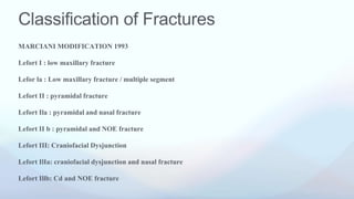
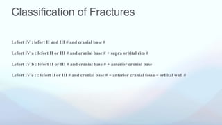
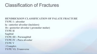
Dr.Surbhi Abrol
Classification and type-1
LEFORT FRACTURE
Classification of Fractures
• CLASSIFICATION OF FRACTURES
• IN 1901, RENE LE FORT, according to the level of injury:
• Lefort I
• Lefort II
• Lefort III
Classification of Fractures
MARCIANI MODIFICATION 1993
Lefort I : low maxillary fracture
Lefor la : Low maxillary fracture / multiple segment
Lefort II : pyramidal fracture
Lefort Ila : pyramidal and nasal fracture
Lefort II b : pyramidal and NOE fracture
Lefort III: Craniofacial Dysjunction
Lefort IlIa: craniofacial dysjunction and nasal fracture
Lefort Illb: Cd and NOE fracture
Classification of Fractures
Lefort IV : lefort II and III # and cranial base #
Lefort IV a : lefort II or III # and cranial base # + supra orbital rim #
Lefort IV b : lefort II or III # and cranial base # + anterior cranial base
Lefort IV c : : lefort Il or III # and cranial base # + anterior cranial fossa + orbital wall #
Classification of Fractures
HENDRICKSON CLASSIFICATION OF PALATE FRACTURE
TYPE 1 : alveolar
la : anterior alveolar (incisiors)
1b : posterior alveolar ( premolar molar)
ТУРЕ II
Sagittal
TYPE III : Parasagittal
TYPE IV : Para alveolar
TYPE V
: Complex
TYPE VI: Transverse
Classification of Fractures
ACCORDING TO ROWE AND WILLIAM (1985)
A. Fracture not involving the occlusion
1. CENTRAL REGION
a. Fracture of the nasal bone or the nasal septum
lateral nasal injuries
anterior nasal injuries
b.. Fracture of the frontal process of the maxilla
c. Fracture type a and b which extend to the Ethmoid bone
d. Fracture type a , b and c, which extend into the frontal bone
2. LATERAL REGION
# involving the zygomatic bone, arch and maxilla excluding dento
Force acting
This type of # occurs from the application of horizontal force just
above the apices of the maxillary teeth.
It results due to the blow from the opposite jaw , which is often
impacted.
This is a horizontal fracture above the level of the nasal floor including the dental component.
The # line runs backward along.
Laterally : lateral margin of the pyriform aperture - lateral wall of maxillary sinus - below the zygomatic butt
Medially: lower third of the nasal septum - lateral margin of the anterior nasal aperture (lateral wall of the no
• The fracture oceurs at the level of the piriform aperture and
involves the anterior and lateral walls of the maxillary sinus, lateral
nasal walls and, pterygoid plates.
• The nasal septum may also be fractured and the nasal cartilage
may be buckled.
• Sagittal fracture(s) of the palate may also be present.
• The pull of the medial and lateral ptergoid muscles may contribute
to displacement of the fractured segment in a posterior and inferior
direction, resulting in an open bite deformity.
• This fracture may present as an impacted, immovable, or free-
floating maxillary segment.
EXAMINATION
Firmly grasping the maxillary arch with the
finger and thumb facially and palatally and
attempting displacement of the maxilla in
three dimensions, as well as compression and
expansion of the maxillary arch.
SIGNS AND SYMPTOMS
Swelling of upper lip and cheek
ECCHYMOSIS: present in maxillary buccal suleus from shearing of soft tissue or periosteal tear.
NASAL BLOCK : mucosal tear in maxillary / ethmoid sinus may include bleeding causing a nasal
block - forcing the patient to undergo oral breathing.
Ocular signs are usually absent. Hypoesthesia of the infraorbital nerve may be caused by the rapid
development of edema.
GUERIN SIGNS: ecchymosis in the palate in the area of greater palatine foramen bilaterally
classical sign.
OCCLUSION:
Undisplaced incomplete Lefort I # usually cause no occlusal disturbance .Complete lefort I#
classically shows varying degree of anterior pen bite. This is from backward and downward
distraction of posterior maxilla resulting from inferior traction of medial pterygoid muscle towards
the mobile maxillary fragment.
TEETH FRACTURE:
Due to impaction of the mandibular teeth against the maxillary counter part, damage to the cusp of
individual maxillary teeth may be seen.
Palatal fracture:
Commonly mid palatal split is associated with Lefort :I evident as
linear mucosal tear in mid palate.
The associated palatal # could be of any of the Hendrickson
Classification pattern, with or without oronasal communication,
depending upon the amount of separation between the fragments
from the effect of bilateral medial pterygoid.
• Cracked-pot sound:
Percussion of the maxillary teeth results in distinctive " cracked pot
sound", similar to the sound produced when a cracked china pot is
INVESTIGATIONS
• Radiographs
• Computed tomography
• 3D CTIMAGING
• MRI
MANUAL/CLOSED REDUCTION
MAXILLA IS HELD
WITH TWO PAIRS OF
ROWE'S DISIMPACTION
FORCEPS.
EACH UNPADDED BLADE IS
PASSED UP A NOSTRIL AND THE
PADDED BLADE ENTERS THE
MOUTH AND GRIPS THE PALATE
HEAD OF OPERATING TABLE
OPERATOR GRASPS THE HANDLE
OF EACH OF THE TWO PAIR OF
FORCEPS AND MANIPULATES THE
(FRAGMENTS INTO PLACE.
(SPECIAL ATTENTION SHOULD BE GIVEN TO CORRECT ANY INFERIOR
DISPLACEMENT OF POSTERIOR ASPECT OF THE MAXILLA- TO RELIEVE "GAGGING
" OF POSTERIOR TEETH AND ANTERIOR OPEN BITE.
A DELIBERATE ROCKING AND
ROTATORY MOVEMENT IN
TRANSVERSE AND SAGITTAL
PLANE IS DONE.
A FORWARD TRACTION IS
APPLIED ONCE THE
FRACTURE IS MOBILE.
EXTERNAL FIXATION
• CRANIOMANDIBULAR
• BOX FRAME
• HALO FRAME
• PLASTER OF PARIS HEAD
• CRANIOMAXILLARY
• SUPRAORBITAL PINS
• ZYGOMATIC PINS
• HALO FRAMES
INTERNAL FIXATION
DIRECT OSTEOSYNTHESIS
• TRANSOSSEOUS WIRING AT # SITE
HIGH LEVEL (FRONTOZYGOMATIC AND FRONTO
NASAL)
MID LEVEL ( ORBITAL RIM / ZYGOMATIC BUTRESS
)
LOW LEVEL (ALVEOLAR/MIDPALATAL)
• MINI PLATES
• TRANSFIXATION WITH KRISCHNER WIRE OR
STEINMAN PIN :
1. TRANSFACIAL
2. ZYGOMATIC SEPTAL
CHANGING TRENDS IN MANAGEMENT
OF MID FACE FRACTURE
Historical approaches
Common to delay surgery for 7-14 days
Closed reduction in most instances
Long periods of Intermaxillary fixation
Small local incisions with restricted access for open reduction (if used
Accuracy of reduction sometimes estimated, esp in vertical dimension
Wire osteosynthesis, wire suspension (craniomandibular) or external
fixation (craniomandibular
Contemporary Approach to treatment
• Earlier one stage repair advocated
• Emphasis on open reduction
• No Intermaxillary fixation (or short period only)
• Wide exposure to all fracture sites
• Anatomical reduction of structural pillars of the face
• Semi rigid miniplates and microplates internal fixation (with primary
bone grafting in some situations
Complications
• Infraorbital nerve
paresthesia
• Enophthalmos
• Infection
• Exposed hardware
• Deviated septum
• Nasal obstruction
• Altered vision
• Nonunion
• Mal-union or
Malocclusion
• Epiphora
• Foreign body
reactions
• Scarring
• Sinusitis
THANK YOU

Presentation 14.pptx

  • 1.
    Dr.Surbhi Abrol Classification andtype-1 LEFORT FRACTURE
  • 2.
    Classification of Fractures •CLASSIFICATION OF FRACTURES • IN 1901, RENE LE FORT, according to the level of injury: • Lefort I • Lefort II • Lefort III
  • 3.
    Classification of Fractures MARCIANIMODIFICATION 1993 Lefort I : low maxillary fracture Lefor la : Low maxillary fracture / multiple segment Lefort II : pyramidal fracture Lefort Ila : pyramidal and nasal fracture Lefort II b : pyramidal and NOE fracture Lefort III: Craniofacial Dysjunction Lefort IlIa: craniofacial dysjunction and nasal fracture Lefort Illb: Cd and NOE fracture
  • 4.
    Classification of Fractures LefortIV : lefort II and III # and cranial base # Lefort IV a : lefort II or III # and cranial base # + supra orbital rim # Lefort IV b : lefort II or III # and cranial base # + anterior cranial base Lefort IV c : : lefort Il or III # and cranial base # + anterior cranial fossa + orbital wall #
  • 5.
    Classification of Fractures HENDRICKSONCLASSIFICATION OF PALATE FRACTURE TYPE 1 : alveolar la : anterior alveolar (incisiors) 1b : posterior alveolar ( premolar molar) ТУРЕ II Sagittal TYPE III : Parasagittal TYPE IV : Para alveolar TYPE V : Complex TYPE VI: Transverse
  • 6.
    Classification of Fractures ACCORDINGTO ROWE AND WILLIAM (1985) A. Fracture not involving the occlusion 1. CENTRAL REGION a. Fracture of the nasal bone or the nasal septum lateral nasal injuries anterior nasal injuries b.. Fracture of the frontal process of the maxilla c. Fracture type a and b which extend to the Ethmoid bone d. Fracture type a , b and c, which extend into the frontal bone 2. LATERAL REGION # involving the zygomatic bone, arch and maxilla excluding dento
  • 9.
    Force acting This typeof # occurs from the application of horizontal force just above the apices of the maxillary teeth. It results due to the blow from the opposite jaw , which is often impacted.
  • 10.
    This is ahorizontal fracture above the level of the nasal floor including the dental component. The # line runs backward along. Laterally : lateral margin of the pyriform aperture - lateral wall of maxillary sinus - below the zygomatic butt Medially: lower third of the nasal septum - lateral margin of the anterior nasal aperture (lateral wall of the no
  • 11.
    • The fractureoceurs at the level of the piriform aperture and involves the anterior and lateral walls of the maxillary sinus, lateral nasal walls and, pterygoid plates. • The nasal septum may also be fractured and the nasal cartilage may be buckled. • Sagittal fracture(s) of the palate may also be present. • The pull of the medial and lateral ptergoid muscles may contribute to displacement of the fractured segment in a posterior and inferior direction, resulting in an open bite deformity. • This fracture may present as an impacted, immovable, or free- floating maxillary segment.
  • 13.
    EXAMINATION Firmly grasping themaxillary arch with the finger and thumb facially and palatally and attempting displacement of the maxilla in three dimensions, as well as compression and expansion of the maxillary arch.
  • 14.
    SIGNS AND SYMPTOMS Swellingof upper lip and cheek ECCHYMOSIS: present in maxillary buccal suleus from shearing of soft tissue or periosteal tear. NASAL BLOCK : mucosal tear in maxillary / ethmoid sinus may include bleeding causing a nasal block - forcing the patient to undergo oral breathing. Ocular signs are usually absent. Hypoesthesia of the infraorbital nerve may be caused by the rapid development of edema. GUERIN SIGNS: ecchymosis in the palate in the area of greater palatine foramen bilaterally classical sign.
  • 15.
    OCCLUSION: Undisplaced incomplete LefortI # usually cause no occlusal disturbance .Complete lefort I# classically shows varying degree of anterior pen bite. This is from backward and downward distraction of posterior maxilla resulting from inferior traction of medial pterygoid muscle towards the mobile maxillary fragment. TEETH FRACTURE: Due to impaction of the mandibular teeth against the maxillary counter part, damage to the cusp of individual maxillary teeth may be seen.
  • 16.
    Palatal fracture: Commonly midpalatal split is associated with Lefort :I evident as linear mucosal tear in mid palate. The associated palatal # could be of any of the Hendrickson Classification pattern, with or without oronasal communication, depending upon the amount of separation between the fragments from the effect of bilateral medial pterygoid. • Cracked-pot sound: Percussion of the maxillary teeth results in distinctive " cracked pot sound", similar to the sound produced when a cracked china pot is
  • 17.
    INVESTIGATIONS • Radiographs • Computedtomography • 3D CTIMAGING • MRI
  • 18.
  • 20.
    MAXILLA IS HELD WITHTWO PAIRS OF ROWE'S DISIMPACTION FORCEPS. EACH UNPADDED BLADE IS PASSED UP A NOSTRIL AND THE PADDED BLADE ENTERS THE MOUTH AND GRIPS THE PALATE HEAD OF OPERATING TABLE OPERATOR GRASPS THE HANDLE OF EACH OF THE TWO PAIR OF FORCEPS AND MANIPULATES THE (FRAGMENTS INTO PLACE.
  • 21.
    (SPECIAL ATTENTION SHOULDBE GIVEN TO CORRECT ANY INFERIOR DISPLACEMENT OF POSTERIOR ASPECT OF THE MAXILLA- TO RELIEVE "GAGGING " OF POSTERIOR TEETH AND ANTERIOR OPEN BITE. A DELIBERATE ROCKING AND ROTATORY MOVEMENT IN TRANSVERSE AND SAGITTAL PLANE IS DONE. A FORWARD TRACTION IS APPLIED ONCE THE FRACTURE IS MOBILE.
  • 22.
    EXTERNAL FIXATION • CRANIOMANDIBULAR •BOX FRAME • HALO FRAME • PLASTER OF PARIS HEAD • CRANIOMAXILLARY • SUPRAORBITAL PINS • ZYGOMATIC PINS • HALO FRAMES
  • 23.
    INTERNAL FIXATION DIRECT OSTEOSYNTHESIS •TRANSOSSEOUS WIRING AT # SITE HIGH LEVEL (FRONTOZYGOMATIC AND FRONTO NASAL) MID LEVEL ( ORBITAL RIM / ZYGOMATIC BUTRESS ) LOW LEVEL (ALVEOLAR/MIDPALATAL) • MINI PLATES • TRANSFIXATION WITH KRISCHNER WIRE OR STEINMAN PIN : 1. TRANSFACIAL 2. ZYGOMATIC SEPTAL
  • 25.
    CHANGING TRENDS INMANAGEMENT OF MID FACE FRACTURE
  • 29.
    Historical approaches Common todelay surgery for 7-14 days Closed reduction in most instances Long periods of Intermaxillary fixation Small local incisions with restricted access for open reduction (if used Accuracy of reduction sometimes estimated, esp in vertical dimension Wire osteosynthesis, wire suspension (craniomandibular) or external fixation (craniomandibular
  • 30.
    Contemporary Approach totreatment • Earlier one stage repair advocated • Emphasis on open reduction • No Intermaxillary fixation (or short period only) • Wide exposure to all fracture sites • Anatomical reduction of structural pillars of the face • Semi rigid miniplates and microplates internal fixation (with primary bone grafting in some situations
  • 35.
    Complications • Infraorbital nerve paresthesia •Enophthalmos • Infection • Exposed hardware • Deviated septum • Nasal obstruction • Altered vision • Nonunion • Mal-union or Malocclusion • Epiphora • Foreign body reactions • Scarring • Sinusitis
  • 36.