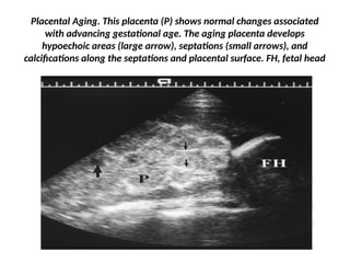
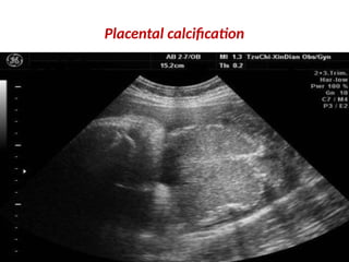
Post-maturity
Postmaturity
• Newborn delivered anytime after 42 weeks gestation.
• Post-term, postmaturity, prolonged pregnancy, and post-
dated pregnancy all refer to postmature birth.
• Less than 6 percent of all babies are born at 42 weeks or
later.
• No harmful effects on the mother; however,
• Fetal malnutrition and jeopardy.
Postmaturity
• After the 42nd week of gestation, the placenta, starts aging
and will eventually fail.
• Near the end of a term pregnancy, placental function
decreases,
• The placenta becomes smaller and less effective,
• Providing fewer nutrients and less oxygen to the fetus.
Aetiopathogenesis
• Post-mature (post term) delivery is much less common than
premature (preterm) delivery.
• Some post-mature pregnancies are because
• The mother is not certain of her last period, so in reality the
baby is not technically post-mature.
• Why a pregnancy continues beyond term is usually not
known.
Aetiopathogenesis
• More commonly seen in primigravida women.
• Once postmature delivery occurs, chance of having
subsequent postmature baby increases .
• Exact cause?
• Fetus with anencephaly have difficulty in initiating labor at
term
– No engagement of head
– Lack of pituitary – adrenal axis
• Trisomy 16 and 18
• Seckel’s syndrome (bird headed syndrome)
Aetiopathogenesis
• Postmature infants are full-grown, mature infants
• Who have stayed in utero beyond the full vigor of the
placenta and
• Have begun to lose weight in utero.
• Normally placenta can support fetal growth upto 42
weeks beyond that it becomes old and dysfunctional.
• This period may be earlier in developing countries, and
fetal growth reaches a plateau much earlier around 38
wks.
Aetiopathogenesis
• Fetus becomes vulnerable to hypoxia and birth asphyxia.
• Vernix and liquor starts decreasing after 36 weeks and vernix
almost disappears around 41 wks.
• Lack of vernix leads to constant exposure to liquor amnii and
• Maceration of skin and desquamation,
• Deepening of creases of soles.
• Skull bones are hardened.
Aetiopathogenesis
• To survive, the fetus begins to
use its own fat and
carbohydrates to provide
energy.
• As a result, its growth rate
slows, and
• Weight may even decrease.
• Post mature babies lack
subcutaneous fat and glycogen
stores.
• They are usually “older
looking” and have a wide-
eyed, alert appearance.
Aetiopathogenesis
• Their skin is dry and peeling, and
• Subcutaneous tissue is diminished; thus the skin appears
wrinkled.
• Initially they may be hypoglycemic and
• Require early feedings to maintain blood glucose levels of 40
mg/dL or higher.
• Need close monitoring for hypoglycemia.
• Some postmature newborns require resuscitation.
Symptoms
• Different babies show different signs.
• Post-mature newborns :
• Appear emaciated, have minimal fat, especially if the
function of the placenta was severely reduced.
• Dry, peeling, loose skin and may either show a brown, green,
or yellow discoloration
• A lot of hair on their head,
• Creases on the baby's palms and soles of their feet are more
pronounced,
• The fingernails and toenails are long.
• The umbilical cord and nails may be stained green if
meconium was present in the amniotic fluid.
Clinical features
• Marked desquamation
• of the skin in a post-
term infant.
• This is a benign
physiologic
desquamation
• with paper-thin peeling;
the underlying skin is
normal.
• The process is more
marked in areas of
irritation.
Treatment
• Postmature newborns may show e/o
• fetal distress may need resuscitation at birth.
• May have MSAF requiring intervention to deliver the baby.
• If develops MAS then appropriate intensive management
• 10% Dext. I.V. or frequent breast milk or formula feedings.
• Application of oil to prevent dryness of desquamating skin.
• If these problems do not occur, then
• Major goal is to provide good nutrition so that postmature
newborns can exhibit catch up growth.
Complications
Fetal and Neonatal Risks
• Reduced placental perfusion— signs of placental
deterioration, once a pregnancy has surpassed the 40 week
gestation period.
• Towards the end of pregnancy
– calcium is deposited on the walls of blood vessels and
– proteins are deposited on the surface of the placenta.
• Thus limits the blood flow, leading to placental insufficiency.
• fetal undernutrition
• Induced labor is strongly encouraged if this happens.
• Oligohydramnios
• Meconium aspiration
Complications
• Prone to develop hypoglycemia after birth
• Lack of adequate oxygen to the fetus, particularly during
labor.
• Fetal distress and HIE AND or MODS.
• Fetal distress may cause MSAF, or even MAS.
Complications
Maternal Risks
• Assisted delivery - higher incidence of
• forceps
• vacuum
• cesarean
• The length of labor may be prolonged.
• The labor is increased because the baby's head is too big to
pass through the mother's pelvis (CPD).
• Shoulder dystocia, becomes an increased risk –
• Difficulty in delivering the shoulders,
• Increased psychological stress to mother
• Higher rates of induction of labour.
Prevention of postmaturity
• Induction of labor after completion of term.
• Good monitoring .
• Readiness for resuscitation.
• Skill to manage hypoxic or asphyxiated baby at delivery.
• Skill to prevent / manage MAS and RDS due to pneumonia
following MAS.
• Monitoring and Prevention of hypoglycemia
Methods of monitoring post-mature babies
• Once a pregnancy is diagnosed to be post-dated, additional
monitoring is being done for the fetal well being.
• Fetal movement recording
• a "kick-chart" by the mother to record the movements of her
baby.
• Regular movements of the baby is the best sign indicating
that it is still in good health.
• Less than 10 movements in 2 hours is not a good sign.
• A reduction in the number of movements could indicate
placental deterioration.
Electronic fetal monitoring
cardiotocography
• To check the baby's heartbeat and is typically monitored
over a 30-minute period.
• Fetal heart irregularity
• Fetal tachycardia
• Fetal bradycardia
Ultrasound scan
• Ultrasound scan evaluates
• The amount of amniotic fluid around the baby.
• The amount of fluid will be low if the placenta is
deteriorating and induction of labor is highly recommended.
• Also helps in monitoring the fetal development .
• USG assisted fetal age of gestation.
Ultrasound scan
Biophysical profile
• A biophysical profile checks for the baby's
• Heart rate,
• Muscle tone,
• Movement,
• Breathing, and
• The amount of amniotic fluid surrounding the baby.
Placental grading
• Gestational-Dates Categories:
– pre-term = <37 weeks ("very pre-term" = <32 weeks).
– normal term range 37-40 weeks.
– post-dates, post term= >40 & up to 42 weeks.
• post-dates, post mature= >42 weeks
Ultrasound scan
• Doppler flow study
• Doppler flow study is a type of ultrasound that measures the
amount of blood flowing in and out of the placenta.
Blood flow in umb.BV during systolic and diastolic
phase
• Umbilical cord displaying
umbilical artery (red) and
umbilical vein (blue), the
gate or sample.
• volume include both signals
(left). Sonogram of the
umbilical artery and vein
(right).
Normal Placental Venous Lakes. Venous lakes (large arrows) appear as
focal echolucent areas just beneath the chorionic membrane C, A, or
within the substance of the placenta (P),
B. Note the swirling blood flow (small arrow) in B. Venous lakes are
incidental finding of no clinical significance.
Placental Aging. This placenta (P) shows normal changes associated
with advancing gestational age. The aging placenta develops
hypoechoic areas (large arrow), septations (small arrows), and
calcifications along the septations and placental surface. FH, fetal head
Placental calcification
UMBILICAL ARTERY DOPPLER. A. A NORMAL UMBILICAL ARTERY
DOPPLER SPECTRUM IS DISPLAYED ABOVE THE BASELINE, WHEREAS
THE UMBILICAL VEIN SPECTRUM IS DISPLAYED BELOW THE BASELINE
INDICATING NORMAL BLOOD FLOW IN OPPOSITE DIRECTIONS.
DISTINCT, MODERATELY HIGH-VELOCITY BLOOD FLOW IS SEEN IN THE
UMBILICAL ARTERY THROUGHOUT DIASTOLE, RESULTING IN A
RESISTANCE INDEX (RI) OF 0.58 AND A SYSTOLE/DIASTOLE (S/D) RATIO
OF 2.36. B. DOPPLER SPECTRUM FROM THE UMBILICAL ARTERY OF A
GROWTH-RETARDED FETUS SHOWS REVERSAL OF BLOOD FLOW
DIRECTION IN DIASTOLE (ARROW). THIS IS A HIGHLY SPECIFIC FINDING
OF SEVERE FETAL DISTRESS.
Expectant Management
• They have two alternatives
• 1) induction of labor
• 2) waits for the onset of labour naturally.
• With additional monitoring of their baby, with regular CTG,
ultrasound and biophysical profile, she continues her
pregnancy
Inducing labor
• There are several reasons
• If there is potential harm to the mother or child
• The mother's water breaks and
• Contractions have not started, the child is post-mature,
• The mother has diabetes/ hypertension/ Oligohydramnios
• Induction of Labor is not always the best choice because it
has its own risks.
• Avoid inducing labor unless it is completely necessary.
Procedure
• There are four common methods of starting contractions.
• 1) stripping the membranes,
• Once this membrane is stripped the hormone prostaglandin
is naturally released into the mother's body and starts the
contractions
• 2) Breaking the mother's water, labor begins in few hrs.
• 3) Giving the hormone prostaglandin , ripens the
cervix(os/gel) under close monitoring of the
mother and fetus .
• 4) Giving the synthetic hormone ( oxytocin ).
Monitoring
• Check fetal well-being and identify problems by proper
monitoring, which includes
• 1. Ultrasound,
• 2. Non-stress testing –NST (how the fetal heart rate responds
to fetal activity), and
• 3. Estimation of the amniotic fluid volume.
• 4. During labor, monitor fetal heart rate to help identify
changes in the heart rate due to low oxygenation.
• 5. Changes in a baby's condition may require a LSCS delivery.
Care after delivery
• Special care of the postmature baby may include:
• Checking for respiratory problems related to meconium
stained liqour and
• Prevention of aspiration.
• Prevention of hypoglycemia .
• Monitoring for Blood glucose.
Prevention of postmaturity
• Accurate pregnancy due dates can help identify babies at
risk for postmaturity.
• Ultrasound examinations early in pregnancy help establish
more accurate dating by measurements taken of the fetus.
• Ultrasound is also important in evaluating the placenta for
signs of aging.
Diagnosis
• Postmaturity is usually diagnosed by
• a combination of assessments, including the following:
• 1) Baby's physical appearance
• 2) Length of the pregnancy
• 3) Baby's assessed gestational age
COMPLICATIONS IN POSTMATURE BABIES
• MAS
• FEEDING
• HYPOGLYCEMIA
PREVENTION
• Ultrasound examinations early in pregnancy help establish
more accurate dating by measurements taken of the fetus.
• Ultrasound is also important in evaluating the placenta for
signs of aging.
• The use of ultrasound to identify the fetus at risk for
postmaturity among postterm pregnancies with placental
grading.
PREVENTION
• The presence of immature placentas (grade 0 or 1) is rare
after 42 weeks of gestation.
• Advanced postmaturity was found with grade 2 and 3
placentas.
• Oligohydramnios is very common (81.8%) among
postmature pregnancies.. Placental grading cannot be used
to predict postmaturity.
WHICH ONE?
Choice should be
ours.
THANX

Postmaturity: Birth after 42 weeks gestation,

  • 1.
  • 2.
    Postmaturity • Newborn deliveredanytime after 42 weeks gestation. • Post-term, postmaturity, prolonged pregnancy, and post- dated pregnancy all refer to postmature birth. • Less than 6 percent of all babies are born at 42 weeks or later. • No harmful effects on the mother; however, • Fetal malnutrition and jeopardy.
  • 3.
    Postmaturity • After the42nd week of gestation, the placenta, starts aging and will eventually fail. • Near the end of a term pregnancy, placental function decreases, • The placenta becomes smaller and less effective, • Providing fewer nutrients and less oxygen to the fetus.
  • 4.
    Aetiopathogenesis • Post-mature (postterm) delivery is much less common than premature (preterm) delivery. • Some post-mature pregnancies are because • The mother is not certain of her last period, so in reality the baby is not technically post-mature. • Why a pregnancy continues beyond term is usually not known.
  • 5.
    Aetiopathogenesis • More commonlyseen in primigravida women. • Once postmature delivery occurs, chance of having subsequent postmature baby increases . • Exact cause? • Fetus with anencephaly have difficulty in initiating labor at term – No engagement of head – Lack of pituitary – adrenal axis • Trisomy 16 and 18 • Seckel’s syndrome (bird headed syndrome)
  • 6.
    Aetiopathogenesis • Postmature infantsare full-grown, mature infants • Who have stayed in utero beyond the full vigor of the placenta and • Have begun to lose weight in utero. • Normally placenta can support fetal growth upto 42 weeks beyond that it becomes old and dysfunctional. • This period may be earlier in developing countries, and fetal growth reaches a plateau much earlier around 38 wks.
  • 7.
    Aetiopathogenesis • Fetus becomesvulnerable to hypoxia and birth asphyxia. • Vernix and liquor starts decreasing after 36 weeks and vernix almost disappears around 41 wks. • Lack of vernix leads to constant exposure to liquor amnii and • Maceration of skin and desquamation, • Deepening of creases of soles. • Skull bones are hardened.
  • 8.
    Aetiopathogenesis • To survive,the fetus begins to use its own fat and carbohydrates to provide energy. • As a result, its growth rate slows, and • Weight may even decrease. • Post mature babies lack subcutaneous fat and glycogen stores. • They are usually “older looking” and have a wide- eyed, alert appearance.
  • 9.
    Aetiopathogenesis • Their skinis dry and peeling, and • Subcutaneous tissue is diminished; thus the skin appears wrinkled. • Initially they may be hypoglycemic and • Require early feedings to maintain blood glucose levels of 40 mg/dL or higher. • Need close monitoring for hypoglycemia. • Some postmature newborns require resuscitation.
  • 10.
    Symptoms • Different babiesshow different signs. • Post-mature newborns : • Appear emaciated, have minimal fat, especially if the function of the placenta was severely reduced. • Dry, peeling, loose skin and may either show a brown, green, or yellow discoloration • A lot of hair on their head, • Creases on the baby's palms and soles of their feet are more pronounced, • The fingernails and toenails are long. • The umbilical cord and nails may be stained green if meconium was present in the amniotic fluid.
  • 11.
    Clinical features • Markeddesquamation • of the skin in a post- term infant. • This is a benign physiologic desquamation • with paper-thin peeling; the underlying skin is normal. • The process is more marked in areas of irritation.
  • 13.
    Treatment • Postmature newbornsmay show e/o • fetal distress may need resuscitation at birth. • May have MSAF requiring intervention to deliver the baby. • If develops MAS then appropriate intensive management • 10% Dext. I.V. or frequent breast milk or formula feedings. • Application of oil to prevent dryness of desquamating skin. • If these problems do not occur, then • Major goal is to provide good nutrition so that postmature newborns can exhibit catch up growth.
  • 14.
    Complications Fetal and NeonatalRisks • Reduced placental perfusion— signs of placental deterioration, once a pregnancy has surpassed the 40 week gestation period. • Towards the end of pregnancy – calcium is deposited on the walls of blood vessels and – proteins are deposited on the surface of the placenta. • Thus limits the blood flow, leading to placental insufficiency. • fetal undernutrition • Induced labor is strongly encouraged if this happens. • Oligohydramnios • Meconium aspiration
  • 15.
    Complications • Prone todevelop hypoglycemia after birth • Lack of adequate oxygen to the fetus, particularly during labor. • Fetal distress and HIE AND or MODS. • Fetal distress may cause MSAF, or even MAS.
  • 16.
    Complications Maternal Risks • Assisteddelivery - higher incidence of • forceps • vacuum • cesarean • The length of labor may be prolonged. • The labor is increased because the baby's head is too big to pass through the mother's pelvis (CPD). • Shoulder dystocia, becomes an increased risk – • Difficulty in delivering the shoulders, • Increased psychological stress to mother • Higher rates of induction of labour.
  • 17.
    Prevention of postmaturity •Induction of labor after completion of term. • Good monitoring . • Readiness for resuscitation. • Skill to manage hypoxic or asphyxiated baby at delivery. • Skill to prevent / manage MAS and RDS due to pneumonia following MAS. • Monitoring and Prevention of hypoglycemia
  • 18.
    Methods of monitoringpost-mature babies • Once a pregnancy is diagnosed to be post-dated, additional monitoring is being done for the fetal well being. • Fetal movement recording • a "kick-chart" by the mother to record the movements of her baby. • Regular movements of the baby is the best sign indicating that it is still in good health. • Less than 10 movements in 2 hours is not a good sign. • A reduction in the number of movements could indicate placental deterioration.
  • 19.
    Electronic fetal monitoring cardiotocography •To check the baby's heartbeat and is typically monitored over a 30-minute period. • Fetal heart irregularity • Fetal tachycardia • Fetal bradycardia
  • 20.
    Ultrasound scan • Ultrasoundscan evaluates • The amount of amniotic fluid around the baby. • The amount of fluid will be low if the placenta is deteriorating and induction of labor is highly recommended. • Also helps in monitoring the fetal development . • USG assisted fetal age of gestation.
  • 21.
    Ultrasound scan Biophysical profile •A biophysical profile checks for the baby's • Heart rate, • Muscle tone, • Movement, • Breathing, and • The amount of amniotic fluid surrounding the baby.
  • 22.
    Placental grading • Gestational-DatesCategories: – pre-term = <37 weeks ("very pre-term" = <32 weeks). – normal term range 37-40 weeks. – post-dates, post term= >40 & up to 42 weeks. • post-dates, post mature= >42 weeks
  • 23.
    Ultrasound scan • Dopplerflow study • Doppler flow study is a type of ultrasound that measures the amount of blood flowing in and out of the placenta.
  • 24.
    Blood flow inumb.BV during systolic and diastolic phase • Umbilical cord displaying umbilical artery (red) and umbilical vein (blue), the gate or sample. • volume include both signals (left). Sonogram of the umbilical artery and vein (right).
  • 25.
    Normal Placental VenousLakes. Venous lakes (large arrows) appear as focal echolucent areas just beneath the chorionic membrane C, A, or within the substance of the placenta (P), B. Note the swirling blood flow (small arrow) in B. Venous lakes are incidental finding of no clinical significance.
  • 26.
    Placental Aging. Thisplacenta (P) shows normal changes associated with advancing gestational age. The aging placenta develops hypoechoic areas (large arrow), septations (small arrows), and calcifications along the septations and placental surface. FH, fetal head
  • 27.
  • 28.
    UMBILICAL ARTERY DOPPLER.A. A NORMAL UMBILICAL ARTERY DOPPLER SPECTRUM IS DISPLAYED ABOVE THE BASELINE, WHEREAS THE UMBILICAL VEIN SPECTRUM IS DISPLAYED BELOW THE BASELINE INDICATING NORMAL BLOOD FLOW IN OPPOSITE DIRECTIONS. DISTINCT, MODERATELY HIGH-VELOCITY BLOOD FLOW IS SEEN IN THE UMBILICAL ARTERY THROUGHOUT DIASTOLE, RESULTING IN A RESISTANCE INDEX (RI) OF 0.58 AND A SYSTOLE/DIASTOLE (S/D) RATIO OF 2.36. B. DOPPLER SPECTRUM FROM THE UMBILICAL ARTERY OF A GROWTH-RETARDED FETUS SHOWS REVERSAL OF BLOOD FLOW DIRECTION IN DIASTOLE (ARROW). THIS IS A HIGHLY SPECIFIC FINDING OF SEVERE FETAL DISTRESS.
  • 29.
    Expectant Management • Theyhave two alternatives • 1) induction of labor • 2) waits for the onset of labour naturally. • With additional monitoring of their baby, with regular CTG, ultrasound and biophysical profile, she continues her pregnancy
  • 30.
    Inducing labor • Thereare several reasons • If there is potential harm to the mother or child • The mother's water breaks and • Contractions have not started, the child is post-mature, • The mother has diabetes/ hypertension/ Oligohydramnios • Induction of Labor is not always the best choice because it has its own risks. • Avoid inducing labor unless it is completely necessary.
  • 31.
    Procedure • There arefour common methods of starting contractions. • 1) stripping the membranes, • Once this membrane is stripped the hormone prostaglandin is naturally released into the mother's body and starts the contractions • 2) Breaking the mother's water, labor begins in few hrs. • 3) Giving the hormone prostaglandin , ripens the cervix(os/gel) under close monitoring of the mother and fetus . • 4) Giving the synthetic hormone ( oxytocin ).
  • 32.
    Monitoring • Check fetalwell-being and identify problems by proper monitoring, which includes • 1. Ultrasound, • 2. Non-stress testing –NST (how the fetal heart rate responds to fetal activity), and • 3. Estimation of the amniotic fluid volume. • 4. During labor, monitor fetal heart rate to help identify changes in the heart rate due to low oxygenation. • 5. Changes in a baby's condition may require a LSCS delivery.
  • 33.
    Care after delivery •Special care of the postmature baby may include: • Checking for respiratory problems related to meconium stained liqour and • Prevention of aspiration. • Prevention of hypoglycemia . • Monitoring for Blood glucose.
  • 34.
    Prevention of postmaturity •Accurate pregnancy due dates can help identify babies at risk for postmaturity. • Ultrasound examinations early in pregnancy help establish more accurate dating by measurements taken of the fetus. • Ultrasound is also important in evaluating the placenta for signs of aging.
  • 35.
    Diagnosis • Postmaturity isusually diagnosed by • a combination of assessments, including the following: • 1) Baby's physical appearance • 2) Length of the pregnancy • 3) Baby's assessed gestational age
  • 36.
    COMPLICATIONS IN POSTMATUREBABIES • MAS • FEEDING • HYPOGLYCEMIA
  • 37.
    PREVENTION • Ultrasound examinationsearly in pregnancy help establish more accurate dating by measurements taken of the fetus. • Ultrasound is also important in evaluating the placenta for signs of aging. • The use of ultrasound to identify the fetus at risk for postmaturity among postterm pregnancies with placental grading.
  • 38.
    PREVENTION • The presenceof immature placentas (grade 0 or 1) is rare after 42 weeks of gestation. • Advanced postmaturity was found with grade 2 and 3 placentas. • Oligohydramnios is very common (81.8%) among postmature pregnancies.. Placental grading cannot be used to predict postmaturity.
  • 39.