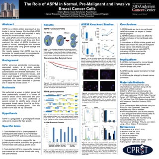
1. The study found that ASPM levels increase as breast cancer progresses from normal to pre-malignant to invasive stages. Knockdown of ASPM inhibited growth and cell division in pre-malignant and invasive breast cancer cells but not in normal breast cells.
2. Immunofluorescence showed higher ASPM levels in invasive breast cancer cells compared to normal breast cells. Knockdown of ASPM reduced ASPM levels in invasive cells but not normal cells.
3. Cell cycle analysis found that ASPM knockdown increased the G2/M phase in invasive breast cancer cells but not normal cells, indicating ASPM is important for cell division in breast cancer.
Relationship between CCL5 and TGFβ1 in breast cancer patients at both systemi...Marion Hartmann
Chemokines are chemotactic cytokines that play an important role in inflammation through promotion of leukocyte motility. Studies have shown
that expression and activation of chemokine receptors promotes growth and
migration of primary tumour cells which leads to metastatic spread. Stromal-epithelial crosstalk in the tumour microenvironment is facilitated through
chemokines and their receptors. The chemokine (C-C motif) ligand 5 (CCL5) and its principle receptor CCR5 has a primary role in inflammation and has
been implicated in breast cancer progression. Previous studies linked elevated CCL5 serum levels with late stage breast cancer, similarly to what
has been described for transforming growth factor beta 1 (TGFβ1), a well established factor in tumourigenesis. TGFβ1 is thought to act as a tumour
suppressor in early stage disease and switch to being potentially tumour promoting in later stage breast cancer.
3D collagen fibrillar microstructure guides pancreatic cancer cell phenotype ...Arun kumar
Pancreatic cancer, one of the deadliest cancers, is characterized by high rates of metastasis
and intense desmoplasia, both of which are associated with changes in fibrillar type I collagen
composition and microstructure. Epithelial to mesenchymal transition (EMT), a critical
step of metastasis, also involves a change in extracellular matrix (ECM) context as cells
detach from basement membrane (BM) and engage interstitial matrix (IM). The objective of
this work was to develop and apply an in-vitro three-dimensional (3D) tumor-ECM model to
define how ECM composition and biophysical properties modulate pancreatic cancer EMT.
Three established pancreatic ductal adenocarcinoma (PDAC) lines were embedded within
3D matrices prepared with type I collagen Oligomer (IM) at various fibril densities to control
matrix stiffness or Oligomer and Matrigel combined at various ratios while maintaining constant
matrix stiffness
Tumor Markers include a wide range of biomacromolecules orchestrated in abundance fixation by a wide assortment of neoplastic cells. The markers could be endogenous results of exceptionally dynamic metabolic threatening cells or the results of recently turned on qualities, which stayed unexpressed in early life or recently obtained antigens at cell and sub-cell levels. The presence of tumor marker and their focus are identified with the beginning and development of dangerous tumors in patients. A perfect tumor marker ought to be profoundly delicate, explicit, dependable with high prognostic worth, organ particularity and it should relate with tumor stages. Be that as it may, none of the tumor markers answered to date has every one of these attributes. Inspite of these impediments, numerous tumor markers have indicated incredible clinical significance in checking adequacy of various methods of treatments during whole course of sickness in malignant growth patients. Moreover, assurance of markers additionally helps in early discovery of malignant growth repeat and in anticipation.
The Phase I Personalized Genomic Vaccine trial at Mount Sinai recently began enrolling patients to receive a long-peptide vaccine targeting tumor neoantigens identified by exome and RNA sequencing. Each patient's vaccine is the result of a computational pipeline encompassing somatic variant calling, coding sequence prediction, variant-specific isoform identification, sequence-based HLA typing, peptide-MHC binding prediction, and a final ranking procedure over vaccine peptides. This talk will review the design considerations that went into our pipeline and introduce three new open source packages we have developed for variant coding sequence prediction, vaccine peptide selection, and peptide-MHC binding affinity prediction.
Correlation between circulating and tumour tissue CCL5 and TGFβ1 in breast ca...Marion Hartmann
This study demonstrates a novel correlation between CCL5 and TGFβ1 in breast cancer which is maintained in early and advanced disease
The mechanisms and controlling influences warrant further investigation and may open avenues for therapeutic manipulation in selected patients
maintrac liquid biopsy on circulating epithelial tumor cells Peter Pachmann
maintrac liquid biopsy on circulating epithelial tumor cells. Microscope based semi-automated discrimination of cancer cells, effectiveness testing of cancer drugs (before treatment) andtherapy monitoring
Wild Type and Mutated BRCA - Differentiation of Breast Cancer - BioGenexVictoria Miller
https://info.biogenex.com/wild-type-and-mutated-brca-differentiation-of-breast-cancer-using-new-mirna-biomarker-panel
breast cancer, wild type breast cancer, BRCA mutated breast cancer , BRCA gene mutation.Wild Type and Mutated BRCA , Differentiation of Breast Cancer, brca mutated breast cancer, brca mutation breast cancer
A reading report for <Exosomal transfer of stroma-derived miR21 confers pacl...星云 王
A reading report for <Exosomal transfer of stroma-derived miR21 confers paclitaxel resistance in ovarian cancer cells through targeting APAF1
>, only for private study use, please do not use it for profit or public.
El Condominio Terras de São José decidió modernizar su tecnología de comunicación entre los equipos de trabajo y de gestión con un sistema de radios digitales MOTOTRBO
El 7 de Abril de 2015, se publicó en el Boletín Oficial del Estado, el Real Decreto 180/2015, de 13 de Marzo, por el que se regula el traslado de residuos en el interior del territorio del Estado. La fecha de entrada en vigor de este Real Decreto fue el 7 de Mayo 2.015.
Volunteering is one of the key ways in which you can enhance your entrepreneurial abilities, team working abilities, leadership qualities, networking abilities and so much more! Most often people fail to understand it fully and make the most of what it can offer. These set of slides give you a detailed insight into what volunteering is and how it can make an impact on your day-to-day life.
Breast cancer research, in vitro model cell line study, MCF-7 human breast cancer cell line with it's details like history, importance, applications, Culture tips, a model responsible for ER targeted therapy etc.
Robert Anders, MD, PhD, Robert L. Ferris, MD, PhD, and Lauren L. Ritterhouse, MD, PhD, prepared useful Practice Aids pertaining to biomarker testing for this CME/MOC activity titled "The Central Role of Biomarker Testing in Piecing Together the Immuno-Oncology Puzzle: Essential Guidance for Pathologists to Maximize the Potential of Cancer Immunotherapies." For the full presentation, monograph, complete CME/MOC information, and to apply for credit, please visit us at http://bit.ly/2QAudYX. CME/MOC credit will be available until December 26, 2019.
HDAC4 and HDAC7 Promote Breast and Ovarian Cancer Cell Migration by Regulatin...CrimsonpublishersCancer
Breast and ovarian cancer have been remained as a highly malignant tumor among women, posing a serious threat to women health worldwide. In this study, we were aimed to investigate the underlying mechanism of breast and ovarian cancer cell migration. Wound healing assay showed that MDA-MB-231and C13* have higher migration potential compare with MCF-7 and OV2078 cells, as well as regulated epithelial-mesenchymal transition (EMT) marker. We found that HDAC4 and HADC7 mRNA are up regulated in MDA-MB-231 and C13* cells. Moreover, target HDAC4 and HDAC7 by TSA or shRNA block MDA-MB-231and C13* migration. These results reveal a new link between HDACs and EMT in the regulation of breast and ovarian cancer migration.
Clinical Biochemistry part III
Tumors and tumor markers, Toxicology, TDM, pregnancy, Paediatric clinical chemistry, screening of newborns for diseases, inborn errors of metabolism, geriatric Clinical chemistry, Nutritional assessment
Tumour Markers are substances present in the tumour, produced by the tumour or by the host as a response to the presence of the tumour, providing information about biological characteristics of the tumour. these tumour markers may specific for the tissue but often get elevated in neoplastic as well non-neoplastic lesions, further Various analytical platforms available for serum tumour markers lack standardisation. These factors add to interpretative challenges in serum tumour markers
1. Abstract
ASPM is a mitotic protein expressed at low
levels in normal tissues. We identified ASPM
as being both mutated and amplified in early
lesions and invasive breast cancer. 1
We used qPCR and immunofluorescence to
validate these results in cell lines, and then
analyzed the effect of ASPM inhibition on
growth of normal, premalignant and invasive
breast cancer cells using growth assays and
cell cycle analysis.
Our results suggest that ASPM may be a
biomarker for breast cancer forming potential,
as well as a putative prophylactic target.
Background
ASPM, abnormal spindle-like microcephaly-
associated protein, is a mitotic spindle
regulatory protein assisting in microtubule
polymerization and centriole stabilization.2
It is
largely expressed in embryonic tissues, and
not in adult tissues.3,6
ASPM inactivation is
associated with human neuropathies. ASPM
upregulation has been observed in gliomas
and in basal-like breast cancer. 6,7
Rationale
We performed a screen to detect genes that
were preferentially mutated in a subset of
invasive breast cancer associated with poor
patient survival1
. We then performed a
second screen to identify early drivers of
aggressive breast cancer from the top thirty-
two preferentially mutates genes. From this
screen ASPM was the top candidate.
Hypothesis
ASPM is upregulated in premalignant breast
cells and is required for their growth.
Conclusions
1.ASPM levels are low in normal breast
cells but increase as stages of breast
cancer progress.
2.ASPM knockdown does not affect the
growth or division of normal breast cells
(hTERT).
3.Knockdown of ASPM inhibits growth and
affects cell division for both pre-malignant
breast cancer cells (DCIS.com) and
invasive breast cancer cells (MCF7),
indicating ASPM is important for
proliferation in these cell types.
Implications
If ASPM is not required by normal breast
cells compared to pre-malignant and
invasive breast cells, then
1.ASPM may be a good biomarker for high
risk lesions
2.ASPM may be a target for breast cancer
prevention
Materials/Methods
1.Transfections were performed using
Dharmafect and Opti-Mem along with the
appropriate siRNA treatment.
2.RNA for qPCR was extracted using the
Qiagen QiaShredder and RNeasy Mini Kits.
cDNA was made using PT 200 Peltier Thermal
Cycler. qRT-PCR was performed using 7900
HT Fast Real-Time PCR System and analyzed
with Sequence Detection Systems (SDS)
software.
3.Cell cycle analysis was performed using the
Accuri C6 Flow Cytometer. Data was
analyzed using FlowJo software.
4.Immunofluorescence used ASPM antibody
(1:500) and goat-anti-rabbit antibody (488) as
the primary and secondary antibodies,
respectively. Slides were mounted using
Vectashield Mounting Medium for
Flouroescence with Dapi. Pictures were taken
using a Nikon Eclipse Ti Microscope.
References
1) Haricharan et al. BCRT. 2014.
2) Xu et al. PlosOne. 2012.
3) Fish et al. PNAS. 2006.
4) Oncomine Database
5) BioGPS Database
6) Higgins et al. BMC Cell Biology. 2010.
7) Zhong et al. Cell Cycle. 2005
ASPM Knockout Studies
The Role of ASPM in Normal, Pre-Malignant and Invasive
Breast Cancer Cells
Kristen Maslar, Svasti Haricharan, Powel Brown
Cancer Prevention & Research Institute of Texas Summer Research Program
Department of Clinical Cancer Prevention
Specific Aims
1. Test whether ASPM is overexpressed in
premalignant cells relative to normal breast
cells using qRT-PCR and immunofluorescence.
2. Test whether ASPM is required for the
growth of pre-invasive, but not for the growth of
normal breast cells using a growth assay.
3. Test whether ASPM is required for mitosis in
pre-invasive but not normal breast cells using
cell cycle analysis.
ASPM Functional Profile
ASPM gene expression
Results
Figure 4: Average ASPM mRNA levels within different types of
cell lines were tested using qRT-PCR. ASPM levels are lowest
in normal breast cells and increase as the disease progresses.
ASPM Baseline Levels
Figure 3: Heat maps for five different studies display ASPM
gene expression ranging from normal breast tissue to invasive
breast cancer tissue. The blue indicates the lowest levels of
ASPM, whereas the yellow indicates highest levels of ASPM4, 5
.
Recurrence-free Survival Curve
Figure 2: This graph represents the probability of recurrence-
free survival for breast cancer based on ASPM levels. Higher
ASPM levels result in a worse recurrence-free survival opposed
to lower survival with lower ASPM levels.
Figure 1: The illustration represents the functional profile of
ASPM. When a cell is not undergoing mitosis, ASPM remains
in the cyptoplasm. When a cell is ready to undergo mitosis,
however, ASPM can cycle into the nucleus to assist in various
mitotic functions, resulting in proliferation and division.
Figure 5: The set of graphs display both siASPM knockdown
and its affect on cell growth. The graphs on the left indicate
siASPM knockdown calculated from qRT-PCR for both hTERT
(normal breast) and DCIS.com (pre-malignant) cells lines. The
graphs on the right display a growth assay in the presence and
absence of siASPM for both hTERT and DCIS.com cell lines.
siLuc siASPM
hTERT
DCIS.com
hTERT
MCF7
Figure 7: Cell cycle analysis was performed to show how the
presence and absence of ASPM affects stages of the cell cycle.
The top images represent graphs of normal breast cells with
and without ASPM, indicating no difference between them. The
bottom graphs represent normal breast cells (hTERT) and ER+
breast cancer cells (MCF7). hTERT cells show no difference in
cell cycle regulation, whereas MCF7 show an increase in the
G2/M phase, indicating ASPM is important for division in breast
cancer cells.
Figure 6: Immunofluorescence displays ASPM within normal
breast cells (hTERT) and invasive breast cancer cells
(MCF7). For the negative control (siLuc), ASPM is more
pronounced in the MCF7 cells than in hTERT cells due to
their different in baseline levels. For the positive control
(siASPM), there is a reduction in ASPM in the MCF7 cells but
no change in the hTERT cells.
siLuc siASPM
No Mitosis Mitosis
2. Title of the Poster Presentation Goes Here
Authors of the Poster Presentation Goes Here
Institutional and/or Graduate School of Biomedical
Sciences Affiliation Goes Here
Title of the Poster Presentation Goes Here
Authors of the Poster Presentation Goes Here
Institutional and/or Graduate School of Biomedical
Sciences Affiliation Goes Here