1. RESEARCH ARTICLE
Protease-Activated Receptor 4 Induces
Bladder Pain through High Mobility Group
Box-1
Dimitrios E. Kouzoukas1,5¤
, Fei Ma1,3
, Katherine L. Meyer-Siegler2
, Karin N. Westlund1,3
,
David E. Hunt1
, Pedro L. Vera1,3,4
*
1 Research and Development, Lexington Veterans Affairs Medical Center, Lexington, Kentucky, United
States of America, 2 Department of Natural Sciences, St. Petersburg College, St. Petersburg, Florida, United
States of America, 3 Department of Physiology, University of Kentucky, Lexington, Kentucky, United States
of America, 4 Department of Surgery, University of Kentucky, Lexington, Kentucky, United States of
America, 5 Saha Cardiovascular Research Center, University of Kentucky, Lexington, Kentucky, United
States of America
¤ Current address: Department of Molecular Pharmacology & Therapeutics, Loyola University Chicago,
Maywood, Illinois, United States of America
* pedro.vera@va.gov
Abstract
Pain is the significant presenting symptom in Interstitial Cystitis/Painful Bladder Syndrome
(IC/PBS). Activation of urothelial protease activated receptor 4 (PAR4) causes pain through
release of urothelial macrophage migration inhibitory factor (MIF). High Mobility Group Box-
1 (HMGB1), a chromatin-binding protein, mediates bladder pain (but not inflammation) in an
experimental model (cyclophosphamide) of cystitis. To determine if PAR4-induced bladder
hypersensitivity depends on HMGB1 downstream, we tested whether: 1) bladder PAR4
stimulation affected urothelial HMGB1 release; 2) blocking MIF inhibited urothelial HMGB1
release; and 3) blocking HMGB1 prevented PAR4-induced bladder hypersensitivity.
HMGB1 release was examined in immortalized human urothelial cultures (UROtsa)
exposed to PAR4-activating peptide (PAR4-AP; 100 μM; 2 hours) or scrambled control pep-
tide. Female C57BL/6 mice, pretreated with a HMGB1 inhibitor (glycyrrhizin: 50 mg/kg; ip)
or vehicle, received intravesical PAR4-AP or a control peptide (100 μM; 1 hour) to determine
1) HMGB1 levels at 1 hour in the intravesical fluid (released HMGB1) and urothelium, and
2) abdominal hypersensitivity to von Frey filament stimulation 24 hours later. We also tested
mice pretreated with a MIF blocker (ISO-1: 20 mg/kg; ip) to determine whether MIF medi-
ated PAR4-induced urothelial HMGB1 release. PAR4-AP triggered HMGB1 release from
human (in vitro) and mice (in vivo) urothelial cells. Intravesical PAR4 activation elicited
abdominal hypersensitivity in mice that was prevented by blocking HMGB1. MIF inhibition
prevented PAR4-mediated HMGB1 release from mouse urothelium. Urothelial MIF and
HGMB1 represent novel targets for therapeutic intervention in bladder pain conditions.
PLOS ONE | DOI:10.1371/journal.pone.0152055 March 24, 2016 1 / 11
OPEN ACCESS
Citation: Kouzoukas DE, Ma F, Meyer-Siegler KL,
Westlund KN, Hunt DE, Vera PL (2016) Protease-
Activated Receptor 4 Induces Bladder Pain through
High Mobility Group Box-1. PLoS ONE 11(3):
e0152055. doi:10.1371/journal.pone.0152055
Editor: Joseph J Barchi, National Cancer Institute at
Frederick, UNITED STATES
Received: November 25, 2015
Accepted: March 8, 2016
Published: March 24, 2016
Copyright: This is an open access article, free of all
copyright, and may be freely reproduced, distributed,
transmitted, modified, built upon, or otherwise used
by anyone for any lawful purpose. The work is made
available under the Creative Commons CC0 public
domain dedication.
Data Availability Statement: All relevant data are
within the paper and its Supporting Information files.
Funding: This work was supported by funding from
the National Institutes of Health (DK0093496; P.L.V.).
The funders had no role in study design, data
collection and analysis, decision to publish, or
preparation of the manuscript.
Competing Interests: The authors have declared
that no competing interests exist.
Abbreviations: CXCR4, C-X-C chemokine receptor
type 4; GZ, glycyrrhizin; H&E, hematoxylin and eosin;
HMGB1, high-mobility group box 1 protein; IC/PBS,
2. Introduction
Bladder pain commonly occurs without obvious bladder pathology and is a cardinal symptom
of interstitial cystitis / painful bladder syndrome (IC/PBS), a chronic condition with unknown
etiology affecting 2.7–6.5% of women in the U.S. [1]. Rodent models of bladder pain histori-
cally relied on producing pain secondary to bladder injury and inflammation [2,3]. However
recent reports [4–7] show that bladder pain can be independent from bladder inflammation
including one where sequestration of high-mobility group box 1 protein (HMGB1, a nuclear
chromatin-binding protein) prevented bladder pain in a cyclophosphamide model of cystitis
without affecting inflammatory indicators [6].
HMGB1 is translocated to the cytoplasm and secreted by active and passive processes dur-
ing pathogenic infection or tissue injury [8]. Extracellular release of HMGB1 mediates both
inflammation, by acting as a proinflammatory cytokine [9], and pain, by directly affecting neu-
ronal activity [10]. Elevated levels are observed in inflammatory pain conditions like rheuma-
toid arthritis [11] and pancreatitis [12]. Blocking HMGB1 with anti-HMGB1 monoclonal
antibodies ameliorates pain behaviors in rodent models of neuropathic and bone cancer pain
[13,14] while injection of recombinant HMGB1 elicits pain behaviors in rodents [10]. Together
these findings indicate HMGB1 plays a key role in mediating pain at multiple sites, including
peripheral (e.g. organ) and central (e.g. spinal cord) levels [10].
Urothelial cells express protease-activated receptors (PARs) that are activated when prote-
ases cleave the tethered ligand [15,16]. Interestingly, IC/PBS patients have elevated urine prote-
ase levels [17,18], which presumably may lead to greater bladder PAR activation. We recently
showed that activation of urothelial PAR4 receptors triggered pain without causing overt
inflammation through a macrophage migration inhibitory factor (MIF)-mediated mechanism
[7]. Urothelial MIF is constitutively expressed and stored for release upon noxious stimuli to
further mediate downstream inflammatory changes and pain in the bladder [19]. Since
HMGB1 release can initiate pain independent of inflammation, we tested the hypothesis that
HMGB1 also mediates pain in our PAR bladder pain model.
To this end, we examined PAR4-induced HMGB1 release in human (SV40-transformed)
urothelial cells (UROtsa). In addition, in female mice receiving intravesical instillation of a
PAR4-activating peptide (AP), we tested (1) urothelial HMGB1 release; (2) HMGB1 inhibitor
antagonism of PAR4-induced bladder hypersensitivity; and (3) MIF inhibitor antagonism of
PAR4-induced HMGB1 release. Our findings revealed that bladder PAR4 receptor activation
elicits HMGB1 release from the urothelia through a MIF-mediated mechanism to cause blad-
der pain.
Materials and Methods
In vitro experiments
UROtsa cells (derived from the urothelium lining of benign human ureter immortalized with
SV40; a gift of Scott H Garrett [20]) were used as an in vitro model of normal urothelium. Cells
were plated in 24-well plates (five replicates per treatment group) at a density of 6 x 10⁴
cells/ml
overnight in DMEM with 10% FBS. Cells were synchronized 1 hour in fresh DMEM (with 0.1%
BSA) before replacing media with DMEM (with 0.1% BSA) containing a human PAR4-activating
peptide (AYPGKF-NH2) or a scrambled control peptide (YAPGKF-NH2) at 100 μM (Peptides
International, Inc., Louisville, KY). Culture medium was collected at 2 hours, and assayed for
HMGB1 by western blotting.
PAR4 Induces Bladder Pain through HMGB1
PLOS ONE | DOI:10.1371/journal.pone.0152055 March 24, 2016 2 / 11
Interstitial Cystitis / Painful Bladder Syndrome; ISO-1,
(S,R)3-(4-hy-droxyphenyl)-4,5-dihydro-5-isoxazole
acetic acid methyl ester; MIF, macrophage migration
inhibitory factor; PAR, protease activated receptor;
PAR4, protease activated receptor 4; PAR4-AP,
PAR4-activating peptide; Pep, peptide; RAGE,
receptor for advanced glycation endproducts; ROI,
region of interest; TLR4, toll-like receptor 4; Veh,
vehicle.
3. In vivo experiments
All animal experiments were approved by Lexington Veterans Affairs Medical Center Institu-
tional Animal Care and Use Committee (VER-11-016-HAF) and performed according to the
guidelines of the National Institutes of Health.
Abdominal mechanical hypersensitivity testing
Abdominal mechanical hypersensitivity was tested in mice (13–17 week-old female C57BL/6;
Jackson Laboratory, Bar Harbor, ME) as previously described [7]. Briefly, von Frey filaments of
ascending bending force (0.008, 0.020 0.040, 0.070 g) were pressed to the lower abdominal
region in trials of 10 before (baseline) and 24 hours after PAR4 peptide instillation to detect
referred bladder pain. Positive response was defined as any one of three behaviors: 1) licking
the abdomen, 2) flinching/jumping, or 3) abdomen withdrawal. Mice responding more than
30% to the weakest filament (0.008 g) during baseline testing were excluded from the study.
The experimental design is illustrated in Fig 1.
Intravesical instillation of PAR4 peptides and bladder collection
Isoflurane-anesthetized mice were transurethrally catheterized (PE10, 11 mm length) and
drained of urine [7]. Fifteen minutes before instillation, mice received either HMGB1 antago-
nist [21], glycyrrhizin (50 mg/kg, ip; Calbiochem, Billerica, MA), glycyrrhizin vehicle control
(10 μM NH4OH in sterile PBS, pH 7.4; ip), or MIF antagonist, (S,R)3-(4-hy-droxyphenyl)-
4,5-dihydro-5-isoxazole acetic acid methyl ester [22] (ISO-1; 20 mg/kg, ip; EMD Bioscience,
San Diego, CA; catalog 475837). Bladders were instilled with either PAR4-activating peptide
(AYPGKF-NH2; 100 μM in PBS; pH 7.4, 150 μl) or a scrambled control peptide
(YAPGKF-NH2; 100 μM in PBS; pH 7.4, 150 μl) and retained for 1 hour. Intravesical fluid was
collected from the catheter tip, treated with protease inhibitors (Halt III; Thermo Sci., Rock-
ford, IL), and stored at -80°C until analysis.
Twenty-four hours after instillation, mice were tested for abdominal mechanical allodynia
(as above) and then anesthetized (isofluorane anesthesia). Bladders were removed, fixed in
10% formalin, and embedded in paraffin for histology (see below).
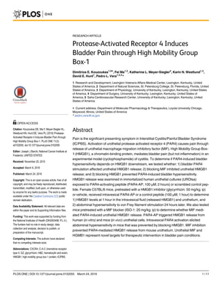
Fig 1. The flow chart demonstrates in vivo study in mice with treatments.
doi:10.1371/journal.pone.0152055.g001
PAR4 Induces Bladder Pain through HMGB1
PLOS ONE | DOI:10.1371/journal.pone.0152055 March 24, 2016 3 / 11
4. Western blotting
Culture medium from human urothelial cells (UROtsa) was diluted 1:1 with 2X Laemmli sam-
ple buffer with 10% beta-mercaptoethanol and proteins separated using a 4–15% Mini-PRO-
TEAN TGX precast polyacrylamide gel (Bio-Rad, Hercules, CA). We also measured HMGB1
levels in mouse intravesical fluid collected after intravesical instillation of PAR4-AP or control
peptide. Intravesical fluid, using equal volume across all samples, was heated to 99°C for 20
minutes in Laemmli buffer (Bio-Rad, Hercules, CA) in non-reducing, denaturing, conditions
before loading in 12% gels (Mini-PROTEAN TGX; Bio-Rad, Hercules, CA). After electropho-
resis, separated proteins were transferred to a polyvinylidene difluoride membrane. For culture
media, the blot was incubated with a secondary antibody (donkey anti-rabbit IR Dye 800 CW;
LI-COR, Lincoln, NE) at a concentration of 1:10,000 for 1 hour. The blot was washed three
times for 10 minutes and then imaged using Odyssey Imager (LI-COR). In order to test the
specificity of the antibody, antibody-blocking peptide (ab18650; Abcam, Cambridge, MA) was
made up in block buffer by mixing 10 μg of antibody with five times excess blocking peptide
(50 μg) overnight at 4°C. The primary antibody block solution was added to the blocked blot
overnight at 4°C. The secondary antibody and the imaging conditions were the same as for the
Western blot. In order to document equal protein loading in all lanes, a duplicate gel was not
transferred but just stained using Coomassie. In addition, the blot was stained right after trans-
fer with the Novex1 Reversible Protein Stain (IB7710; Thermo Sci., Rockford, IL), photo-
graphed, destained and blocked overnight.
HMGB1 protein bands in the intravesical fluid were visualized using a rabbit polyclonal pri-
mary antibody (ab18256; Abcam, Cambridge, MA; 1:4000 or 1 mg/ml for culture media), a bio-
tinylated anti-rabbit secondary antibody (Vector Labs, Burlingame, CA; 1:400), streptavidin-
HRP conjugates and chemiluminescent substrate (Pierce, Rockford, IL). Band densitometry
was performed using ImageJ (NIH, Bethesda, MD).
Histology and immunohistochemistry
Bladder paraffin sections (5 μm) were processed for routine hematoxylin and eosin (H&E)
staining or immunohistochemistry. H&E-stained sections were evaluated by a pathologist
blinded to the experimental treatment and scored for edema and inflammation according to
the following scale: 0, No edema and no infiltrating cells; 1, Mild submucosal edema and no
inflammatory cells; 2, Moderate edema and several inflammatory cells; 3, Frank edema, vascu-
lar congestion and many inflammatory cells.
For immunohistochemistry, batch-stained paraffin sections (N = 6/group) were blocked
(5% goat serum, 0.2% Triton X-100 in PBS, 30 min at room temp.), then incubated overnight
at 4°C with either rabbit polyclonal anti-HMGB1 antibody (1:100; ab18256; Abcam, Cam-
bridge, MA) or monoclonal anti-cleaved caspase-3 (as a marker for apoptosis; 1:400 Asp175,
clone 5A1E #9664; Cell Signaling Technology, Danvers, MA). Immunoreactivity was detected
with goat anti-rabbit TRITC-labeled secondary antibody (1:100 in PBS with 1% goat serum,
0.2% Triton X-100; 1 hour at room temp.; Jackson ImmunoResearch, Inc., West Grove, PA)
before cover-slipping (Vectashield, Vector Laboratories, Burlingame, CA).
Computer-assisted densitometry of HMGB1 immunostaining intensity was performed on
images captured using a LEICA DMI4000B microscope equipped with the LAS V4 program
and ImageJ (NIH, Bethesda, MD). For each bladder, intensity values represent an average of
mean grey values recorded from region-of-interests (ROIs) drawn around the urothelium in
three fields of view in the same section for comparisons between groups. Cleaved caspase-3
immunostained nuclei were counted from 3 random fields/animal.
PAR4 Induces Bladder Pain through HMGB1
PLOS ONE | DOI:10.1371/journal.pone.0152055 March 24, 2016 4 / 11
5. Statistical analyses
HMGB1 levels from human urothelial cell (UROtsa) culture media and intravesical fluid col-
lected from mouse bladders exposed to PAR-AP were analyzed using planned comparisons
(Student’s t-tests). Bonferroni-corrected one-tailed paired t-tests comparing positive response
frequency (%) at baseline to that at 24 hours after treatment evaluated significant changes in
abdominal hypersensitivity. Differences in immunofluorescence intensity were assessed in
planned comparisons (Student’s t-tests).
All data are presented as mean ± SE, with statistical differences of p 0.05 considered sig-
nificant. All statistical analyses were performed using SPSS Statistics (IBM, New York, NY).
Results
PAR4 activation induced HMGB1 release
Activation of urothelial PAR4 receptor elicited HMGB1 release in vitro and in vivo. HMGB1
western blotting of media collected from human immortalized urothelial cultures (UROtsa) 2
hours after receiving PAR4-AP revealed a single clear band whereas no band was detected in
the media of cultures receiving control peptide (Fig 2A). The specificity of HMGB1 could be
verified by the presence of a single band by HMGB1 primary antibody in the blot and con-
firmed by an absorption control experiment where pre-incubating the antibody with blocking
peptide (ab18650; Abcam, Cambridge, MA) obtained resulted in no bands detected (data not
shown). The protein loading was equal in all lanes and these were the same samples used for
the above Western blot to be served as loading controls. Commassie staining of the gel (S1 Fig)
and protein staining (Novex1 Reversible Protein Stain; Thermo Sci., Rockford, IL) of the blot
(S2 Fig) confirmed that equal amounts of protein were loaded in all lanes.
Similarly in mice, HMGB1 western blotting of intravesical fluid (using the same antibody)
collected 1 hour after bladder PAR4-AP instillation revealed a single band (at the expected
molecular weight 28 kDa) in every sample whereas only a very faint band was visible in samples
from mice receiving intravesical control peptide (Fig 2B and 2C). Immunostaining of cleaved
caspase-3, an apoptosis marker, revealed minimal expression in umbrella cells of either group,
and no difference was found between treatments (Fig 2D; Control peptide, mean = 3.0 ± 2.0;
PAR4-AP, mean = 3.9 ± 0.9 nuclei/mouse).
Fig 2. PAR4 activation elicits HMGB1 release in vitro and in vivo. Western blotting determined HMGB1
levels in culture media collected from human immortalized urothelial cultures (UROtsa) exposed to PAR4-AP
for 2 hours (A; representative blot), and in intravesical fluid collected from mice receiving intravesical
PAR4-AP for 1 hour (B). Densitometry revealed significantly more HMGB1 release from bladders receiving
intravesical PAR4-AP than those receiving control peptide (C; ** p = 0.011; Wilcoxon rank test). Caspase-3
immunostaining (arrows) revealed few labeled nuclei in each treatment group (D).
doi:10.1371/journal.pone.0152055.g002
PAR4 Induces Bladder Pain through HMGB1
PLOS ONE | DOI:10.1371/journal.pone.0152055 March 24, 2016 5 / 11
6. HMGB1 antagonist prevented PAR4-induced bladder hypersensitivity. We measured
positive responses to von Frey filaments applied to the abdominal / perineal area before and 24
hours after instilling solutions into the bladder containing a control peptide or PAR-4AP. After
24 hours, frequency of positive responses remained the same for control peptide (Fig 3A) but
increased significantly for PAR4-AP treatment (Fig 3B). PAR4-AP increased positive responses
to the firmest filament (0.070 g) from 33.8 ± 3.8% before treatment (baseline) to 66.3 ± 7.1% 24
hours after treatment (p 0.05), indicating PAR4-AP elevated abdominal mechanical sensitiv-
ity. Pretreatment with the HMGB1 antagonist, glycyrrhizin, completely prevented the PAR4--
induced hypersensitivity (Fig 3C).
We evaluated urothelial morphology, bladder edema and inflammatory infiltrates 24 hours
after intravesical administration of PAR4-AP. Urothelial morphology was normal in all three
groups and there was no evidence of inflammatory infiltrates in any of the treatment groups
(Fig 3D–3F). Subtle stromal reactive changes (submucosal fibrosis with lamina propria expan-
sion; Fig 3E; mean score = 0.75, SEM = 0.11) were noted only in six of eight mice treated with
vehicle and PAR4-AP (mean overall score = 0.75, SEM = 0.1). These changes were not present
Fig 3. HMGB1 mediates urothelial PAR4-induced mechanical hypersensitivity without inflammation.
Responses to abdominal mechanical stimulation with von Frey filaments before (baseline) and 24 hours after
each treatment are shown (A-C). Intravesical PAR4-AP (B) increases abdominal hypersensitivity over
corresponding baseline values (* p 0.025 after Bonferroni correction for multiple one-tailed paired t-tests).
HMGB1 inhibitor, glycyrrhizin (50 mg/kg, ip), abolished PAR4-mediated responses (C). HE stained paraffin
bladder sections showed normal urothelial morphology in all treatment groups (D-F). No inflammatory cells
were observed in any of the treatment groups. Submucosal fibrosis with lamina propria expansion (arrow in
panel E) was observed in animals treated with PAR4-AP and vehicle, but not in the other groups.
doi:10.1371/journal.pone.0152055.g003
PAR4 Induces Bladder Pain through HMGB1
PLOS ONE | DOI:10.1371/journal.pone.0152055 March 24, 2016 6 / 11
7. in the groups receiving control peptide or the group receiving glycyrrhizin pre-treatment and
PAR4-AP treatment (mean overall score = 0, both groups).
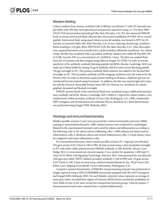
MIF antagonist prevented PAR4-induced urothelial HMGB1 intensity
decrease
Since we previously reported that MIF mediates PAR4-induced bladder hypersensitivity [7],
we investigated whether MIF mediates bladder pain by modulating HMGB1 release. Strong
HMGB1 immunofluorescence was detected in urothelial nuclei of mice treated with control
peptide (Fig 4A), whereas intravesical PAR4-AP administration reduced urothelial HMGB1
immunofluorescence (Fig 4B) after 1 hour of exposure. Pretreatment with MIF antagonist,
ISO-1, prior to intravesical PAR4 instillation, completely prevented this reduction of urothelial
HMGB1 immunofluorescence (Fig 4C). Control slides where primary antisera were omitted
had no immunofluorescence (not shown). Quantitative image analysis of the urothelia (Fig
4D) showed that PAR4-AP administration reduced urothelial HMGB1 by 41.7% compared to
controls (p 0.01), whereas ISO-1 pretreatment blocked the reduction induced by bladder
PAR4-AP infusion and actually elevated HGMB1 levels (Fig 4C and 4D; p 0.001).
Discussion
Our results demonstrate a novel finding: activation of urothelial PAR4 receptors elicits
HGMB1 release from both human and mouse urothelial cells. This conclusion is supported by
the increased levels of HMGB1 seen in the intravesical fluid of mice and in the culture media of
human urothelial cells following exposure to PAR4-AP. We also documented a decrease in
nuclear HMGB1 in the urothelium of mice after exposure to PAR4-AP which complements
the intravesical findings.
The present study, not only confirms our earlier findings [7] that PAR4 activation induced
bladder pain but also extends them by showing that such bladder pain can be prevented by
administration of a HMGB1 antagonist (glycyrrhizin). Our findings are in agreement with the
Fig 4. Urothelial PAR4-induced HMGB1 intensity decrease is mediated through MIF. Panels (A-C) show
urothelial HMGB1 immunofluorescence 1 hour after intravesical exposure to control peptide, PAR4-AP, or
PAR4-AP after pretreatment with MIF antagonist, ISO-1 (ip). White arrows identify the intravesical surface of
the urothelium. Less urothelial HMGB1 immunofluorescent labeling is apparent in PAR4-AP exposed
bladders (B) than in control peptide-treated bladders (A). Pretreatment with MIF-1 antagonist ISO-1
prevented this decrease (C). Quantitative image analysis (D) revealed that average urothelial HMGB1
immunofluorescence significantly decreased after intravesical PAR4-AP administration in comparison to
control peptide-treated animals (** p 0.01), indicating urothelial release of HMGB1. In contrast, ISO-1
pretreatment elevated urothelial HMGB1 immunofluorescence from PAR4-AP administration (***
p 0.001), suggesting MIF antagonism blocks HMGB1 release.
doi:10.1371/journal.pone.0152055.g004
PAR4 Induces Bladder Pain through HMGB1
PLOS ONE | DOI:10.1371/journal.pone.0152055 March 24, 2016 7 / 11
8. observations of Tanaka et al. [6] who reported that HGMB1 mediated pain caused by a chemi-
cal model (cyclophosphamide) of cystitis in mice.
Lastly, since we showed that PAR4 activation induced MIF release and bladder pain that
were blocked by a MIF antagonist (ISO1) [7], we tested whether MIF was upstream of HMGB1
release in our model. In fact, our current results show that a MIF antagonist (ISO1) also pre-
vents PAR4-induced urothelial HMGB1 release. Thus, the current study provides a mechanism
for our findings that PAR4 activation induces bladder pain mediated by MIF [7] by providing a
mechanism (release of urothelial HMGB1) for this pain.
Our current findings using a model that results in no urothelial damage or frank inflamma-
tion provide a mechanism for MIF-mediated pain that we reported using other models of blad-
der inflammation (cyclophosphamide cystitis) [23]. Thus, our current and past experimental
observations [7,23] indicate that MIF likely plays a pivotal role in mediating bladder pain, at
least in experimental models of bladder pain and cystitis.
Therefore, we propose that activation of urothelial PAR4 by proteases (either in the urine or
produced by local inflammatory events) is likely to elicit urothelial MIF release. Released MIF
then binds to urothelial MIF receptors [7] resulting in HMGB1 release from urothelial cells. In
turn, released HMGB1 interacts with receptors localized at nerve endings or on the urothelium
relaying signals to the nerve endings to elicit bladder pain [6,24,25].
HMGB1, is a ubiquitous nuclear non-histone DNA-binding protein that signals tissue dam-
age when passively released from cells during apoptosis [26]. Cleaved caspase-3 staining showed a
few cells stained in top layer of urothelium as a regular program death of umbrella cells in all
groups. Since no immunohistochemical difference (cleaved caspase-3 staining) was observed in the
urothelium of PAR4-AP treated mice, we consider it unlikely that PAR4-AP treatment is causing
apoptotic changes in the urothelium that account for HMGB1 release. Endogenous HMGB1 can
also be actively released from cells as a result of inflammatory stimuli (e.g. LPS, TNF) to mediate
further inflammation and also pain (for a review see Kato Svensson, 2015 [8]).
The physiological activity of HMGB1 depends on the redox-state of its 3 cysteine groups
[27]. In the fully reduced state (all-thiol), HMGB1 is a chemoattractant acting through the
receptor for advanced glycation endproducts (RAGE). In the partially reduced state (disulfide),
HGMB1 induces cytokine expression and mediates inflammation through binding with TLR4
receptor. Fully oxidized HGMB1 has no known physiological activity.
Recent evidence shows that HGMB1 can mediate pain by acting at the organ level (as is the
case in this study) [6] and also at spinal levels [28,29], but the mechanisms for inducing pain
are still being investigated. All-thiol HGMB1 mediates dorsal root ganglia neuronal excitability
in vitro through RAGE receptors while the disulfide form mediates nociception at the spinal
cord level in vivo [24]. Tanaka reported that bladder pain from cyclophosphamide injection
was prevented by systemic administration of a RAGE antagonist [6] but the HMGB1 redox
form was not investigated. Therefore, the redox form of HGMB1 that mediates bladder pain in
our model (urothelial PAR4 activation) and the receptor for HGMB1-mediated bladder pain,
remain to be investigated.
Urothelial basal and intermediate cells co-express MIF, a cytokine involved in pain and
inflammatory processes [19], along with PAR1 and PAR4 receptors [16,30]. We previously
showed that, when stimulated, PAR1 and PAR4 receptors elicit urothelial MIF release to medi-
ate additional inflammatory changes and bladder pain [7,19,23,30]. The exact mechanism
whereby MIF is acting as a nociceptive molecule is not known, but it is likely to involve MIF
binding to one of its receptors (CD74, CXCR2 or C-X-C chemokine receptor type 4 (CXCR4)
[31]). Interestingly, we previously showed that an antagonist of CXCR4 reduced bladder pain
after PAR [7]. Whether, CXCR4 antagonism or antagonism of any of the other MIF receptors
can prevent PAR4 induced HMGB1 release is not known but will be investigated.
PAR4 Induces Bladder Pain through HMGB1
PLOS ONE | DOI:10.1371/journal.pone.0152055 March 24, 2016 8 / 11
9. In summary, our study shows that bladder pain may be modulated by disrupting several dis-
tinct molecules. Preventing urothelial PAR4 from becoming activated represents the highest
point in the activation cascade sequence. Preventing the released MIF from binding one (or sev-
eral) of MIF’s receptors after PAR4 activation may prevent HMGB1 release. Finally blocking
released HMGB1 from binding to its receptors may block pain. The contribution of intracellular
signaling pathways is unclear and needs to be studied further. These events offer multiple control
points that may offer therapeutic targets to relieve bladder pain in clinical conditions such as IC/
PBS. Whether these molecules are also elevated in clinical conditions remains to be determined.
IC/PBS patients show elevated urine protease levels [17,18], which presumably may lead to
greater bladder PAR activation, and in turn, bladder hypersensitivity. Our model of intravesical
administration of PAR peptides to induce bladder pain may be replicating this process [7].
Conclusions
Intravesical stimulation of bladder PAR4 receptors induced bladder pain in this study by elicit-
ing urothelial HMGB1 release through a MIF-mediated mechanism. These findings suggest
that MIF, acting upstream of HMGB1, plays a key role in mediating bladder pain. Both mole-
cules represent novel targets for therapeutic intervention in bladder pain conditions. Future
studies will examine the contribution of specific receptors activated by MIF and HMGB1 in
mediating bladder pain.
Supporting Information
S1 Fig. Commassie-stained gel shows equivalent protein in each lane. Human epithelial cells
(UROtsa) culture media (from samples used in Fig 2A) were loaded on a gel, electrophoresed
and stained for protein using a Commassie procedure.
(JPG)
S2 Fig. Protein-stained gel shows equivalent protein in each lane. Human epithelial cells
(UROtsa) culture media (from samples used in Fig 2A) were loaded on a gel, eletrophoresed
and stained for protein.
(JPG)
S1 File. Raw data used for the analyses and figures.
(ZIP)
Acknowledgments
Judy Glass and Xiu Xu provided excellent technical assistance. This material is the result of
work supported with resources and the use of facilities at the Lexington (Kentucky) Veterans
Affairs Medical Center.
Author Contributions
Conceived and designed the experiments: DK FM KLMS PLV. Performed the experiments: DK
FM. Analyzed the data: DK FM PLV KNH DEH. Wrote the paper: DK FM KLMS KNH DEH
PLV.
References
1. Berry SH, Elliott MN, Suttorp M, Bogart LM, Stoto MA, Eggers P, et al. (2011) Prevalence of symptoms
of bladder pain syndrome/interstitial cystitis among adult females in the United States. J Urol 186: 540–
544. doi: 10.1016/j.juro.2011.03.132 PMID: 21683389
PAR4 Induces Bladder Pain through HMGB1
PLOS ONE | DOI:10.1371/journal.pone.0152055 March 24, 2016 9 / 11
10. 2. Fry CH, Daneshgari F, Thor K, Drake M, Eccles R, Kanai AJ, et al. (2010) Animal models and their use
in understanding lower urinary tract dysfunction. Neurourol Urodyn 29: 603–608. doi: 10.1002/nau.
20903 PMID: 20432320
3. Westropp JL, Buffington CA (2002) In vivo models of interstitial cystitis. J Urol 167: 694–702. PMID:
11792956
4. Guerios SD, Wang ZY, Bjorling DE (2006) Nerve growth factor mediates peripheral mechanical hyper-
sensitivity that accompanies experimental cystitis in mice. Neurosci Lett 392: 193–197. PMID:
16203088
5. Rudick CN, Billips BK, Pavlov VI, Yaggie RE, Schaeffer AJ, Klumpp DJ (2010) Host-pathogen interac-
tions mediating pain of urinary tract infection. J Infect Dis 201: 1240–1249. doi: 10.1086/651275 PMID:
20225955
6. Tanaka J, Yamaguchi K, Ishikura H, Tsubota M, Sekiguchi F, Seki Y, et al. (2014) Bladder pain relief by
HMGB1 neutralization and soluble thrombomodulin in mice with cyclophosphamide-induced cystitis.
Neuropharmacology 79: 112–118. doi: 10.1016/j.neuropharm.2013.11.003 PMID: 24262631
7. Kouzoukas DE, Meyer-Siegler KL, Ma F, Westlund KN, Hunt DE, Vera PL (2015) Macrophage Migra-
tion Inhibitory Factor Mediates PAR-Induced Bladder Pain. PLoS One 10: e0127628. doi: 10.1371/
journal.pone.0127628 PMID: 26020638
8. Kato J, Svensson CI (2015) Role of extracellular damage-associated molecular pattern molecules
(DAMPs) as mediators of persistent pain. Prog Mol Biol Transl Sci 131: 251–279. doi: 10.1016/bs.
pmbts.2014.11.014 PMID: 25744676
9. Yang H, Wang H, Czura CJ, Tracey KJ (2005) The cytokine activity of HMGB1. J Leukoc Biol 78: 1–8.
PMID: 15734795
10. Agalave NM, Svensson CI (2015) Extracellular High-Mobility Group Box 1 Protein (HMGB1) as a Medi-
ator of Persistent Pain. Mol Med 20: 569–578. doi: 10.2119/molmed.2014.00176 PMID: 25222915
11. Kokkola R, Sundberg E, Ulfgren AK, Palmblad K, Li J, Wang H, et al. (2002) High mobility group
box chromosomal protein 1: a novel proinflammatory mediator in synovitis. Arthritis Rheum 46: 2598–
2603. PMID: 12384917
12. Yasuda T, Ueda T, Takeyama Y, Shinzeki M, Sawa H, Nakajima T, et al. (2006) Significant increase of
serum high-mobility group box chromosomal protein 1 levels in patients with severe acute pancreatitis.
Pancreas 33: 359–363. PMID: 17079940
13. Tong W, Wang W, Huang J, Ren N, Wu SX, Li YQ (2010) Spinal high-mobility group box 1 contributes
to mechanical allodynia in a rat model of bone cancer pain. Biochem Biophys Res Commun 395: 572–
576. doi: 10.1016/j.bbrc.2010.04.086 PMID: 20399746
14. Shibasaki M, Sasaki M, Miura M, Mizukoshi K, Ueno H, Hashimoto S, et al. (2010) Induction of high
mobility group box-1 in dorsal root ganglion contributes to pain hypersensitivity after peripheral nerve
injury. Pain 149: 514–521. doi: 10.1016/j.pain.2010.03.023 PMID: 20392563
15. Zhao P, Metcalf M, Bunnett NW (2014) Biased signaling of protease-activated receptors. Front Endocri-
nol (Lausanne) 5: 67.
16. D'Andrea MR, Saban MR, Nguyen NB, Andrade-Gordon P, Saban R (2003) Expression of protease-
activated receptor-1, -2, -3, and -4 in control and experimentally inflamed mouse bladder. Am J Pathol
162: 907–923. PMID: 12598324
17. Boucher W, el-Mansoury M, Pang X, Sant GR, Theoharides TC (1995) Elevated mast cell tryptase in
the urine of patients with interstitial cystitis. Br J Urol 76: 94–100. PMID: 7648070
18. Kuromitsu S, Yokota H, Hiramoto M, Morita S, Mita H, Yamada T (2008) Increased concentration of
neutrophil elastase in urine from patients with interstitial cystitis. Scand J Urol Nephrol 42: 455–461.
doi: 10.1080/00365590802025881 PMID: 18609268
19. Meyer-Siegler KL, Ordorica RC, Vera PL (2004) Macrophage migration inhibitory factor is upregulated
in an endotoxin-induced model of bladder inflammation in rats. J Interferon Cytokine Res 24: 55–63.
PMID: 14980085
20. Rossi MR, Masters JR, Park S, Todd JH, Garrett SH, Sens MA, et al. (2001) The immortalized UROtsa
cell line as a potential cell culture model of human urothelium. Environ Health Perspect 109: 801–808.
PMID: 11564615
21. Mollica L, De Marchis F, Spitaleri A, Dallacosta C, Pennacchini D, Zamai M, et al. (2007) Glycyrrhizin
binds to high-mobility group box 1 protein and inhibits its cytokine activities. Chem Biol 14: 431–441.
PMID: 17462578
22. Al-Abed Y, Dabideen D, Aljabari B, Valster A, Messmer D, Ochani M, et al. (2005) ISO-1 binding to the
tautomerase active site of MIF inhibits its pro-inflammatory activity and increases survival in severe
sepsis. J Biol Chem 280: 36541–36544. PMID: 16115897
PAR4 Induces Bladder Pain through HMGB1
PLOS ONE | DOI:10.1371/journal.pone.0152055 March 24, 2016 10 / 11
11. 23. Vera PL, Iczkowski KA, Howard DJ, Jiang L, Meyer-Siegler KL (2010) Antagonism of macrophage
migration inhibitory factor decreases cyclophosphamide cystitis in mice. Neurourol Urodyn 29: 1451–
1457. doi: 10.1002/nau.20878 PMID: 20127836
24. Allette YM, Due MR, Wilson SM, Feldman P, Ripsch MS, Khanna R, et al. (2014) Identification of a
functional interaction of HMGB1 with Receptor for Advanced Glycation End-products in a model of neu-
ropathic pain. Brain Behav Immun 42: 169–177. doi: 10.1016/j.bbi.2014.06.199 PMID: 25014009
25. Roundy LM, Jia W, Zhang J, Ye X, Prestwich GD, Oottamasathien S (2013) LL-37 induced cystitis and
the receptor for advanced glycation end-products (RAGE) pathway. Adv Biosci Biotechnol 4: 1–8.
PMID: 24883227
26. Harris HE, Andersson U, Pisetsky DS (2012) HMGB1: a multifunctional alarmin driving autoimmune
and inflammatory disease. Nat Rev Rheumatol 8: 195–202. doi: 10.1038/nrrheum.2011.222 PMID:
22293756
27. Venereau E, Casalgrandi M, Schiraldi M, Antoine DJ, Cattaneo A, De Marchis F, et al. (2012) Mutually
exclusive redox forms of HMGB1 promote cell recruitment or proinflammatory cytokine release. J Exp
Med 209: 1519–1528. doi: 10.1084/jem.20120189 PMID: 22869893
28. Agalave NM, Larsson M, Abdelmoaty S, Su J, Baharpoor A, Lundback P, et al. (2014) Spinal HMGB1
induces TLR4-mediated long-lasting hypersensitivity and glial activation and regulates pain-like behav-
ior in experimental arthritis. Pain 155: 1802–1813. doi: 10.1016/j.pain.2014.06.007 PMID: 24954167
29. Wang YS, Li YY, Wang LH, Kang Y, Zhang J, Liu ZQ, et al. (2015) Tanshinone IIA Attenuates Chronic
Pancreatitis-Induced Pain in Rats via Downregulation of HMGB1 and TRL4 Expression in the Spinal
Cord. Pain Physician 18: E615–628. PMID: 26218952
30. Vera PL, Wolfe TE, Braley AE, Meyer-Siegler KL (2010) Thrombin induces macrophage migration
inhibitory factor release and upregulation in urothelium: a possible contribution to bladder inflammation.
PLoS One 5: e15904. doi: 10.1371/journal.pone.0015904 PMID: 21209875
31. Schober A, Bernhagen J, Weber C (2008) Chemokine-like functions of MIF in atherosclerosis. J Mol
Med (Berl) 86: 761–770.
PAR4 Induces Bladder Pain through HMGB1
PLOS ONE | DOI:10.1371/journal.pone.0152055 March 24, 2016 11 / 11