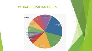
2. LEUKEMIAS
Hematological malignancies- abnormal clonal proliferation- immature cells
LYMPHOCYTIC
MYELOGENOUS
Both forms presenting in 2 forms-
A. Acute- 1. AML
M:F rate= 1:1 (5th decade) to 1:1.8 (8th decade)
Risk factor= Immunosuppression, Trisomy 21, HTLV-1, Chemicals
S/S= Fever, bleeding gums, respiratory infections, organomegaly, Lymphadenopathy,
neurological
2. ALL(80%)
Peak age= 3-7 years ( 2nd peak- >40 yrs)
Bone lesions common, mediastinal mass
5 yr survival rate
B. Chronic- CML, CLL
3. B-cell lymphoblastic
leukemia in a 9-year-old
boy (Diffuse periosteal
reaction)
Leukostasis in a 9-year-old
girl with AML
Diffuse bilateral opacities
Leukemic pulmonary
infiltration
Nodules(AML)
4. Acute myeloblastic leukemia with an orbital granulocytic
sarcoma in a 1-year-old girl with a left orbital mass
ALL in a 5-year-old girl
Axial contrast-enhanced CT scan of the orbit
shows an enlarged enhancing left optic nerve
5. Non-Hodgkin’s Lymphoma
Malignant solid tumor of immune system
Undifferentiated lymphoid cells
Spread: aggressive, diffuse,
unpredictable
Lymphoid tissue; BM and CNS infiltration
Extra-nodal
Incidence
6% childhood cancer
60% of childhood lymphomas
Peak age of 5-15; M:F ratio of 2.5:1
Examples- DLBCL
Burkitts lymphoma
Anaplastic lymphoma
Follicular
MALT
NHLwith transcalvarial infiltration in an 18-year-
old boy with a left-sided calvarial mass
6. DLBCL in 27 yr old male= proximal tibia
show poorly defined permeative lucency
Cutaneous T-cell lymphoma in a 64-year-
old man, skin surface irregularity and a
dermally based soft-tissue mass (arrow)
obliterates the fat plane between the
skin and underlying Achilles tendon.
T-cell lymphoma .
diffuse replacement of bone marrow in the
sacrum, iliac bones, and lower lumbar vertebra,
with low T1 signal intensity
Leptomeningeal seeding by non-
Hodgkin lymphoma in a 4-year-old
boy with right arm and leg paresis
7. Hodgkin’s Disease
Immune system malignancy
Involving B or T lymphocytes
Reed-Sternberg cells (CD 20+)
Spread: slow, predictable, with extension to
contiguous lymph nodes
Infiltration to non-lymphoid organs is rare
Rarely extranodal
B symptoms- Fever of >38C for 3 days,
drenching night sweats, 10% weight loss
Incidence
Hodgkin’s 14% of all lymphomas
Bimodal peaks, at 15-35 and >50; rare < 5
M:F ratio of 3:1; variation r/t geography and SES,
and increased in immunologic disorders, HIV, EBV
9. Medulloblastoma
most common malignant posterior fossa tumor
63.6% of all CNS embryonal tumors
Aged 0–19 years
M-F ratio of 1.7:1.0
Extensive desmoplastic and nodular histologic features
10-year overall survival rate
four molecular subgroups of these tumors: wingless (Wnt),
SHH, group 3, and group 4
CT= Heart / Pear shaped hyperdense midline vermian mass
Medulloblastoma in a 14-year-old girl who presented with
headache
11. ASTROCYTOMA
MC pediatric brain tumor
Half being found in posterior fossa
Cerebellar astrocytoma make up 40% of pilocytic astrocytoma
Mean age= 7 yrs old
Rarely found in <1 yr old
M:F= 2:1
Association with NF-1= indolent course
Commonly involve white matter
12. CT= Hypodense or isodense
calcification(10-20%)
1
2
MRI= T1- Hypointense
T2- Hyperintense with discrete margins
13. ASKIN TUMOR
Type of pPNET
Recently named as Ewing sarcoma of chest wall
Affects children and young adults
Large extrapulmonary invasive soft tissue masses
Arises in osseous structures – ribs, sternum, clavicle
Presents as painful warm masses
MRI = T1- iso-or hyperintense to muscle
T2- heterogenous high signal
T1C+(Gd)- heterogenous enhancement
CT= heterogenous
attenuation and areas of
cystic degeneration
14. Hepatoblastoma
Most common malignant hepatic tumor
• Majority present under 2 years
• No association with cirrhosis
• Increased risk with beckwith wiedemann syndrome,
affected siblings, familial polyposis coli and trisomy 18.
usually present with an abdominal mass
Highly vascular
Lung is the most frequent site of metastases
15. USG= single or multiple hyperechoic
masses with distortion of the adjacent
vascular architecture
CT=heterogeneous low attenuation lesion are seen with
areas of necrosis and hemorrhage and often containing
coarse calcification
16. Infantile haemangioendothelioma
Common benign hepatic mass in newborn.
May be multifocal or solitary.
Colour Doppler sonographic evaluation will show
increased flow.
USG = either hypoechoic or hyperechoic or
may have mixed echogenicity
17. CT = enhancement is typical
MRI = low signal on T1 and high
on T2 with large vascular signal
voids
18. Wilm’s tumour
Arises from the primitive metanephric epithelium
Bilateral synchrous tumours occur in 5-10%.
Nephroblastomatosis as precursor
Increased incidence sporadic aniridia,hemihypertrophy,
Beckwith-wiedemann syndorme,Drash
syndrome,Horseshoe kidney,Family history.
Mostly present as asymptomatic mass
• Abdominal pain
• Haematuria
• Fever
• Hypertension
20. Neuroblastoma
Malignant tumor of neural crest cells
Commonest extracranial solid malignant tumor
Approximately 70% originate in the abdomen of
which 2/3rd arise in adrenal,20% in the chest and
10% in the head and neck
May present as palpable abdominal mass or non
specific symptoms
50 to 60% of all neuroblastoma cases present with
metastases
21. USG=hyperechoic mass in the adrenal
or central retroperitonuem often with
flecks of calcification
CT=calcification with
low attenuation mass
22. RETINOBLASTOMA
Most common intraocular neoplasm
Curable
Bilateral (30-40% cases)
Mutation of RB gene( 55% cases), rest sporadic
Median age of D/x = 18-24 months
Most common presentation = Leukocoria
USG = multiple areas of floating debris in vitreous
CT = contrast enhanced retrolental mass with calcifications
MRI = T1- hyperintense wrt vitreous
T2- hypointense wrt vitreous
DWI- restricted diffusion
A portion of the mass is densely
calcified. Calcification is
detectable with CT in at least 80-
90% of retinoblastomas
24. Rhabdomyosarcoma
• Commonest pediatric soft tissue
sarcoma
• Pelvis most frequent site of origin
• In boys mostly arises from the prostate
or bladder base
• In girls from the urinary bladder, uterus
or vagina
• These are aggressive tumors
• Invasion of adjacent viscera and pelvic
wall
• Distant spread to lymph nodes, lung
and bone
Nasopharyngeal rhabdomyosarcoma
25. USG = shows heterogeneous
well-defined irregular mass of
low to medium echogenicity
CT = soft tissue density
some enhancement with contrast
26. Primary small cell bone neoplasm
Derived from reticulocyte- occurs in areas of red marrow;
long bones, axial skeleton
Diaphyseal- older; Metaphyseal younger
Age: 1st –2nd decade; 96% Caucasian
Medullary tumor; infiltrative; periosteal reaction-
spiculated, lamellated- many layers present