SlideShare a Scribd company logo
Iranian Journal of Immunology (IJI)
iji@sums.ac.ir
Furue M, et al. Iran J Immunol. 2019; 16(2):97-107.
https://doi.org/10.22034/iji.2019.80253.
Iran.J.Immunol. VOL.16 NO.2 June 2019 97
REVIEW ARTICLE
Pathogenesis of Atopic Dermatitis: Current
Paradigm
Masutaka Furue1,2,3*
, Dugarmaa Ulzii1
, Yen Hai Vu1
, Gaku Tsuji1
, Makiko Kido-
Nakahara1
, Takeshi Nakahara1,2
1
Department of Dermatology, Faculty of Medical Sciences, 2
Division of Skin Surface Sensing, Department
of Dermatology, Faculty of Medical Sciences, Kyushu University, 3
Research and Clinical Center for Yusho
and Dioxin, Kyushu University Hospital, Fukuoka, Japan
ABSTRACT
Atopic dermatitis (AD) is characterized by skin inflammation, barrier dysfunction and
chronic pruritus. In this review, recent advances in the pathogenesis of AD are
summarized. Clinical efficacy of the anti-IL-4 receptor antibody dupilumab implies that
type 2 cytokines IL-4 and IL-13 have pivotal roles in atopic inflammation. The expression
of IL-4 and IL-13 as well as type 2 chemokines such as CCL17, CCL22 and CCL26 is
increased in the lesional skin of AD. In addition, IL-4 and IL-13 down-regulate the
expression of filaggrin in keratinocytes and exacerbate epidermal barrier dysfunction.
Keratinocytes in barrier-disrupted epidermis produce large amounts of thymic stromal
lymphopoietin, IL-25 and IL-33, conducing to type 2 immune deviation via
OX40L/OX40 signaling. IL-31, produced by type 2 T cells, is a cardinal pruritogenic
cytokine. IL-4 and IL-13 also amplify the IL-31-mediated sensory nerve signal. These
molecules are particularly important targets for future drug development for AD.
Received: 2019-02-06, Revised: 2019-03-18, Accepted: 2019-05-25.
Citation: Furue M, Ulzii D, Vu Y H, Tsuji G, Kido-Nakahara M, Nakahara T. Pathogenesis of Atopic Dermatitis: Current Paradigm. Iran J
Immunol. 2019; 16(2):97-107. doi: 10.22034/iji.2019.80253.
Keywords: Atopic Dermatitis, IL-4, IL-13, IL-31, OX40
---------------------------------------------------------------------------------------------------------------------------------------------------------------
*Corresponding author: Dr. Masutaka Furue, Department of Dermatology, Faculty of Medical Sciences, Kyushu
University, Japan, e-mail: furue@dermatol.med.kyushu-u.ac.jp
Furue M, et al.
Iran.J.Immunol. VOL.16 NO.2 June 2019 98
INTRODUCTION
Atopic dermatitis (AD) is a common eczematous skin disorder, the prevalence or
incidence of which in the first 5 years of childhood is 10% to 16.5%. It is generally
considered to have increased worldwide, at least from the 1980s to early 2000s (1).
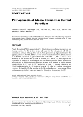
Clinical features of AD include skin inflammation, barrier disruption and chronic pruritus
(Figure 1) (2). Abnormal microbial colonization, such as Staphylococcus aureus, is also
associated with barrier dysfunction (3). The co-occurrence of autoimmune diseases is
slightly increased in patients with AD (4). Since the discovery of type 2 helper T (Th2)
cells by Mosmann et al. (5), type 2 cytokines such as interleukin (IL)-4 and IL-13 are
highlighted as major players in allergic inflammation in AD (6-9). The pathogenic
importance of IL-4 and IL-13 has recently been suggested by an excellent treatment
response of AD to the anti-IL-4 receptor α (IL4R) antibody dupilumab, which inhibits
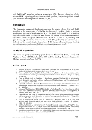
both IL-4 and IL-13 signals (Figure 2) (10).
Figure 1. Characteristics of atopic dermatitis.
Although the mechanisms responsible for barrier dysfunction in AD are multidirectional
and interconnected, research has underscored loss-of-function mutations and/or a type 2
immune response-induced decrease in filaggrin (FLG) (11-13). In addition to the genetic
loss of FLG, type 2 cytokines IL-4 and IL-13 potently inhibit FLG expression (11,14-16).
In addition, keratinocytes in the barrier-disrupted skin accelerate the type 2 immune
response by producing thymic stromal lymphopoietin (TSLP), IL-25 and IL-33, which
are type 2-associated epithelial cytokines (17). Chronic pruritus and pruritus-induced
sleep disturbances markedly deteriorate the life quality of the patients (18-23). Among
various pruritogens, atopic itch is likely to be mediated by IL-31, which is produced by
Th2 cells (24). In parallel, the anti-IL-31 receptor antibody nemolizumab potently
improves pruritus in AD patients as early as one week following its administration (Figure
2) (25,26). In this review, the focus is on type 2 immune deviation as the major driving
force of AD development.
Pathogenesis of atopic dermatitis
Iran.J.Immunol. VOL.16 NO.2 June 2019 99
Figure 2. Pathogenesis of atopic dermatitis. Barrier-disrupted keratinocytes are potent
producers of immunoregulatory cytokines such as TSLP, IL-25 and IL-33. These cytokines
induce a type 2 immune response. TSLP and IL-25 activate dendritic cells (DCs) to express
OX40L. Most allergens possess protease activity. The protease allergens cleave full-length
IL-33 to the active form and activate group 2 innate lymphoid cells (ILC2s) to express OX40L.
Ligation of OX40L/OX40 initiates type 2 immune differentiation of T cells. IL-17C is also
involved in keratinocyte proliferation and is a potential inducer of type 2 inflammation. Th2
cells produce IL-4, IL-13, IL-31 and IL-5. IL-4 and IL-13 inhibit barrier function by
downregulating FLG expression via IL-24 induction or inhibition of OVOL1 function in
keratinocytes. IL-22 induces keratinocyte proliferation and downregulates FLG expression.
IL-31 stimulates sensory nerves and mediates pruritus. IL-4 and IL-13 amplify IL-31-induced
and histamine-induced pruritus. Pruritus evokes scratching, which aggravates barrier
disruption. IL-31 also downregulates FLG expression. The barrier-disrupted epidermis is
susceptible to colonization of Staphylococcus aureus, which further worsens barrier
disruption. IL-4 and IL-13 stimulate B cells to produce IgE, which binds to mast cells and
induces their degranulation upon binding to allergens. Mast cells are a major source of
histamine. IL-4 and IL-13 upregulate the production of CCL17, CCL22 and CCL26. These
chemokines as well as IL-5 recruit Th2 cells and eosinophils. Thus, interconnected vicious
cycles develop full-blown AD. FLG; filaggrin, Th2 cell; T helper type 2 cell, TSLP; thymic
stromal lymphopoietin.
Furue M, et al.
Iran.J.Immunol. VOL.16 NO.2 June 2019 100
IL-4R Signaling Plays a Crucial Role in Atopic Dermatitis.
The anti-IL4R antibody dupilumab significantly improved skin lesions and pruritus in
patients with moderate to severe AD in two randomized, placebo-controlled clinical trials
(10). The severity of AD is authentically evaluated using Eczema Area and Severity Index
(EASI). At 16-week post-treatment, a reduction in the EASI of at least 75% was observed
in 51% and 44% of patients in the dupilumab monotherapy groups, and in only 15% and
12% of patients in placebo groups, respectively (10). Dupilumab also provides clinically
meaningful improvement in patient-reported outcome measures (27). IL-4 and IL-13 have
historically been the focus of much attention in AD. As IL4R receives signals from both
IL-4 and IL-13, the therapeutic success of dupilumab testifies to the pivotal roles of IL-4
and IL-13 in the pathogenesis of AD (Figure 2), a notion supported by a series of studies.
Gene expression of IL-4 and IL-13 is upregulated in the lesional skin of pediatric and
adult AD patients compared to that in the normal skin of healthy controls (7-9). Type 2
predominance is likely to be progressive from nonlesional to lesional skin and from acute
to chronic lesions in AD (7,8). Type 2 predominance is corroborated in peripheral blood
Th cells (28). IL-13-producing Th2 cells are significantly increased in the skin-homing
cutaneous lymphocyte antigen (CLA)+ Th cell population in both pediatric and adult AD
patients in comparison to those in the healthy controls (28). CCL17, CCL18, CCL22 and
CCL26 are type 2 chemokines overexpressed in the lesional skin of AD (6,29) (Figure 2).
CCL17, CCL18 and CCL22 are chemoattractive to Th2 cells and are mainly produced by
dendritic cells and other dermal cells activated by IL-4 and IL-13 (6,29,30). Concerning
CCL17, platelets are probably the important source (31), and serum CCL17 levels became
undetectable in a certain patient with AD comorbid with idiopathic thrombocytopenic
purpura (32). CCL26 is a potent chemotactic factor for eosinophils and is generated by
endothelial cells stimulated by IL-4 and IL-13 (6,29). The expression levels of these type
2 chemokines are normalized by dupilumab and topical steroids (29,33). Serum levels of
CCL17 and CCL22 are significantly augmented in patients with AD compared to those
in healthy controls and are associated with disease severity in AD (34,35). Interstitial
fluids contain significantly higher levels of IL-13 and CCL17 in the lesional skin of AD
patients compared to those in healthy individuals (36). Increased levels of CCL17 and
CCL22 are further reported in tape-stripped stratum corneum in AD (37). IL-5 is also a
type 2 cytokine crucial for eosinophil growth, differentiation and migration (38). Gene
expression of IL-5 is upregulated in the lesional skin of pediatric and adult AD patients
compared to the normal skin of healthy controls (7-9). Administration of the anti-IL-5
antibody mepolizumab significantly reduces circulating eosinophils; however, no
significant improvement is observed in the severity score or pruritus in patients with AD
(38). These clinical results suggest that IL-5 plays a major role in the recruitment of
eosinophils, but its pathogenic significance is limited in AD.
IL-22 Perpetuates the Chronicity of Atopic Dermatitis.
In addition to IL-4 and IL-13, the increased expression of IL-22 is associated with type 2
dominance in AD, and is also progressive from nonlesional to lesional skin and from
acute to chronic lesions (7,8). Serum levels of IL-22 are significantly correlated with
serum levels of CCL17 (39). IL-22 is a potent inducer of keratinocyte proliferation (40).
An increased number of circulating IL-22+CLA+ Th cells were detected in adult patients
but not in pediatric patients with AD (28). Notably, the anti-IL-22 antibody fezakinumab
exhibits clear efficacy for AD and is more efficacious in severe AD patients compared
with non-severe AD patients (41). Patients expressing higher baseline levels of IL-22
Pathogenesis of atopic dermatitis
Iran.J.Immunol. VOL.16 NO.2 June 2019 101
show a better treatment response to fezakinumab (42). Topical steroids and tacrolimus
are effective in downregulating the gene expression of IL-13 and IL-22 and improving
skin symptoms (33,43,44). These results underline the critical role of IL-22 in AD,
particularly in the chronic phase (41).
IL-4R Signaling Downregulates Filaggrin Expression.
Skin barrier formation is a sophisticated biochemical sequence composed of epidermal
differentiation molecules such as FLG, intercellular lipids and corneocyte adhesion (12).
The expression of FLG is downregulated in the lesional and nonlesional skin of AD
compared with the normal skin of healthy individuals (11,14). Among the 31 susceptible
gene loci reported by meta-analysis of genome-wide association studies, FLG, OVOL1
and IL13 are the top 3 significant genes associated with AD (13). The most potent risk
factors are null mutations of the FLG gene in AD (13). However, FLG mutations are not
found in all AD patients, are less common in Southern Europeans (45), and are even
absent in certain African countries (46,47), suggesting that FLG mutations only partly
explain FLG protein downregulation in AD. OVOL1 is an upstream transcription factor
for FLG expression (14,48) (Figure 2). Activation of OVOL1 induces its cytoplasmic-to-
nuclear translocation and upregulates FLG expression (14,48). Notably, type 2 cytokines
IL-4 and IL-13 consistently inhibit FLG expression by interfering with OVOL1 signaling
(11,14-16,48,49). IL-4 signaling also disrupts barrier permeability (50) and modulates E-
cadherin distribution (51). Therefore, the IL-4/IL-13-induced FLG downregulation is
likely to be more meaningful compared with the genetic mutation of FLG. IL-13 also
stimulates keratinocytes to produce IL-24, inhibiting FLG expression in autocrine and/or
paracrine fashions (52). Other cytokines, such as IL-20, IL-22, IL-25, IL-31 and IL-33,
also entail the downregulation of FLG expression (53-56); however, the molecular
mechanisms leading to FLG downregulation by these cytokines are not fully fathomed.
IL-20 and IL-24 are partly responsible for IL-31-mediated FLG downregulation (54).
Skin Barrier Dysfunction Induces Type 2 Immune Deviation by Producing TSLP,
IL-24, IL-25 and IL-33.
Epicutaneous application of picryl chloride or mite antigen on barrier-disrupted skin,
upregulates IL-4 and IgE expression in the regional lymph node, compared to
sensitization through barrier-intact skin (57). Barrier disruption by tape stripping
upregulates the expression of CCL17, CCL22 and CCL5 in epidermal cells, and induces
the recruitment of IL-4-producing cells and eosinophils (58). Current studies suggest the
crucial roles of TSLP, IL-25 and IL-33 in the type 2 immune deviation induced by barrier
dysfunction (17,59) (Figure 2). In the lesional skin of AD, the expression of TSLP, IL-25
and IL-33 is upregulated by epidermal keratinocytes (7,60,61). Tape stripped skin
expresses an increased level of TSLP (62). TSLP upregulates the expression of OX40L
in murine dendritic cells, and TSLP-treated dendritic cells induce OX40+ T cells to
produce IL-4, IL-5 and IL-13 (63,64). OX40L/OX40 interaction works as a type 2
immune checkpoint (64,65). Additionally, TSLP-treated human dendritic cells are
preferentially prime naïve T cells producing IL-4, IL-5 and IL-13 (61). In addition, TSLP
upregulates the production of CCL17 and CCL22 by human dendritic cells (61). IL-25,
also called IL-17E, is a member of the IL-17 family (66). Th2 immune responses
characterized by eosinophilia, increased serum levels of IgE, and an elevated expression
of IL-4, IL-5, and IL-13, are observed in transgenic mice that overexpress IL-25 (67).
Intranasal administration of IL-25 upregulates the expression of IL-4, IL-5, IL-13 and
Furue M, et al.
Iran.J.Immunol. VOL.16 NO.2 June 2019 102
eotaxin with marked eosinophilia in lung tissues (68). IL-25 further upregulates OX40L
in dendritic cells and promotes IL-4 and IL-13 production in T cells (69). Among other
IL-17 family members, mention can be made of IL-17C, potentially important in AD
because the anti-IL-17C antibody may be efficacious in the treatment of AD (70). IL-17C
may be related to epidermal proliferation and thickening (71). In contrast, the blockade
of the IL-17A pathways may exacerbate AD (72). IL-33 is a tissue-derived IL-1 family
cytokine which also facilitates type 2 immune responses (73). Barrier disruption by tape
stripping induces IL-33 production in keratinocytes (74). IL-33 expression by
keratinocytes is markedly augmented by herpes infection, which, more often than not,
aggravates AD and results in distinct eruptions, known as Kaposi’s varicelliform
eruptions (75). House dust mites activate keratinocytes via toll-like receptor 6 and induce
IL-25 and IL-33 production (76). Many potent allergens such as house dust mites, fungi
and pollens have intrinsic protease activity (73,77,78). Full-length IL-33 released from
epithelial cells is cleaved to the mature active form by various sorts of protease allergens,
and cleaved IL-33 generates IL-5 and IL-13 by immune cells, and recruits eosinophils
(73). Therefore, IL-33 acts as a sensitive sensor of external protease allergens (73). IL-
33, but not TSLP, upregulates OX40L in group 2 innate lymphoid cells (ILC2s) and
stimulates OX40+ T cells towards type 2 immune deviation (65). IL-25 also induces
OX40L in ILC2, but to a lesser extent compared with IL-33 (63). Mouse and human
ILC2s are phenotypically comparable, lineage negative, and non T-, non B-lymphocytes
and are high producers of IL-13 and IL-5, but not IL-4 (79,80). ILC2 resides in the skin
and is increased in number in AD lesion (80). IL-33 as well as IL-25 stimulates ILC2 and
promotes Th2 response by enhancing their release of IL-13 and IL-5 (80). In Schistosoma
mansoni infection, individual ablation of TSLP, IL-25, or IL-33 had no impact on the
development of IL-4- and IL-13-dependent inflammation or fibrosis (81). However,
simultaneous disruption of all three cytokines ameliorates the type 2 deviation (81).
Further studies are warranted to elucidate the significance and redundancy of TSLP, IL-
25 and IL-33. Notably, a clinical trial of the anti-TSLP antibody tezepelumab failed to
show the efficacy of AD (82), while the anti-IL-33 antibody ANB020 was efficacious in
all 12 AD patients in a phase 2a clinical trial (83).
Atopic Pruritus and IL-31, IL-4 and IL-13.
Pruritus is the fundamental symptom of AD (21). Histamine is virtually not a key
pruritogenic mediator of AD because the antipruritic effect of antihistamine is limited,
and is only appreciated by patients when antihistamine is used in combination with topical
steroids (84). IL-31 is also a Th2-associated cytokine (24,85) (Figure 2) whose expression
is increased in the lesional skin of AD (29). IL-31 promotes the elongation and branching
of sensory nerve fibers (86). Administration of IL-31 induces pruritus in mammals such
as mice, dogs, monkeys and humans (24). A single shot of the anti-IL-31 receptor
antibody nemolizumab significantly and rapidly reduces pruritus in patients with AD
(25,26). Monthly injection of nemolizumab effectively reduces pruritis in AD for at least
52 weeks (87). The anti-canine IL-31 antibody lokivetmab is already commercially
available in the treatment of dogs with canine AD (88). These results point to the crucial
role of IL-31 in atopic pruritus. Murine and human sensory neurons express IL-4 and IL-
13 receptors, but a simple injection of IL-4 and/or IL-13 does not induce acute itch (89).
However, IL-4 and IL-13-pretreated neurons respond to subthreshold concentrations of
histamine or IL-31, suggesting that IL-4 and IL-13 amplify histamine- and IL-31-induced
pruritus (89). Both IL-31 receptor and IL-4 receptor α activate downstream JAK1/JAK2
Pathogenesis of atopic dermatitis
Iran.J.Immunol. VOL.16 NO.2 June 2019 103
and JAK1/JAK3 signaling pathways, respectively (24). Targeted disruption of the
neuronal JAK1 signaling pathway reduces chronic pruritus, corroborating the success of
JAK inhibitors in treating chronic pruritus (89,90).
DISCUSSION
The therapeutic success of dupilumab underpins the pivotal role of IL-4 and IL-13
signaling in the pathogenesis of AD (29). Another type 2 cytokine, IL-31, is a potent
pruritogenic mediator of atopic pruritus. IL-4, IL-13 and IL-31 inhibit FLG expression
and conduce to barrier dysfunction. The vicious itch-scratch cycle further exacerbates
epidermal barrier disruption which releases TSLP, IL-25 and IL-33, initiating and
perpetuating type 2 immune deviation. The IL-4/IL-13 signal further exacerbates IL-31-
induced pruritus by reducing the threshold of IL-31. Current advances in understanding
the pathogenic mechanism may facilitate new drug development in AD.
ACKNOWLEDGEMENTS
This work was partly supported by grants from The Ministry of Health, Labour, and
Welfare in Japan (H30-Shokuhin-Shitei-005) and The Leading Advanced Projects for
Medical Innovation in Japan (LEAP).
REFERENCES
1. Williams H, Stewart A, von Mutius E, Cookson W, Anderson HR. Is eczema really on the increase
worldwide? J Allergy Clin Immunol. 2008; 121:947-954.
2. Furue M, Chiba T, Tsuji G, Ulzii D, Kido-Nakahara M, Nakahara T, et al. Atopic dermatitis:
immune deviation, barrier dysfunction, IgE autoreactivity and new therapies. Allergol Int. 2017;
66:398-403.
3. Furue M, Iida K, Imaji M, Nakahara T. Microbiome analysis of forehead skin in patients with
atopic dermatitis and healthy subjects: Implication of Staphylococcus and Corynebacterium. J
Dermatol. 2018; 45:876-877.
4. Cipriani F, Marzatico A, Ricci G. Autoimmune diseases involving skin and intestinal mucosa are
more frequent in adolescents and young adults suffering from atopic dermatitis. J Dermatol. 2017;
44:1341-1348.
5. Mosmann TR, Cherwinski H, Bond MW, Giedlin MA, Coffman RL. Two types of murine helper
T cell clone. I. Definition according to profiles of lymphokine activities and secreted proteins. J
Immunol. 1986; 136:2348-2357.
6. Esaki H, Ewald DA, Ungar B, Rozenblit M, Zheng X, Xu H, et al. Identification of novel immune
and barrier genes in atopic dermatitis by means of laser capture microdissection. J Allergy Clin
Immunol. 2015; 135:153-163.
7. Esaki H, Brunner PM, Renert-Yuval Y, Czarnowicki T, Huynh T, Tran G, et al. Early-onset
pediatric atopic dermatitis is T(H)2 but also T(H)17 polarized in skin. J Allergy Clin Immunol.
2016; 138:1639-1651.
8. Gittler JK, Shemer A, Suárez-Fariñas M, Fuentes-Duculan J, Gulewicz KJ, Wang CQ, et al.
Progressive activation of T (H)2/T(H)22 cytokines and selective epidermal proteins characterizes
acute and chronic atopic dermatitis. J Allergy Clin Immunol. 2012;130:1344-1354
9. Hamid Q, Boguniewicz M, Leung DY. Differential in situ cytokine gene expression in acute versus
chronic atopic dermatitis. J Clin Invest. 1994; 94:870-876.
Furue M, et al.
Iran.J.Immunol. VOL.16 NO.2 June 2019 104
10. Simpson EL, Bieber T, Guttman-Yassky E, Beck LA, Blauvelt A, Cork MJ, et al. Two phase 3
trials of dupilumab versus placebo in atopic dermatitis. N Engl J Med. 2016; 375:2335-2348.
11. Van den Bogaard EH, Bergboer JG, Vonk-Bergers M, van Vlijmen-Willems IM, Hato SV, van
der Valk PG, et al. Coal tar induces AHR-dependent skin barrier repair in atopic dermatitis. J Clin
Invest. 2013; 123:917-927.
12. Egawa G, Kabashima K. Barrier dysfunction in the skin allergy. Allergol Int. 2018; 67:3-11.
13. Paternoster L, Standl M, Waage J, Baurecht H, Hotze M, Strachan DP, et al. Multi-ancestry
genome-wide association study of 21,000 cases and 95,000 controls identifies new risk loci for
atopic dermatitis. Nat Genet. 2015; 47:1449-1456.
14. Tsuji G, Hashimoto-Hachiya A, Kiyomatsu-Oda M, Takemura M, Ohno F, Ito T, et al. Aryl
hydrocarbon receptor activation restores filaggrin expression via OVOL1 in atopic dermatitis. Cell
Death Dis. 2017; 8:e2931.
15. Hirano A, Goto M, Mitsui T, Hashimoto-Hachiya A, Tsuji G, Furue M. Antioxidant Artemisia
princeps extract enhances the expression of filaggrin and loricrin via the AHR/OVOL1 pathway.
Int J Mol Sci. 2017; 18:E1948.
16. Takei K, Mitoma C, Hashimoto-Hachiya A, Uchi H, Takahara M, Tsuji G, et al. Antioxidant
soybean tar Glyteer rescues T-helper-mediated downregulation of filaggrin expression via aryl
hydrocarbon receptor. J Dermatol. 2015; 42:171-180.
17. Han H, Roan F, Ziegler SF. The atopic march: current insights into skin barrier dysfunction and
epithelial cell-derived cytokines. Immunol Rev. 2017; 278:116-130.
18. Arima K, Gupta S, Gadkari A, Hiragun T, Kono T, Katayama I, et al. Burden of atopic dermatitis
in Japanese adults: Analysis of data from the 2013 National Health and Wellness Survey. J
Dermatol. 2018; 45:390-396.
19. Cai L, Kaneko S, Morita E. Changes in salivary chromogranin A levels in adults with atopic
dermatitis are correlated with changes in their condition. J Dermatol. 2018; 45:554-559.
20. Jung HJ, Bae JY, Kim JE, Na CH, Park GH, Bae YI, et al. Survey of disease awareness, treatment
behavior and treatment satisfaction in patients with atopic dermatitis in Korea: A multicenter
study. J Dermatol. 2018; 45:1172-1180.
21. Kido-Nakahara M, Furue M, Ulzii D, Nakahara T. Itch in Atopic Dermatitis. Immunol Allergy
Clin North Am. 2017; 37:113-122.
22. Takeuchi S, Oba J, Esaki H, Furue M. Non-corticosteroid adherence and itch severity influence
perception of itch in atopic dermatitis. J Dermatol. 2018; 45:158-164.
23. Wei W, Anderson P, Gadkari A, Blackburn S, Moon R, Piercy J, et al. Extent and consequences
of inadequate disease control among adults with a history of moderate to severe atopic dermatitis.
J Dermatol. 2018; 45:150-157.
24. Furue M, Yamamura K, Kido-Nakahara M, Nakahara T, Fukui Y. Emerging role of interleukin-
31 and interleukin-31 receptor in pruritus in atopic dermatitis. Allergy. 2018; 73:29-36.
25. Nemoto O, Furue M, Nakagawa H, Shiramoto M, Hanada R, Matsuki S, et al. The first trial of
CIM331, a humanized antihuman interleukin-31 receptor A antibody, in healthy volunteers and
patients with atopic dermatitis to evaluate safety, tolerability and pharmacokinetics of a single
dose in a randomized, double-blind, placebo-controlled study. Br J Dermatol. 2016; 174:296-304.
26. Ruzicka T, Hanifin JM, Furue M, Pulka G, Mlynarczyk I, Wollenberg A, et al. Anti-interleukin-
31 receptor A antibody for atopic dermatitis. N Engl J Med. 2017; 376:826-835.
27. Simpson EL, Gadkari A, Worm M, Soong W, Blauvelt A, Eckert L, et al. Dupilumab therapy
provides clinically meaningful improvement in patient-reported outcomes (PROs): A phase IIb,
randomized, placebo-controlled, clinical trial in adult patients with moderate to severe atopic
dermatitis (AD). J Am Acad Dermatol. 2016; 75:506-515.
28. Czarnowicki T, Esaki H, Gonzalez J, Malajian D, Shemer A, Noda S, et al. Early pediatric atopic
dermatitis shows only a cutaneous lymphocyte antigen (CLA)(+) TH2/TH1 cell imbalance,
whereas adults acquire CLA(+) TH22/TC22 cell subsets. J Allergy Clin Immunol. 2015; 136:941-
951.
29. Guttman-Yassky E, Bissonnette R, Ungar B, Suárez-Fariñas M, Ardeleanu M, Esaki H, et al.
Dupilumab progressively improves systemic and cutaneous abnormalities in atopic dermatitis
patients. J Allergy Clin Immunol. 2019; 143:155-172.
30. Takemura M, Nakahara T, Hashimoto-Hachiya A, Furue M, Tsuji G. Glyteer, soybean tar, impairs
IL-4/Stat6 signaling in murine bone marrow-derived dendritic cells: The basis of its therapeutic
effect on atopic dermatitis. Int J Mol Sci. 2018; 19: E1169.
Pathogenesis of atopic dermatitis
Iran.J.Immunol. VOL.16 NO.2 June 2019 105
31. Fujisawa T, Fujisawa R, Kato Y, Nakayama T, Morita A, Katsumata H, et al. Presence of high
contents of thymus and activation-regulated chemokine in platelets and elevated plasma levels of
thymus and activation-regulated chemokine and macrophage-derived chemokine in patients with
atopic dermatitis. J Allergy Clin Immunol. 2002; 110:139-146.
32. Ozawa M, Sasahara Y, Aiba S. Case of atopic dermatitis concurrent with idiopathic
thrombocytopenic purpura, whose serum thymus and activation-regulated chemokine level
remained undetectable. J Dermatol. 2018; 45:606-608.
33. Guttman-Yassky E, Ungar B, Malik K, Dickstein D, Suprun M, Estrada YD, et al. Molecular
signatures order the potency of topically applied anti-inflammatory drugs in patients with atopic
dermatitis. J Allergy Clin Immunol. 2017; 140:1032-1042.
34. Kakinuma T, Nakamura K, Wakugawa M, Mitsui H, Tada Y, Saeki H, et al. Thymus and
activation-regulated chemokine in atopic dermatitis: Serum thymus and activation-regulated
chemokine level is closely related with disease activity. J Allergy Clin Immunol. 2001; 107:535-
541.
35. Kakinuma T, Nakamura K, Wakugawa M, Mitsui H, Tada Y, Saeki H, et al. Serum macrophage-
derived chemokine (MDC) levels are closely related with the disease activity of atopic dermatitis.
Clin Exp Immunol. 2002; 127:270-273.
36. Szegedi K, Lutter R, Res PC, Bos JD, Luiten RM, Kezic S, et al. Cytokine profiles in interstitial
fluid from chronic atopic dermatitis skin. J Eur Acad Dermatol Venereol. 2015; 29:2136-2144.
37. Hulshof L, Hack DP, Hasnoe QCJ, Dontje B, Jakasa I, Riethmüller C, et al. A minimally invasive
tool to study immune response and skin barrier in children with atopic dermatitis. Br J Dermatol.
2019; 180:621-630.
38. Oldhoff JM, Darsow U, Werfel T, Katzer K, Wulf A, Laifaoui J, et al. Anti-IL-5 recombinant
humanized monoclonal antibody (mepolizumab) for the treatment of atopic dermatitis. Allergy.
2005; 60:693-696.
39. Hayashida S, Uchi H, Takeuchi S, Esaki H, Moroi Y, Furue M. Significant correlation of serum
IL-22 levels with CCL17 levels in atopic dermatitis. J Dermatol Sci. 2011; 61:78-79.
40. Mitra A, Raychaudhuri SK, Raychaudhuri SP. IL-22 induced cell proliferation is regulated by
PI3K/Akt/mTOR signaling cascade. Cytokine. 2012; 60:38-42.
41. Guttman-Yassky E, Brunner PM, Neumann AU, Khattri S, Pavel AB, Malik K, et al. Efficacy and
safety of fezakinumab (an IL-22 monoclonal antibody) in adults with moderate-to-severe atopic
dermatitis inadequately controlled by conventional treatments: A randomized, double-blind, phase
2a trial. J Am Acad Dermatol. 2018; 78:872-881.
42. Brunner PM, Pavel AB, Khattri S, Leonard A, Malik K, Rose S, et al. Baseline IL22 expression
in atopic dermatitis patients stratifies tissue responses to fezakinumab. J Allergy Clin Immunol.
2019; 143:142-154.
43. Nakahara T, Morimoto H, Murakami N, Furue M. Mechanistic insights into topical tacrolimus for
the treatment of atopic dermatitis. Pediatr Allergy Immunol. 2018; 29:233-238.
44. Ohtsuki M, Morimoto H, Nakagawa H. Tacrolimus ointment for the treatment of adult and
pediatric atopic dermatitis: Review on safety and benefits. J Dermatol. 2018; 45:936-942.
45. Cascella R, Foti Cuzzola V, Lepre T, Galli E, Moschese V, et al. Full sequencing of the FLG gene
in Italian patients with atopic eczema: evidence of new mutations, but lack of an association. J
Invest Dermatol. 2011; 131:982-984.
46. Thawer-Esmail F, Jakasa I, Todd G, Wen Y, Brown SJ, Kroboth K, et al. South African amaXhosa
patients with atopic dermatitis have decreased levels of filaggrin breakdown products but no loss-
of-function mutations in filaggrin. J Allergy Clin Immunol. 2014; 133:280-282.
47. Winge MC, Bilcha KD, Liedén A, Shibeshi D, Sandilands A, Wahlgren CF, et al. Novel filaggrin
mutation but no other loss-of-function variants found in Ethiopian patients with atopic dermatitis.
Br J Dermatol. 2011; 165:1074-1080.
48. Hashimoto-Hachiya A, Tsuji G, Murai M, Yan X, Furue M. Upregulation of FLG, LOR, and IVL
expression by Rhodiola crenulata root extract via aryl hydrocarbon receptor: Differential
involvement of OVOL1. Int J Mol Sci. 2018; 19: E1654.
49. Tsuji G, Ito T, Chiba T, Mitoma C, Nakahara T, Uchi H, et al. The role of the OVOL1-OVOL2
axis in normal and diseased human skin. J Dermatol Sci. 2018; 90:227-231.
50. Kobayashi J, Inai T, Morita K, Moroi Y, Urabe K, Shibata Y, et al. Reciprocal regulation of
permeability through a cultured keratinocyte sheet by IFN-gamma and IL-4. Cytokine. 2004;
28:186-189.
Furue M, et al.
Iran.J.Immunol. VOL.16 NO.2 June 2019 106
51. Fujii-Maeda S, Kajiwara K, Ikizawa K, Shinazawa M, Yu B, Koga T, et al. Reciprocal regulation
of thymus and activation-regulated chemokine/macrophage-derived chemokine production by
interleukin (IL)-4/IL-13 and interferon-gamma in HaCaT keratinocytes is mediated by alternations
in E-cadherin distribution. J Invest Dermatol. 2004; 122:20-28.
52. Mitamura Y, Nunomura S, Nanri Y, Ogawa M, Yoshihara T, Masuoka M, et al. The IL-
13/periostin/IL-24 pathway causes epidermal barrier dysfunction in allergic skin inflammation.
Allergy. 2018; 73:1881-1891.
53. Gutowska-Owsiak D, Schaupp AL, Salimi M, Taylor S, Ogg GS. Interleukin-22 downregulates
filaggrin expression and affects expression of profilaggrin processing enzymes. Br J Dermatol.
2011; 165:492-498.
54. Cornelissen C, Marquardt Y, Czaja K, Wenzel J, Frank J, Lüscher-Firzlaff J, et al. IL-31 regulates
differentiation and filaggrin expression in human organotypic skin models. J Allergy Clin
Immunol. 2012; 129:426-433.
55. Hvid M, Vestergaard C, Kemp K, Christensen GB, Deleuran B, et al. IL-25 in atopic dermatitis: a
possible link between inflammation and skin barrier dysfunction? J Invest Dermatol. 2011;
131:150-157.
56. Seltmann J, Roesner LM, von Hesler FW, Wittmann M, Werfel T. IL-33 impacts on the skin
barrier by downregulating the expression of filaggrin. J Allergy Clin Immunol. 2015; 135:1659-
1661.
57. Kondo H, Ichikawa Y, Imokawa G. Percutaneous sensitization with allergens through barrier-
disrupted skin elicits a Th2-dominant cytokine response. Eur J Immunol. 1998; 28:769-779.
58. Onoue A, Kabashima K, Kobayashi M, Mori T, Tokura Y. Induction of eosinophil- and Th2-
attracting epidermal chemokines and cutaneous late-phase reaction in tape-stripped skin. Exp
Dermatol. 2009; 18:1036-1043.
59. Hammad H, Lambrecht BN. Barrier epithelial cells and the control of type 2 immunity. Immunity.
2015; 43:29-40.
60. Aktar MK, Kido-Nakahara M, Furue M, Nakahara T. Mutual upregulation of endothelin-1 and IL-
25 in atopic dermatitis. Allergy. 2015; 70:846-854.
61. Soumelis V, Reche PA, Kanzler H, Yuan W, Edward G, Homey B, et al. Human epithelial cells
trigger dendritic cell mediated allergic inflammation by producing TSLP. Nat Immunol. 2002;
3:673-680.
62. Oyoshi MK, Larson RP, Ziegler SF, Geha RS. Mechanical injury polarizes skin dendritic cells to
elicit a T(H)2 response by inducing cutaneous thymic stromal lymphopoietin expression. J Allergy
Clin Immunol. 2010; 126:976-984.
63. Gilliet M, Soumelis V, Watanabe N, Hanabuchi S, Antonenko S, de Waal-Malefyt R, et al. Human
dendritic cells activated by TSLP and CD40L induce proallergic cytotoxic T cells. J Exp Med.
2003; 197:1059-1063.
64. Ito T, Wang YH, Duramad O, Hori T, Delespesse GJ, Watanabe N, et al. TSLP-activated dendritic
cells induce an inflammatory T helper type 2 cell response through OX40 ligand. J Exp Med.
2005; 202:1213-1223.
65. Halim TYF, Rana BMJ, Walker JA, Kerscher B, Knolle MD, Jolin HE, et al. Tissue-restricted
adaptive type 2 immunity is orchestrated by expression of the costimulatory molecule OX40L on
group 2 innate lymphoid cells. Immunity. 2018; 48:1195-1207.
66. Kolls JK, Lindén A. Interleukin-17 family members and inflammation. Immunity. 2004; 21:467-
476.
67. Pan G, French D, Mao W, Maruoka M, Risser P, Lee J, et al. Forced expression of murine IL-17E
induces growth retardation, jaundice, a Th2-biased response, and multiorgan inflammation in
mice. J Immunol. 2001; 167:6559-6567.
68. Hurst SD, Muchamuel T, Gorman DM, Gilbert JM, Clifford T, Kwan S, et al. New IL-17 family
members promote Th1 or Th2 responses in the lung: in vivo function of the novel cytokine IL-25.
J Immunol. 2002; 169:443-453.
69. Zheng R, Chen FH, Gao WX, Wang D, Yang QT, Wang K, et al. The T(H)2-polarizing function
of atopic interleukin 17 receptor B-positive dendritic cells up-regulated by lipopolysaccharide.
Ann Allergy Asthma Immunol. 2017; 118:474-482.
70. Guttman-Yassky E, Krueger JG. IL-17C: A unique epithelial cytokine with potential for targeting
across the spectrum of atopic dermatitis and psoriasis. J Invest Dermatol. 2018; 138:1467-1469.
71. Ramirez-Carrozzi V, Sambandam A, Luis E, Lin Z, Jeet S, Lesch J, et al. IL-17C regulates the
Pathogenesis of atopic dermatitis
Iran.J.Immunol. VOL.16 NO.2 June 2019 107
innate immune function of epithelial cells in an autocrine manner. Nat Immunol. 2011; 12:1159-
1166.
72. Ishiuji Y, Umezawa Y, Asahina A, Fukuta H, Aizawa N, Yanaba K, et al. Exacerbation of atopic
dermatitis symptoms by ustekinumab in psoriatic patients with elevated serum immunoglobulin E
levels: Report of two cases. J Dermatol. 2018; 45:732-734.
73. Cayrol C, Duval A, Schmitt P, Roga S, Camus M, Stella A, et al. Environmental allergens induce
allergic inflammation through proteolytic maturation of IL-33. Nat Immunol. 2018; 19:375-385.
74. Dickel H, Gambichler T, Kamphowe J, Altmeyer P, Skrygan M. Standardized tape stripping prior
to patch testing induces upregulation of Hsp90, Hsp70, IL-33, TNF-α and IL-8/CXCL8 mRNA:
new insights into the involvement of 'alarmins'. Contact Dermatitis. 2010; 63:215-222.
75. Jin M, Komine M, Tsuda H, Oshio T, Ohtsuki M. Interleukin-33 is expressed in the lesional
epidermis in herpes virus infection but not in verruca vulgaris. J Dermatol. 2018; 45:855-857.
76. Jang YH, Choi JK, Jin M, Choi YA, Ryoo ZY, Lee HS, et al. House dust mite increases pro-Th2
cytokines IL-25 and IL-33 via the activation of TLR1/6 signaling. J Invest Dermatol. 2017;
137:2354-2261.
77. Palm NW, Rosenstein RK, Medzhitov R. Allergic host defences. Nature. 2012; 484:465-472.
78. Sokol CL, Barton GM, Farr AG, Medzhitov R. A mechanism for the initiation of allergen-induced
T helper type 2 responses. Nat Immunol. 2008; 9:310-318.
79. Hurrell BP, Shafiei Jahani P, Akbari O. Social Networking of Group Two Innate Lymphoid Cells
in Allergy and Asthma. Front Immunol. 2018; 9:2694.
80. Salimi M, Barlow JL, Saunders SP, Xue L, Gutowska-Owsiak D, Wang X, et al. A role for IL-25
and IL-33-driven type-2 innate lymphoid cells in atopic dermatitis. J Exp Med. 2013; 210:2939-
2950.
81. Vannella KM, Ramalingam TR, Borthwick LA, Barron L, Hart KM, Thompson RW, et al.
Combinatorial targeting of TSLP, IL-25, and IL-33 in type 2 cytokine-driven inflammation and
fibrosis. Sci Transl Med. 2016; 8:337ra65.
82. https://www.fiercebiotech.com/biotech/after-asthma-success-astrazeneca-and-amgen-stezepelu-
mab-misses-atopic-dermatitis?utm_source=internal&utm_medium=rss.
83. https://globenewswire.com/news-release/2018/02/17/1361150/0/en/AnaptysBio-Presents-Updat-
ed-Data-from-ANB020-Phase-2a-Atopic-Dermatitis-Trial-at-AAD-Annual-Meeting.html.
84. Kawashima M, Tango T, Noguchi T, Inagi M, Nakagawa H, Harada S. Addition of fexofenadine
to a topical corticosteroid reduces the pruritus associated with atopic dermatitis in a 1-week
randomized, multicentre, double-blind, placebo-controlled, parallel-group study. Br J Dermatol.
2003; 148:1212-1221.
85. Yamamura K, Uruno T, Shiraishi A, Tanaka Y, Ushijima M, Nakahara T, et al. The transcription
factor EPAS1 links DOCK8 deficiency to atopic skin inflammation via IL-31 induction. Nat
Commun. 2017; 8:13946.
86. Feld M, Garcia R, Buddenkotte J, Katayama S, Lewis K, Muirhead G, et al. The pruritus- and
TH2-associated cytokine IL-31 promotes growth of sensory nerves. J Allergy Clin Immunol. 2016;
138:500-508.
87. Kabashima K, Furue M, Hanifin JM, Pulka G, Wollenberg A, Galus R, et al. Nemolizumab in
patients with moderate-to-severe atopic dermatitis: Randomized, phase II, long-term extension
study. J Allergy Clin Immunol. 2018; 142:1121-1130.
88. Souza CP, Rosychuk RAW, Contreras ET, Schissler JR, Simpson AC. A retrospective analysis
of the use of lokivetmab in the management of allergic pruritus in a referral population of 135
dogs in the western USA. Vet. Dermatol. 2018; 29:489-e164.
89. Oetjen LK, Mack MR, Feng J, Whelan TM, Niu H, Guo CJ, et al. Sensory neurons co-opt classical
immune signaling pathways to mediate chronic itch. Cell. 2017; 171:217-228.
90. Nakagawa H, Nemoto O, Yamada H, Nagata T, Ninomiya N. Phase 1 studies to assess the safety,
tolerability and pharmacokinetics of JTE-052 (a novel Janus kinase inhibitor) ointment in Japanese
healthy volunteers and patients with atopic dermatitis. J Dermatol. 2018; 45:701-709.

More Related Content

Similar to pathogenesis dermatitis atopik.pdf

The Negative Clinical Consequences Due to the Lack of the Elaboration of a Sc...
The Negative Clinical Consequences Due to the Lack of the Elaboration of a Sc...The Negative Clinical Consequences Due to the Lack of the Elaboration of a Sc...
The Negative Clinical Consequences Due to the Lack of the Elaboration of a Sc...
asclepiuspdfs
 
IMMUNITY AGAINST HELMINTHS
IMMUNITY AGAINST HELMINTHSIMMUNITY AGAINST HELMINTHS
IMMUNITY AGAINST HELMINTHS
Dr. Aquil Mohmad
 
Communicable Disease_ Review Malaria paper
Communicable Disease_ Review Malaria paperCommunicable Disease_ Review Malaria paper
Communicable Disease_ Review Malaria paperThant Zin
 
vascular insulin and IGF-1 in diabetic wounds
vascular insulin and IGF-1 in diabetic woundsvascular insulin and IGF-1 in diabetic wounds
vascular insulin and IGF-1 in diabetic woundsSaeed Aghdam
 
The biology of innate lymphoid cells
The biology of innate lymphoid cellsThe biology of innate lymphoid cells
The biology of innate lymphoid cells
Luis Miguel Rodríguez Torres
 
Cytokines in diseases
Cytokines in diseasesCytokines in diseases
Cytokines in diseases
Dr.M.Prasad Naidu
 
Cytokine and COVID19 A Literature Review
Cytokine and COVID19 A Literature ReviewCytokine and COVID19 A Literature Review
Cytokine and COVID19 A Literature Review
ijtsrd
 
cytokine-class.ppt
cytokine-class.pptcytokine-class.ppt
cytokine-class.ppt
JenniferSZiegen
 
Non Specific Immune Defense
Non Specific Immune DefenseNon Specific Immune Defense
Non Specific Immune Defenselevouge777
 
Apps covid 19 immunological and toxicological implication innate immune senso...
Apps covid 19 immunological and toxicological implication innate immune senso...Apps covid 19 immunological and toxicological implication innate immune senso...
Apps covid 19 immunological and toxicological implication innate immune senso...
M. Luisetto Pharm.D.Spec. Pharmacology
 
Clinical aspects and molecular diagnostics of skin aging
Clinical aspects and molecular diagnostics of skin agingClinical aspects and molecular diagnostics of skin aging
Clinical aspects and molecular diagnostics of skin agingM.YOUSRY Abdel-Mawla
 
Chitin gama delta linfocitos
Chitin gama delta linfocitosChitin gama delta linfocitos
Chitin gama delta linfocitos
UFRJ
 
joURNAL READING BIOMARKER.pptx
joURNAL READING BIOMARKER.pptxjoURNAL READING BIOMARKER.pptx
joURNAL READING BIOMARKER.pptx
joganks1
 
Immunity & Inflammation
Immunity & InflammationImmunity & Inflammation
Immunity & Inflammation
Dr. Mariyam Momin
 
COVID-19 treatment
COVID-19 treatmentCOVID-19 treatment
COVID-19 treatment
MdNazmulIslamTanmoy
 
In vivo effects of Interleukin 2 on Lymphocyte Subpopulations in a Patient wi...
In vivo effects of Interleukin 2 on Lymphocyte Subpopulations in a Patient wi...In vivo effects of Interleukin 2 on Lymphocyte Subpopulations in a Patient wi...
In vivo effects of Interleukin 2 on Lymphocyte Subpopulations in a Patient wi...
Elke Stein
 
Immunology Pathway of During Autoimmune Disease: A Review Article
Immunology Pathway of During Autoimmune Disease: A Review ArticleImmunology Pathway of During Autoimmune Disease: A Review Article
Immunology Pathway of During Autoimmune Disease: A Review Article
komalicarol
 
Lary nel abao nc-nga mice revised report
Lary nel abao nc-nga mice revised reportLary nel abao nc-nga mice revised report
Lary nel abao nc-nga mice revised report
Obihiro University of Agriculture and Veterinary Medicine
 
Cytokines
CytokinesCytokines
Cytokines
jyoti arora
 
Cytokines in diseases
Cytokines in diseasesCytokines in diseases
Cytokines in diseases
Dr.M.Prasad Naidu
 

Similar to pathogenesis dermatitis atopik.pdf (20)

The Negative Clinical Consequences Due to the Lack of the Elaboration of a Sc...
The Negative Clinical Consequences Due to the Lack of the Elaboration of a Sc...The Negative Clinical Consequences Due to the Lack of the Elaboration of a Sc...
The Negative Clinical Consequences Due to the Lack of the Elaboration of a Sc...
 
IMMUNITY AGAINST HELMINTHS
IMMUNITY AGAINST HELMINTHSIMMUNITY AGAINST HELMINTHS
IMMUNITY AGAINST HELMINTHS
 
Communicable Disease_ Review Malaria paper
Communicable Disease_ Review Malaria paperCommunicable Disease_ Review Malaria paper
Communicable Disease_ Review Malaria paper
 
vascular insulin and IGF-1 in diabetic wounds
vascular insulin and IGF-1 in diabetic woundsvascular insulin and IGF-1 in diabetic wounds
vascular insulin and IGF-1 in diabetic wounds
 
The biology of innate lymphoid cells
The biology of innate lymphoid cellsThe biology of innate lymphoid cells
The biology of innate lymphoid cells
 
Cytokines in diseases
Cytokines in diseasesCytokines in diseases
Cytokines in diseases
 
Cytokine and COVID19 A Literature Review
Cytokine and COVID19 A Literature ReviewCytokine and COVID19 A Literature Review
Cytokine and COVID19 A Literature Review
 
cytokine-class.ppt
cytokine-class.pptcytokine-class.ppt
cytokine-class.ppt
 
Non Specific Immune Defense
Non Specific Immune DefenseNon Specific Immune Defense
Non Specific Immune Defense
 
Apps covid 19 immunological and toxicological implication innate immune senso...
Apps covid 19 immunological and toxicological implication innate immune senso...Apps covid 19 immunological and toxicological implication innate immune senso...
Apps covid 19 immunological and toxicological implication innate immune senso...
 
Clinical aspects and molecular diagnostics of skin aging
Clinical aspects and molecular diagnostics of skin agingClinical aspects and molecular diagnostics of skin aging
Clinical aspects and molecular diagnostics of skin aging
 
Chitin gama delta linfocitos
Chitin gama delta linfocitosChitin gama delta linfocitos
Chitin gama delta linfocitos
 
joURNAL READING BIOMARKER.pptx
joURNAL READING BIOMARKER.pptxjoURNAL READING BIOMARKER.pptx
joURNAL READING BIOMARKER.pptx
 
Immunity & Inflammation
Immunity & InflammationImmunity & Inflammation
Immunity & Inflammation
 
COVID-19 treatment
COVID-19 treatmentCOVID-19 treatment
COVID-19 treatment
 
In vivo effects of Interleukin 2 on Lymphocyte Subpopulations in a Patient wi...
In vivo effects of Interleukin 2 on Lymphocyte Subpopulations in a Patient wi...In vivo effects of Interleukin 2 on Lymphocyte Subpopulations in a Patient wi...
In vivo effects of Interleukin 2 on Lymphocyte Subpopulations in a Patient wi...
 
Immunology Pathway of During Autoimmune Disease: A Review Article
Immunology Pathway of During Autoimmune Disease: A Review ArticleImmunology Pathway of During Autoimmune Disease: A Review Article
Immunology Pathway of During Autoimmune Disease: A Review Article
 
Lary nel abao nc-nga mice revised report
Lary nel abao nc-nga mice revised reportLary nel abao nc-nga mice revised report
Lary nel abao nc-nga mice revised report
 
Cytokines
CytokinesCytokines
Cytokines
 
Cytokines in diseases
Cytokines in diseasesCytokines in diseases
Cytokines in diseases
 

More from warisanuhurridha2

Modul Anatomi Kulit Terkait Bedah Skalpel_K1.2.3.docx
Modul Anatomi Kulit Terkait Bedah Skalpel_K1.2.3.docxModul Anatomi Kulit Terkait Bedah Skalpel_K1.2.3.docx
Modul Anatomi Kulit Terkait Bedah Skalpel_K1.2.3.docx
warisanuhurridha2
 
tranexamid acid for angioedema.pdf
tranexamid acid for angioedema.pdftranexamid acid for angioedema.pdf
tranexamid acid for angioedema.pdf
warisanuhurridha2
 
Omalizumab 4_230724_120258.pdf
Omalizumab 4_230724_120258.pdfOmalizumab 4_230724_120258.pdf
Omalizumab 4_230724_120258.pdf
warisanuhurridha2
 
Fitz Urticaria & Angioedema (wecompress.com).docx
Fitz Urticaria & Angioedema (wecompress.com).docxFitz Urticaria & Angioedema (wecompress.com).docx
Fitz Urticaria & Angioedema (wecompress.com).docx
warisanuhurridha2
 
recurrent angioedema.pptx
recurrent angioedema.pptxrecurrent angioedema.pptx
recurrent angioedema.pptx
warisanuhurridha2
 
tugas DERMATITIS ATOPIK.pptx
tugas DERMATITIS ATOPIK.pptxtugas DERMATITIS ATOPIK.pptx
tugas DERMATITIS ATOPIK.pptx
warisanuhurridha2
 
CPC Morbus Hansen Indeterminate.pptx
CPC Morbus Hansen Indeterminate.pptxCPC Morbus Hansen Indeterminate.pptx
CPC Morbus Hansen Indeterminate.pptx
warisanuhurridha2
 
After thank you Refractory Dermatitis Contributed by Pityriasis Versicolor.pptx
After thank you Refractory Dermatitis Contributed by Pityriasis Versicolor.pptxAfter thank you Refractory Dermatitis Contributed by Pityriasis Versicolor.pptx
After thank you Refractory Dermatitis Contributed by Pityriasis Versicolor.pptx
warisanuhurridha2
 
treatment EN.pdf
treatment EN.pdftreatment EN.pdf
treatment EN.pdf
warisanuhurridha2
 
admin,+1+Gambaran+Dermoskopik+Pasien+Alopesia.pdf
admin,+1+Gambaran+Dermoskopik+Pasien+Alopesia.pdfadmin,+1+Gambaran+Dermoskopik+Pasien+Alopesia.pdf
admin,+1+Gambaran+Dermoskopik+Pasien+Alopesia.pdf
warisanuhurridha2
 
02 INSTRUMEN SUPERVISI SD SMP SMA SMK ok.pdf
02 INSTRUMEN SUPERVISI SD SMP SMA SMK ok.pdf02 INSTRUMEN SUPERVISI SD SMP SMA SMK ok.pdf
02 INSTRUMEN SUPERVISI SD SMP SMA SMK ok.pdf
warisanuhurridha2
 
Fitz Pioderma Translate .docx
Fitz Pioderma Translate .docxFitz Pioderma Translate .docx
Fitz Pioderma Translate .docx
warisanuhurridha2
 
att lapsus pedi.pptx
att lapsus pedi.pptxatt lapsus pedi.pptx
att lapsus pedi.pptx
warisanuhurridha2
 

More from warisanuhurridha2 (13)

Modul Anatomi Kulit Terkait Bedah Skalpel_K1.2.3.docx
Modul Anatomi Kulit Terkait Bedah Skalpel_K1.2.3.docxModul Anatomi Kulit Terkait Bedah Skalpel_K1.2.3.docx
Modul Anatomi Kulit Terkait Bedah Skalpel_K1.2.3.docx
 
tranexamid acid for angioedema.pdf
tranexamid acid for angioedema.pdftranexamid acid for angioedema.pdf
tranexamid acid for angioedema.pdf
 
Omalizumab 4_230724_120258.pdf
Omalizumab 4_230724_120258.pdfOmalizumab 4_230724_120258.pdf
Omalizumab 4_230724_120258.pdf
 
Fitz Urticaria & Angioedema (wecompress.com).docx
Fitz Urticaria & Angioedema (wecompress.com).docxFitz Urticaria & Angioedema (wecompress.com).docx
Fitz Urticaria & Angioedema (wecompress.com).docx
 
recurrent angioedema.pptx
recurrent angioedema.pptxrecurrent angioedema.pptx
recurrent angioedema.pptx
 
tugas DERMATITIS ATOPIK.pptx
tugas DERMATITIS ATOPIK.pptxtugas DERMATITIS ATOPIK.pptx
tugas DERMATITIS ATOPIK.pptx
 
CPC Morbus Hansen Indeterminate.pptx
CPC Morbus Hansen Indeterminate.pptxCPC Morbus Hansen Indeterminate.pptx
CPC Morbus Hansen Indeterminate.pptx
 
After thank you Refractory Dermatitis Contributed by Pityriasis Versicolor.pptx
After thank you Refractory Dermatitis Contributed by Pityriasis Versicolor.pptxAfter thank you Refractory Dermatitis Contributed by Pityriasis Versicolor.pptx
After thank you Refractory Dermatitis Contributed by Pityriasis Versicolor.pptx
 
treatment EN.pdf
treatment EN.pdftreatment EN.pdf
treatment EN.pdf
 
admin,+1+Gambaran+Dermoskopik+Pasien+Alopesia.pdf
admin,+1+Gambaran+Dermoskopik+Pasien+Alopesia.pdfadmin,+1+Gambaran+Dermoskopik+Pasien+Alopesia.pdf
admin,+1+Gambaran+Dermoskopik+Pasien+Alopesia.pdf
 
02 INSTRUMEN SUPERVISI SD SMP SMA SMK ok.pdf
02 INSTRUMEN SUPERVISI SD SMP SMA SMK ok.pdf02 INSTRUMEN SUPERVISI SD SMP SMA SMK ok.pdf
02 INSTRUMEN SUPERVISI SD SMP SMA SMK ok.pdf
 
Fitz Pioderma Translate .docx
Fitz Pioderma Translate .docxFitz Pioderma Translate .docx
Fitz Pioderma Translate .docx
 
att lapsus pedi.pptx
att lapsus pedi.pptxatt lapsus pedi.pptx
att lapsus pedi.pptx
 

Recently uploaded

R3 Stem Cells and Kidney Repair A New Horizon in Nephrology.pptx
R3 Stem Cells and Kidney Repair A New Horizon in Nephrology.pptxR3 Stem Cells and Kidney Repair A New Horizon in Nephrology.pptx
R3 Stem Cells and Kidney Repair A New Horizon in Nephrology.pptx
R3 Stem Cell
 
the IUA Administrative Board and General Assembly meeting
the IUA Administrative Board and General Assembly meetingthe IUA Administrative Board and General Assembly meeting
the IUA Administrative Board and General Assembly meeting
ssuser787e5c1
 
GURGAON Call Girls ❤8901183002❤ #ℂALL# #gIRLS# In GURGAON ₹,2500 Cash Payment...
GURGAON Call Girls ❤8901183002❤ #ℂALL# #gIRLS# In GURGAON ₹,2500 Cash Payment...GURGAON Call Girls ❤8901183002❤ #ℂALL# #gIRLS# In GURGAON ₹,2500 Cash Payment...
GURGAON Call Girls ❤8901183002❤ #ℂALL# #gIRLS# In GURGAON ₹,2500 Cash Payment...
ranishasharma67
 
HEAT WAVE presented by priya bhojwani..pptx
HEAT WAVE presented by priya bhojwani..pptxHEAT WAVE presented by priya bhojwani..pptx
HEAT WAVE presented by priya bhojwani..pptx
priyabhojwani1200
 
CHAPTER 1 SEMESTER V - ROLE OF PEADIATRIC NURSE.pdf
CHAPTER 1 SEMESTER V - ROLE OF PEADIATRIC NURSE.pdfCHAPTER 1 SEMESTER V - ROLE OF PEADIATRIC NURSE.pdf
CHAPTER 1 SEMESTER V - ROLE OF PEADIATRIC NURSE.pdf
Sachin Sharma
 
Jaipur ❤cALL gIRLS 89O1183002 ❤ℂall Girls IN JaiPuR ESCORT SERVICE
Jaipur ❤cALL gIRLS 89O1183002 ❤ℂall Girls IN JaiPuR ESCORT SERVICEJaipur ❤cALL gIRLS 89O1183002 ❤ℂall Girls IN JaiPuR ESCORT SERVICE
Jaipur ❤cALL gIRLS 89O1183002 ❤ℂall Girls IN JaiPuR ESCORT SERVICE
ranishasharma67
 
CONSTRUCTION OF TEST IN MANAGEMENT .docx
CONSTRUCTION OF TEST IN MANAGEMENT .docxCONSTRUCTION OF TEST IN MANAGEMENT .docx
CONSTRUCTION OF TEST IN MANAGEMENT .docx
PGIMS Rohtak
 
Demystifying-Gene-Editing-The-Promise-and-Peril-of-CRISPR.pdf
Demystifying-Gene-Editing-The-Promise-and-Peril-of-CRISPR.pdfDemystifying-Gene-Editing-The-Promise-and-Peril-of-CRISPR.pdf
Demystifying-Gene-Editing-The-Promise-and-Peril-of-CRISPR.pdf
SasikiranMarri
 
How many patients does case series should have In comparison to case reports.pdf
How many patients does case series should have In comparison to case reports.pdfHow many patients does case series should have In comparison to case reports.pdf
How many patients does case series should have In comparison to case reports.pdf
pubrica101
 
India Clinical Trials Market: Industry Size and Growth Trends [2030] Analyzed...
India Clinical Trials Market: Industry Size and Growth Trends [2030] Analyzed...India Clinical Trials Market: Industry Size and Growth Trends [2030] Analyzed...
India Clinical Trials Market: Industry Size and Growth Trends [2030] Analyzed...
Kumar Satyam
 
Navigating Women's Health: Understanding Prenatal Care and Beyond
Navigating Women's Health: Understanding Prenatal Care and BeyondNavigating Women's Health: Understanding Prenatal Care and Beyond
Navigating Women's Health: Understanding Prenatal Care and Beyond
Aboud Health Group
 
POLYCYSTIC OVARIAN SYNDROME (PCOS)......
POLYCYSTIC OVARIAN SYNDROME (PCOS)......POLYCYSTIC OVARIAN SYNDROME (PCOS)......
POLYCYSTIC OVARIAN SYNDROME (PCOS)......
Ameena Kadar
 
Navigating Challenges: Mental Health, Legislation, and the Prison System in B...
Navigating Challenges: Mental Health, Legislation, and the Prison System in B...Navigating Challenges: Mental Health, Legislation, and the Prison System in B...
Navigating Challenges: Mental Health, Legislation, and the Prison System in B...
Guillermo Rivera
 
10 Ideas for Enhancing Your Meeting Experience
10 Ideas for Enhancing Your Meeting Experience10 Ideas for Enhancing Your Meeting Experience
10 Ideas for Enhancing Your Meeting Experience
ranishasharma67
 
Nursing Care of Client With Acute And Chronic Renal Failure.ppt
Nursing Care of Client With Acute And Chronic Renal Failure.pptNursing Care of Client With Acute And Chronic Renal Failure.ppt
Nursing Care of Client With Acute And Chronic Renal Failure.ppt
Rommel Luis III Israel
 
Global launch of the Healthy Ageing and Prevention Index 2nd wave – alongside...
Global launch of the Healthy Ageing and Prevention Index 2nd wave – alongside...Global launch of the Healthy Ageing and Prevention Index 2nd wave – alongside...
Global launch of the Healthy Ageing and Prevention Index 2nd wave – alongside...
ILC- UK
 
.Metabolic.disordersYYSSSFFSSSSSSSSSSDDD
.Metabolic.disordersYYSSSFFSSSSSSSSSSDDD.Metabolic.disordersYYSSSFFSSSSSSSSSSDDD
.Metabolic.disordersYYSSSFFSSSSSSSSSSDDD
samahesh1
 
A Community health , health for prisoners
A Community health  , health for prisonersA Community health  , health for prisoners
A Community health , health for prisoners
Ahmed Elmi
 
Empowering ACOs: Leveraging Quality Management Tools for MIPS and Beyond
Empowering ACOs: Leveraging Quality Management Tools for MIPS and BeyondEmpowering ACOs: Leveraging Quality Management Tools for MIPS and Beyond
Empowering ACOs: Leveraging Quality Management Tools for MIPS and Beyond
Health Catalyst
 
The Docs PPG - 30.05.2024.pptx..........
The Docs PPG - 30.05.2024.pptx..........The Docs PPG - 30.05.2024.pptx..........
The Docs PPG - 30.05.2024.pptx..........
TheDocs
 

Recently uploaded (20)

R3 Stem Cells and Kidney Repair A New Horizon in Nephrology.pptx
R3 Stem Cells and Kidney Repair A New Horizon in Nephrology.pptxR3 Stem Cells and Kidney Repair A New Horizon in Nephrology.pptx
R3 Stem Cells and Kidney Repair A New Horizon in Nephrology.pptx
 
the IUA Administrative Board and General Assembly meeting
the IUA Administrative Board and General Assembly meetingthe IUA Administrative Board and General Assembly meeting
the IUA Administrative Board and General Assembly meeting
 
GURGAON Call Girls ❤8901183002❤ #ℂALL# #gIRLS# In GURGAON ₹,2500 Cash Payment...
GURGAON Call Girls ❤8901183002❤ #ℂALL# #gIRLS# In GURGAON ₹,2500 Cash Payment...GURGAON Call Girls ❤8901183002❤ #ℂALL# #gIRLS# In GURGAON ₹,2500 Cash Payment...
GURGAON Call Girls ❤8901183002❤ #ℂALL# #gIRLS# In GURGAON ₹,2500 Cash Payment...
 
HEAT WAVE presented by priya bhojwani..pptx
HEAT WAVE presented by priya bhojwani..pptxHEAT WAVE presented by priya bhojwani..pptx
HEAT WAVE presented by priya bhojwani..pptx
 
CHAPTER 1 SEMESTER V - ROLE OF PEADIATRIC NURSE.pdf
CHAPTER 1 SEMESTER V - ROLE OF PEADIATRIC NURSE.pdfCHAPTER 1 SEMESTER V - ROLE OF PEADIATRIC NURSE.pdf
CHAPTER 1 SEMESTER V - ROLE OF PEADIATRIC NURSE.pdf
 
Jaipur ❤cALL gIRLS 89O1183002 ❤ℂall Girls IN JaiPuR ESCORT SERVICE
Jaipur ❤cALL gIRLS 89O1183002 ❤ℂall Girls IN JaiPuR ESCORT SERVICEJaipur ❤cALL gIRLS 89O1183002 ❤ℂall Girls IN JaiPuR ESCORT SERVICE
Jaipur ❤cALL gIRLS 89O1183002 ❤ℂall Girls IN JaiPuR ESCORT SERVICE
 
CONSTRUCTION OF TEST IN MANAGEMENT .docx
CONSTRUCTION OF TEST IN MANAGEMENT .docxCONSTRUCTION OF TEST IN MANAGEMENT .docx
CONSTRUCTION OF TEST IN MANAGEMENT .docx
 
Demystifying-Gene-Editing-The-Promise-and-Peril-of-CRISPR.pdf
Demystifying-Gene-Editing-The-Promise-and-Peril-of-CRISPR.pdfDemystifying-Gene-Editing-The-Promise-and-Peril-of-CRISPR.pdf
Demystifying-Gene-Editing-The-Promise-and-Peril-of-CRISPR.pdf
 
How many patients does case series should have In comparison to case reports.pdf
How many patients does case series should have In comparison to case reports.pdfHow many patients does case series should have In comparison to case reports.pdf
How many patients does case series should have In comparison to case reports.pdf
 
India Clinical Trials Market: Industry Size and Growth Trends [2030] Analyzed...
India Clinical Trials Market: Industry Size and Growth Trends [2030] Analyzed...India Clinical Trials Market: Industry Size and Growth Trends [2030] Analyzed...
India Clinical Trials Market: Industry Size and Growth Trends [2030] Analyzed...
 
Navigating Women's Health: Understanding Prenatal Care and Beyond
Navigating Women's Health: Understanding Prenatal Care and BeyondNavigating Women's Health: Understanding Prenatal Care and Beyond
Navigating Women's Health: Understanding Prenatal Care and Beyond
 
POLYCYSTIC OVARIAN SYNDROME (PCOS)......
POLYCYSTIC OVARIAN SYNDROME (PCOS)......POLYCYSTIC OVARIAN SYNDROME (PCOS)......
POLYCYSTIC OVARIAN SYNDROME (PCOS)......
 
Navigating Challenges: Mental Health, Legislation, and the Prison System in B...
Navigating Challenges: Mental Health, Legislation, and the Prison System in B...Navigating Challenges: Mental Health, Legislation, and the Prison System in B...
Navigating Challenges: Mental Health, Legislation, and the Prison System in B...
 
10 Ideas for Enhancing Your Meeting Experience
10 Ideas for Enhancing Your Meeting Experience10 Ideas for Enhancing Your Meeting Experience
10 Ideas for Enhancing Your Meeting Experience
 
Nursing Care of Client With Acute And Chronic Renal Failure.ppt
Nursing Care of Client With Acute And Chronic Renal Failure.pptNursing Care of Client With Acute And Chronic Renal Failure.ppt
Nursing Care of Client With Acute And Chronic Renal Failure.ppt
 
Global launch of the Healthy Ageing and Prevention Index 2nd wave – alongside...
Global launch of the Healthy Ageing and Prevention Index 2nd wave – alongside...Global launch of the Healthy Ageing and Prevention Index 2nd wave – alongside...
Global launch of the Healthy Ageing and Prevention Index 2nd wave – alongside...
 
.Metabolic.disordersYYSSSFFSSSSSSSSSSDDD
.Metabolic.disordersYYSSSFFSSSSSSSSSSDDD.Metabolic.disordersYYSSSFFSSSSSSSSSSDDD
.Metabolic.disordersYYSSSFFSSSSSSSSSSDDD
 
A Community health , health for prisoners
A Community health  , health for prisonersA Community health  , health for prisoners
A Community health , health for prisoners
 
Empowering ACOs: Leveraging Quality Management Tools for MIPS and Beyond
Empowering ACOs: Leveraging Quality Management Tools for MIPS and BeyondEmpowering ACOs: Leveraging Quality Management Tools for MIPS and Beyond
Empowering ACOs: Leveraging Quality Management Tools for MIPS and Beyond
 
The Docs PPG - 30.05.2024.pptx..........
The Docs PPG - 30.05.2024.pptx..........The Docs PPG - 30.05.2024.pptx..........
The Docs PPG - 30.05.2024.pptx..........
 

pathogenesis dermatitis atopik.pdf

  • 1. Iranian Journal of Immunology (IJI) iji@sums.ac.ir Furue M, et al. Iran J Immunol. 2019; 16(2):97-107. https://doi.org/10.22034/iji.2019.80253. Iran.J.Immunol. VOL.16 NO.2 June 2019 97 REVIEW ARTICLE Pathogenesis of Atopic Dermatitis: Current Paradigm Masutaka Furue1,2,3* , Dugarmaa Ulzii1 , Yen Hai Vu1 , Gaku Tsuji1 , Makiko Kido- Nakahara1 , Takeshi Nakahara1,2 1 Department of Dermatology, Faculty of Medical Sciences, 2 Division of Skin Surface Sensing, Department of Dermatology, Faculty of Medical Sciences, Kyushu University, 3 Research and Clinical Center for Yusho and Dioxin, Kyushu University Hospital, Fukuoka, Japan ABSTRACT Atopic dermatitis (AD) is characterized by skin inflammation, barrier dysfunction and chronic pruritus. In this review, recent advances in the pathogenesis of AD are summarized. Clinical efficacy of the anti-IL-4 receptor antibody dupilumab implies that type 2 cytokines IL-4 and IL-13 have pivotal roles in atopic inflammation. The expression of IL-4 and IL-13 as well as type 2 chemokines such as CCL17, CCL22 and CCL26 is increased in the lesional skin of AD. In addition, IL-4 and IL-13 down-regulate the expression of filaggrin in keratinocytes and exacerbate epidermal barrier dysfunction. Keratinocytes in barrier-disrupted epidermis produce large amounts of thymic stromal lymphopoietin, IL-25 and IL-33, conducing to type 2 immune deviation via OX40L/OX40 signaling. IL-31, produced by type 2 T cells, is a cardinal pruritogenic cytokine. IL-4 and IL-13 also amplify the IL-31-mediated sensory nerve signal. These molecules are particularly important targets for future drug development for AD. Received: 2019-02-06, Revised: 2019-03-18, Accepted: 2019-05-25. Citation: Furue M, Ulzii D, Vu Y H, Tsuji G, Kido-Nakahara M, Nakahara T. Pathogenesis of Atopic Dermatitis: Current Paradigm. Iran J Immunol. 2019; 16(2):97-107. doi: 10.22034/iji.2019.80253. Keywords: Atopic Dermatitis, IL-4, IL-13, IL-31, OX40 --------------------------------------------------------------------------------------------------------------------------------------------------------------- *Corresponding author: Dr. Masutaka Furue, Department of Dermatology, Faculty of Medical Sciences, Kyushu University, Japan, e-mail: furue@dermatol.med.kyushu-u.ac.jp
  • 2. Furue M, et al. Iran.J.Immunol. VOL.16 NO.2 June 2019 98 INTRODUCTION Atopic dermatitis (AD) is a common eczematous skin disorder, the prevalence or incidence of which in the first 5 years of childhood is 10% to 16.5%. It is generally considered to have increased worldwide, at least from the 1980s to early 2000s (1). Clinical features of AD include skin inflammation, barrier disruption and chronic pruritus (Figure 1) (2). Abnormal microbial colonization, such as Staphylococcus aureus, is also associated with barrier dysfunction (3). The co-occurrence of autoimmune diseases is slightly increased in patients with AD (4). Since the discovery of type 2 helper T (Th2) cells by Mosmann et al. (5), type 2 cytokines such as interleukin (IL)-4 and IL-13 are highlighted as major players in allergic inflammation in AD (6-9). The pathogenic importance of IL-4 and IL-13 has recently been suggested by an excellent treatment response of AD to the anti-IL-4 receptor α (IL4R) antibody dupilumab, which inhibits both IL-4 and IL-13 signals (Figure 2) (10). Figure 1. Characteristics of atopic dermatitis. Although the mechanisms responsible for barrier dysfunction in AD are multidirectional and interconnected, research has underscored loss-of-function mutations and/or a type 2 immune response-induced decrease in filaggrin (FLG) (11-13). In addition to the genetic loss of FLG, type 2 cytokines IL-4 and IL-13 potently inhibit FLG expression (11,14-16). In addition, keratinocytes in the barrier-disrupted skin accelerate the type 2 immune response by producing thymic stromal lymphopoietin (TSLP), IL-25 and IL-33, which are type 2-associated epithelial cytokines (17). Chronic pruritus and pruritus-induced sleep disturbances markedly deteriorate the life quality of the patients (18-23). Among various pruritogens, atopic itch is likely to be mediated by IL-31, which is produced by Th2 cells (24). In parallel, the anti-IL-31 receptor antibody nemolizumab potently improves pruritus in AD patients as early as one week following its administration (Figure 2) (25,26). In this review, the focus is on type 2 immune deviation as the major driving force of AD development.
  • 3. Pathogenesis of atopic dermatitis Iran.J.Immunol. VOL.16 NO.2 June 2019 99 Figure 2. Pathogenesis of atopic dermatitis. Barrier-disrupted keratinocytes are potent producers of immunoregulatory cytokines such as TSLP, IL-25 and IL-33. These cytokines induce a type 2 immune response. TSLP and IL-25 activate dendritic cells (DCs) to express OX40L. Most allergens possess protease activity. The protease allergens cleave full-length IL-33 to the active form and activate group 2 innate lymphoid cells (ILC2s) to express OX40L. Ligation of OX40L/OX40 initiates type 2 immune differentiation of T cells. IL-17C is also involved in keratinocyte proliferation and is a potential inducer of type 2 inflammation. Th2 cells produce IL-4, IL-13, IL-31 and IL-5. IL-4 and IL-13 inhibit barrier function by downregulating FLG expression via IL-24 induction or inhibition of OVOL1 function in keratinocytes. IL-22 induces keratinocyte proliferation and downregulates FLG expression. IL-31 stimulates sensory nerves and mediates pruritus. IL-4 and IL-13 amplify IL-31-induced and histamine-induced pruritus. Pruritus evokes scratching, which aggravates barrier disruption. IL-31 also downregulates FLG expression. The barrier-disrupted epidermis is susceptible to colonization of Staphylococcus aureus, which further worsens barrier disruption. IL-4 and IL-13 stimulate B cells to produce IgE, which binds to mast cells and induces their degranulation upon binding to allergens. Mast cells are a major source of histamine. IL-4 and IL-13 upregulate the production of CCL17, CCL22 and CCL26. These chemokines as well as IL-5 recruit Th2 cells and eosinophils. Thus, interconnected vicious cycles develop full-blown AD. FLG; filaggrin, Th2 cell; T helper type 2 cell, TSLP; thymic stromal lymphopoietin.
  • 4. Furue M, et al. Iran.J.Immunol. VOL.16 NO.2 June 2019 100 IL-4R Signaling Plays a Crucial Role in Atopic Dermatitis. The anti-IL4R antibody dupilumab significantly improved skin lesions and pruritus in patients with moderate to severe AD in two randomized, placebo-controlled clinical trials (10). The severity of AD is authentically evaluated using Eczema Area and Severity Index (EASI). At 16-week post-treatment, a reduction in the EASI of at least 75% was observed in 51% and 44% of patients in the dupilumab monotherapy groups, and in only 15% and 12% of patients in placebo groups, respectively (10). Dupilumab also provides clinically meaningful improvement in patient-reported outcome measures (27). IL-4 and IL-13 have historically been the focus of much attention in AD. As IL4R receives signals from both IL-4 and IL-13, the therapeutic success of dupilumab testifies to the pivotal roles of IL-4 and IL-13 in the pathogenesis of AD (Figure 2), a notion supported by a series of studies. Gene expression of IL-4 and IL-13 is upregulated in the lesional skin of pediatric and adult AD patients compared to that in the normal skin of healthy controls (7-9). Type 2 predominance is likely to be progressive from nonlesional to lesional skin and from acute to chronic lesions in AD (7,8). Type 2 predominance is corroborated in peripheral blood Th cells (28). IL-13-producing Th2 cells are significantly increased in the skin-homing cutaneous lymphocyte antigen (CLA)+ Th cell population in both pediatric and adult AD patients in comparison to those in the healthy controls (28). CCL17, CCL18, CCL22 and CCL26 are type 2 chemokines overexpressed in the lesional skin of AD (6,29) (Figure 2). CCL17, CCL18 and CCL22 are chemoattractive to Th2 cells and are mainly produced by dendritic cells and other dermal cells activated by IL-4 and IL-13 (6,29,30). Concerning CCL17, platelets are probably the important source (31), and serum CCL17 levels became undetectable in a certain patient with AD comorbid with idiopathic thrombocytopenic purpura (32). CCL26 is a potent chemotactic factor for eosinophils and is generated by endothelial cells stimulated by IL-4 and IL-13 (6,29). The expression levels of these type 2 chemokines are normalized by dupilumab and topical steroids (29,33). Serum levels of CCL17 and CCL22 are significantly augmented in patients with AD compared to those in healthy controls and are associated with disease severity in AD (34,35). Interstitial fluids contain significantly higher levels of IL-13 and CCL17 in the lesional skin of AD patients compared to those in healthy individuals (36). Increased levels of CCL17 and CCL22 are further reported in tape-stripped stratum corneum in AD (37). IL-5 is also a type 2 cytokine crucial for eosinophil growth, differentiation and migration (38). Gene expression of IL-5 is upregulated in the lesional skin of pediatric and adult AD patients compared to the normal skin of healthy controls (7-9). Administration of the anti-IL-5 antibody mepolizumab significantly reduces circulating eosinophils; however, no significant improvement is observed in the severity score or pruritus in patients with AD (38). These clinical results suggest that IL-5 plays a major role in the recruitment of eosinophils, but its pathogenic significance is limited in AD. IL-22 Perpetuates the Chronicity of Atopic Dermatitis. In addition to IL-4 and IL-13, the increased expression of IL-22 is associated with type 2 dominance in AD, and is also progressive from nonlesional to lesional skin and from acute to chronic lesions (7,8). Serum levels of IL-22 are significantly correlated with serum levels of CCL17 (39). IL-22 is a potent inducer of keratinocyte proliferation (40). An increased number of circulating IL-22+CLA+ Th cells were detected in adult patients but not in pediatric patients with AD (28). Notably, the anti-IL-22 antibody fezakinumab exhibits clear efficacy for AD and is more efficacious in severe AD patients compared with non-severe AD patients (41). Patients expressing higher baseline levels of IL-22
  • 5. Pathogenesis of atopic dermatitis Iran.J.Immunol. VOL.16 NO.2 June 2019 101 show a better treatment response to fezakinumab (42). Topical steroids and tacrolimus are effective in downregulating the gene expression of IL-13 and IL-22 and improving skin symptoms (33,43,44). These results underline the critical role of IL-22 in AD, particularly in the chronic phase (41). IL-4R Signaling Downregulates Filaggrin Expression. Skin barrier formation is a sophisticated biochemical sequence composed of epidermal differentiation molecules such as FLG, intercellular lipids and corneocyte adhesion (12). The expression of FLG is downregulated in the lesional and nonlesional skin of AD compared with the normal skin of healthy individuals (11,14). Among the 31 susceptible gene loci reported by meta-analysis of genome-wide association studies, FLG, OVOL1 and IL13 are the top 3 significant genes associated with AD (13). The most potent risk factors are null mutations of the FLG gene in AD (13). However, FLG mutations are not found in all AD patients, are less common in Southern Europeans (45), and are even absent in certain African countries (46,47), suggesting that FLG mutations only partly explain FLG protein downregulation in AD. OVOL1 is an upstream transcription factor for FLG expression (14,48) (Figure 2). Activation of OVOL1 induces its cytoplasmic-to- nuclear translocation and upregulates FLG expression (14,48). Notably, type 2 cytokines IL-4 and IL-13 consistently inhibit FLG expression by interfering with OVOL1 signaling (11,14-16,48,49). IL-4 signaling also disrupts barrier permeability (50) and modulates E- cadherin distribution (51). Therefore, the IL-4/IL-13-induced FLG downregulation is likely to be more meaningful compared with the genetic mutation of FLG. IL-13 also stimulates keratinocytes to produce IL-24, inhibiting FLG expression in autocrine and/or paracrine fashions (52). Other cytokines, such as IL-20, IL-22, IL-25, IL-31 and IL-33, also entail the downregulation of FLG expression (53-56); however, the molecular mechanisms leading to FLG downregulation by these cytokines are not fully fathomed. IL-20 and IL-24 are partly responsible for IL-31-mediated FLG downregulation (54). Skin Barrier Dysfunction Induces Type 2 Immune Deviation by Producing TSLP, IL-24, IL-25 and IL-33. Epicutaneous application of picryl chloride or mite antigen on barrier-disrupted skin, upregulates IL-4 and IgE expression in the regional lymph node, compared to sensitization through barrier-intact skin (57). Barrier disruption by tape stripping upregulates the expression of CCL17, CCL22 and CCL5 in epidermal cells, and induces the recruitment of IL-4-producing cells and eosinophils (58). Current studies suggest the crucial roles of TSLP, IL-25 and IL-33 in the type 2 immune deviation induced by barrier dysfunction (17,59) (Figure 2). In the lesional skin of AD, the expression of TSLP, IL-25 and IL-33 is upregulated by epidermal keratinocytes (7,60,61). Tape stripped skin expresses an increased level of TSLP (62). TSLP upregulates the expression of OX40L in murine dendritic cells, and TSLP-treated dendritic cells induce OX40+ T cells to produce IL-4, IL-5 and IL-13 (63,64). OX40L/OX40 interaction works as a type 2 immune checkpoint (64,65). Additionally, TSLP-treated human dendritic cells are preferentially prime naïve T cells producing IL-4, IL-5 and IL-13 (61). In addition, TSLP upregulates the production of CCL17 and CCL22 by human dendritic cells (61). IL-25, also called IL-17E, is a member of the IL-17 family (66). Th2 immune responses characterized by eosinophilia, increased serum levels of IgE, and an elevated expression of IL-4, IL-5, and IL-13, are observed in transgenic mice that overexpress IL-25 (67). Intranasal administration of IL-25 upregulates the expression of IL-4, IL-5, IL-13 and
  • 6. Furue M, et al. Iran.J.Immunol. VOL.16 NO.2 June 2019 102 eotaxin with marked eosinophilia in lung tissues (68). IL-25 further upregulates OX40L in dendritic cells and promotes IL-4 and IL-13 production in T cells (69). Among other IL-17 family members, mention can be made of IL-17C, potentially important in AD because the anti-IL-17C antibody may be efficacious in the treatment of AD (70). IL-17C may be related to epidermal proliferation and thickening (71). In contrast, the blockade of the IL-17A pathways may exacerbate AD (72). IL-33 is a tissue-derived IL-1 family cytokine which also facilitates type 2 immune responses (73). Barrier disruption by tape stripping induces IL-33 production in keratinocytes (74). IL-33 expression by keratinocytes is markedly augmented by herpes infection, which, more often than not, aggravates AD and results in distinct eruptions, known as Kaposi’s varicelliform eruptions (75). House dust mites activate keratinocytes via toll-like receptor 6 and induce IL-25 and IL-33 production (76). Many potent allergens such as house dust mites, fungi and pollens have intrinsic protease activity (73,77,78). Full-length IL-33 released from epithelial cells is cleaved to the mature active form by various sorts of protease allergens, and cleaved IL-33 generates IL-5 and IL-13 by immune cells, and recruits eosinophils (73). Therefore, IL-33 acts as a sensitive sensor of external protease allergens (73). IL- 33, but not TSLP, upregulates OX40L in group 2 innate lymphoid cells (ILC2s) and stimulates OX40+ T cells towards type 2 immune deviation (65). IL-25 also induces OX40L in ILC2, but to a lesser extent compared with IL-33 (63). Mouse and human ILC2s are phenotypically comparable, lineage negative, and non T-, non B-lymphocytes and are high producers of IL-13 and IL-5, but not IL-4 (79,80). ILC2 resides in the skin and is increased in number in AD lesion (80). IL-33 as well as IL-25 stimulates ILC2 and promotes Th2 response by enhancing their release of IL-13 and IL-5 (80). In Schistosoma mansoni infection, individual ablation of TSLP, IL-25, or IL-33 had no impact on the development of IL-4- and IL-13-dependent inflammation or fibrosis (81). However, simultaneous disruption of all three cytokines ameliorates the type 2 deviation (81). Further studies are warranted to elucidate the significance and redundancy of TSLP, IL- 25 and IL-33. Notably, a clinical trial of the anti-TSLP antibody tezepelumab failed to show the efficacy of AD (82), while the anti-IL-33 antibody ANB020 was efficacious in all 12 AD patients in a phase 2a clinical trial (83). Atopic Pruritus and IL-31, IL-4 and IL-13. Pruritus is the fundamental symptom of AD (21). Histamine is virtually not a key pruritogenic mediator of AD because the antipruritic effect of antihistamine is limited, and is only appreciated by patients when antihistamine is used in combination with topical steroids (84). IL-31 is also a Th2-associated cytokine (24,85) (Figure 2) whose expression is increased in the lesional skin of AD (29). IL-31 promotes the elongation and branching of sensory nerve fibers (86). Administration of IL-31 induces pruritus in mammals such as mice, dogs, monkeys and humans (24). A single shot of the anti-IL-31 receptor antibody nemolizumab significantly and rapidly reduces pruritus in patients with AD (25,26). Monthly injection of nemolizumab effectively reduces pruritis in AD for at least 52 weeks (87). The anti-canine IL-31 antibody lokivetmab is already commercially available in the treatment of dogs with canine AD (88). These results point to the crucial role of IL-31 in atopic pruritus. Murine and human sensory neurons express IL-4 and IL- 13 receptors, but a simple injection of IL-4 and/or IL-13 does not induce acute itch (89). However, IL-4 and IL-13-pretreated neurons respond to subthreshold concentrations of histamine or IL-31, suggesting that IL-4 and IL-13 amplify histamine- and IL-31-induced pruritus (89). Both IL-31 receptor and IL-4 receptor α activate downstream JAK1/JAK2
  • 7. Pathogenesis of atopic dermatitis Iran.J.Immunol. VOL.16 NO.2 June 2019 103 and JAK1/JAK3 signaling pathways, respectively (24). Targeted disruption of the neuronal JAK1 signaling pathway reduces chronic pruritus, corroborating the success of JAK inhibitors in treating chronic pruritus (89,90). DISCUSSION The therapeutic success of dupilumab underpins the pivotal role of IL-4 and IL-13 signaling in the pathogenesis of AD (29). Another type 2 cytokine, IL-31, is a potent pruritogenic mediator of atopic pruritus. IL-4, IL-13 and IL-31 inhibit FLG expression and conduce to barrier dysfunction. The vicious itch-scratch cycle further exacerbates epidermal barrier disruption which releases TSLP, IL-25 and IL-33, initiating and perpetuating type 2 immune deviation. The IL-4/IL-13 signal further exacerbates IL-31- induced pruritus by reducing the threshold of IL-31. Current advances in understanding the pathogenic mechanism may facilitate new drug development in AD. ACKNOWLEDGEMENTS This work was partly supported by grants from The Ministry of Health, Labour, and Welfare in Japan (H30-Shokuhin-Shitei-005) and The Leading Advanced Projects for Medical Innovation in Japan (LEAP). REFERENCES 1. Williams H, Stewart A, von Mutius E, Cookson W, Anderson HR. Is eczema really on the increase worldwide? J Allergy Clin Immunol. 2008; 121:947-954. 2. Furue M, Chiba T, Tsuji G, Ulzii D, Kido-Nakahara M, Nakahara T, et al. Atopic dermatitis: immune deviation, barrier dysfunction, IgE autoreactivity and new therapies. Allergol Int. 2017; 66:398-403. 3. Furue M, Iida K, Imaji M, Nakahara T. Microbiome analysis of forehead skin in patients with atopic dermatitis and healthy subjects: Implication of Staphylococcus and Corynebacterium. J Dermatol. 2018; 45:876-877. 4. Cipriani F, Marzatico A, Ricci G. Autoimmune diseases involving skin and intestinal mucosa are more frequent in adolescents and young adults suffering from atopic dermatitis. J Dermatol. 2017; 44:1341-1348. 5. Mosmann TR, Cherwinski H, Bond MW, Giedlin MA, Coffman RL. Two types of murine helper T cell clone. I. Definition according to profiles of lymphokine activities and secreted proteins. J Immunol. 1986; 136:2348-2357. 6. Esaki H, Ewald DA, Ungar B, Rozenblit M, Zheng X, Xu H, et al. Identification of novel immune and barrier genes in atopic dermatitis by means of laser capture microdissection. J Allergy Clin Immunol. 2015; 135:153-163. 7. Esaki H, Brunner PM, Renert-Yuval Y, Czarnowicki T, Huynh T, Tran G, et al. Early-onset pediatric atopic dermatitis is T(H)2 but also T(H)17 polarized in skin. J Allergy Clin Immunol. 2016; 138:1639-1651. 8. Gittler JK, Shemer A, Suárez-Fariñas M, Fuentes-Duculan J, Gulewicz KJ, Wang CQ, et al. Progressive activation of T (H)2/T(H)22 cytokines and selective epidermal proteins characterizes acute and chronic atopic dermatitis. J Allergy Clin Immunol. 2012;130:1344-1354 9. Hamid Q, Boguniewicz M, Leung DY. Differential in situ cytokine gene expression in acute versus chronic atopic dermatitis. J Clin Invest. 1994; 94:870-876.
  • 8. Furue M, et al. Iran.J.Immunol. VOL.16 NO.2 June 2019 104 10. Simpson EL, Bieber T, Guttman-Yassky E, Beck LA, Blauvelt A, Cork MJ, et al. Two phase 3 trials of dupilumab versus placebo in atopic dermatitis. N Engl J Med. 2016; 375:2335-2348. 11. Van den Bogaard EH, Bergboer JG, Vonk-Bergers M, van Vlijmen-Willems IM, Hato SV, van der Valk PG, et al. Coal tar induces AHR-dependent skin barrier repair in atopic dermatitis. J Clin Invest. 2013; 123:917-927. 12. Egawa G, Kabashima K. Barrier dysfunction in the skin allergy. Allergol Int. 2018; 67:3-11. 13. Paternoster L, Standl M, Waage J, Baurecht H, Hotze M, Strachan DP, et al. Multi-ancestry genome-wide association study of 21,000 cases and 95,000 controls identifies new risk loci for atopic dermatitis. Nat Genet. 2015; 47:1449-1456. 14. Tsuji G, Hashimoto-Hachiya A, Kiyomatsu-Oda M, Takemura M, Ohno F, Ito T, et al. Aryl hydrocarbon receptor activation restores filaggrin expression via OVOL1 in atopic dermatitis. Cell Death Dis. 2017; 8:e2931. 15. Hirano A, Goto M, Mitsui T, Hashimoto-Hachiya A, Tsuji G, Furue M. Antioxidant Artemisia princeps extract enhances the expression of filaggrin and loricrin via the AHR/OVOL1 pathway. Int J Mol Sci. 2017; 18:E1948. 16. Takei K, Mitoma C, Hashimoto-Hachiya A, Uchi H, Takahara M, Tsuji G, et al. Antioxidant soybean tar Glyteer rescues T-helper-mediated downregulation of filaggrin expression via aryl hydrocarbon receptor. J Dermatol. 2015; 42:171-180. 17. Han H, Roan F, Ziegler SF. The atopic march: current insights into skin barrier dysfunction and epithelial cell-derived cytokines. Immunol Rev. 2017; 278:116-130. 18. Arima K, Gupta S, Gadkari A, Hiragun T, Kono T, Katayama I, et al. Burden of atopic dermatitis in Japanese adults: Analysis of data from the 2013 National Health and Wellness Survey. J Dermatol. 2018; 45:390-396. 19. Cai L, Kaneko S, Morita E. Changes in salivary chromogranin A levels in adults with atopic dermatitis are correlated with changes in their condition. J Dermatol. 2018; 45:554-559. 20. Jung HJ, Bae JY, Kim JE, Na CH, Park GH, Bae YI, et al. Survey of disease awareness, treatment behavior and treatment satisfaction in patients with atopic dermatitis in Korea: A multicenter study. J Dermatol. 2018; 45:1172-1180. 21. Kido-Nakahara M, Furue M, Ulzii D, Nakahara T. Itch in Atopic Dermatitis. Immunol Allergy Clin North Am. 2017; 37:113-122. 22. Takeuchi S, Oba J, Esaki H, Furue M. Non-corticosteroid adherence and itch severity influence perception of itch in atopic dermatitis. J Dermatol. 2018; 45:158-164. 23. Wei W, Anderson P, Gadkari A, Blackburn S, Moon R, Piercy J, et al. Extent and consequences of inadequate disease control among adults with a history of moderate to severe atopic dermatitis. J Dermatol. 2018; 45:150-157. 24. Furue M, Yamamura K, Kido-Nakahara M, Nakahara T, Fukui Y. Emerging role of interleukin- 31 and interleukin-31 receptor in pruritus in atopic dermatitis. Allergy. 2018; 73:29-36. 25. Nemoto O, Furue M, Nakagawa H, Shiramoto M, Hanada R, Matsuki S, et al. The first trial of CIM331, a humanized antihuman interleukin-31 receptor A antibody, in healthy volunteers and patients with atopic dermatitis to evaluate safety, tolerability and pharmacokinetics of a single dose in a randomized, double-blind, placebo-controlled study. Br J Dermatol. 2016; 174:296-304. 26. Ruzicka T, Hanifin JM, Furue M, Pulka G, Mlynarczyk I, Wollenberg A, et al. Anti-interleukin- 31 receptor A antibody for atopic dermatitis. N Engl J Med. 2017; 376:826-835. 27. Simpson EL, Gadkari A, Worm M, Soong W, Blauvelt A, Eckert L, et al. Dupilumab therapy provides clinically meaningful improvement in patient-reported outcomes (PROs): A phase IIb, randomized, placebo-controlled, clinical trial in adult patients with moderate to severe atopic dermatitis (AD). J Am Acad Dermatol. 2016; 75:506-515. 28. Czarnowicki T, Esaki H, Gonzalez J, Malajian D, Shemer A, Noda S, et al. Early pediatric atopic dermatitis shows only a cutaneous lymphocyte antigen (CLA)(+) TH2/TH1 cell imbalance, whereas adults acquire CLA(+) TH22/TC22 cell subsets. J Allergy Clin Immunol. 2015; 136:941- 951. 29. Guttman-Yassky E, Bissonnette R, Ungar B, Suárez-Fariñas M, Ardeleanu M, Esaki H, et al. Dupilumab progressively improves systemic and cutaneous abnormalities in atopic dermatitis patients. J Allergy Clin Immunol. 2019; 143:155-172. 30. Takemura M, Nakahara T, Hashimoto-Hachiya A, Furue M, Tsuji G. Glyteer, soybean tar, impairs IL-4/Stat6 signaling in murine bone marrow-derived dendritic cells: The basis of its therapeutic effect on atopic dermatitis. Int J Mol Sci. 2018; 19: E1169.
  • 9. Pathogenesis of atopic dermatitis Iran.J.Immunol. VOL.16 NO.2 June 2019 105 31. Fujisawa T, Fujisawa R, Kato Y, Nakayama T, Morita A, Katsumata H, et al. Presence of high contents of thymus and activation-regulated chemokine in platelets and elevated plasma levels of thymus and activation-regulated chemokine and macrophage-derived chemokine in patients with atopic dermatitis. J Allergy Clin Immunol. 2002; 110:139-146. 32. Ozawa M, Sasahara Y, Aiba S. Case of atopic dermatitis concurrent with idiopathic thrombocytopenic purpura, whose serum thymus and activation-regulated chemokine level remained undetectable. J Dermatol. 2018; 45:606-608. 33. Guttman-Yassky E, Ungar B, Malik K, Dickstein D, Suprun M, Estrada YD, et al. Molecular signatures order the potency of topically applied anti-inflammatory drugs in patients with atopic dermatitis. J Allergy Clin Immunol. 2017; 140:1032-1042. 34. Kakinuma T, Nakamura K, Wakugawa M, Mitsui H, Tada Y, Saeki H, et al. Thymus and activation-regulated chemokine in atopic dermatitis: Serum thymus and activation-regulated chemokine level is closely related with disease activity. J Allergy Clin Immunol. 2001; 107:535- 541. 35. Kakinuma T, Nakamura K, Wakugawa M, Mitsui H, Tada Y, Saeki H, et al. Serum macrophage- derived chemokine (MDC) levels are closely related with the disease activity of atopic dermatitis. Clin Exp Immunol. 2002; 127:270-273. 36. Szegedi K, Lutter R, Res PC, Bos JD, Luiten RM, Kezic S, et al. Cytokine profiles in interstitial fluid from chronic atopic dermatitis skin. J Eur Acad Dermatol Venereol. 2015; 29:2136-2144. 37. Hulshof L, Hack DP, Hasnoe QCJ, Dontje B, Jakasa I, Riethmüller C, et al. A minimally invasive tool to study immune response and skin barrier in children with atopic dermatitis. Br J Dermatol. 2019; 180:621-630. 38. Oldhoff JM, Darsow U, Werfel T, Katzer K, Wulf A, Laifaoui J, et al. Anti-IL-5 recombinant humanized monoclonal antibody (mepolizumab) for the treatment of atopic dermatitis. Allergy. 2005; 60:693-696. 39. Hayashida S, Uchi H, Takeuchi S, Esaki H, Moroi Y, Furue M. Significant correlation of serum IL-22 levels with CCL17 levels in atopic dermatitis. J Dermatol Sci. 2011; 61:78-79. 40. Mitra A, Raychaudhuri SK, Raychaudhuri SP. IL-22 induced cell proliferation is regulated by PI3K/Akt/mTOR signaling cascade. Cytokine. 2012; 60:38-42. 41. Guttman-Yassky E, Brunner PM, Neumann AU, Khattri S, Pavel AB, Malik K, et al. Efficacy and safety of fezakinumab (an IL-22 monoclonal antibody) in adults with moderate-to-severe atopic dermatitis inadequately controlled by conventional treatments: A randomized, double-blind, phase 2a trial. J Am Acad Dermatol. 2018; 78:872-881. 42. Brunner PM, Pavel AB, Khattri S, Leonard A, Malik K, Rose S, et al. Baseline IL22 expression in atopic dermatitis patients stratifies tissue responses to fezakinumab. J Allergy Clin Immunol. 2019; 143:142-154. 43. Nakahara T, Morimoto H, Murakami N, Furue M. Mechanistic insights into topical tacrolimus for the treatment of atopic dermatitis. Pediatr Allergy Immunol. 2018; 29:233-238. 44. Ohtsuki M, Morimoto H, Nakagawa H. Tacrolimus ointment for the treatment of adult and pediatric atopic dermatitis: Review on safety and benefits. J Dermatol. 2018; 45:936-942. 45. Cascella R, Foti Cuzzola V, Lepre T, Galli E, Moschese V, et al. Full sequencing of the FLG gene in Italian patients with atopic eczema: evidence of new mutations, but lack of an association. J Invest Dermatol. 2011; 131:982-984. 46. Thawer-Esmail F, Jakasa I, Todd G, Wen Y, Brown SJ, Kroboth K, et al. South African amaXhosa patients with atopic dermatitis have decreased levels of filaggrin breakdown products but no loss- of-function mutations in filaggrin. J Allergy Clin Immunol. 2014; 133:280-282. 47. Winge MC, Bilcha KD, Liedén A, Shibeshi D, Sandilands A, Wahlgren CF, et al. Novel filaggrin mutation but no other loss-of-function variants found in Ethiopian patients with atopic dermatitis. Br J Dermatol. 2011; 165:1074-1080. 48. Hashimoto-Hachiya A, Tsuji G, Murai M, Yan X, Furue M. Upregulation of FLG, LOR, and IVL expression by Rhodiola crenulata root extract via aryl hydrocarbon receptor: Differential involvement of OVOL1. Int J Mol Sci. 2018; 19: E1654. 49. Tsuji G, Ito T, Chiba T, Mitoma C, Nakahara T, Uchi H, et al. The role of the OVOL1-OVOL2 axis in normal and diseased human skin. J Dermatol Sci. 2018; 90:227-231. 50. Kobayashi J, Inai T, Morita K, Moroi Y, Urabe K, Shibata Y, et al. Reciprocal regulation of permeability through a cultured keratinocyte sheet by IFN-gamma and IL-4. Cytokine. 2004; 28:186-189.
  • 10. Furue M, et al. Iran.J.Immunol. VOL.16 NO.2 June 2019 106 51. Fujii-Maeda S, Kajiwara K, Ikizawa K, Shinazawa M, Yu B, Koga T, et al. Reciprocal regulation of thymus and activation-regulated chemokine/macrophage-derived chemokine production by interleukin (IL)-4/IL-13 and interferon-gamma in HaCaT keratinocytes is mediated by alternations in E-cadherin distribution. J Invest Dermatol. 2004; 122:20-28. 52. Mitamura Y, Nunomura S, Nanri Y, Ogawa M, Yoshihara T, Masuoka M, et al. The IL- 13/periostin/IL-24 pathway causes epidermal barrier dysfunction in allergic skin inflammation. Allergy. 2018; 73:1881-1891. 53. Gutowska-Owsiak D, Schaupp AL, Salimi M, Taylor S, Ogg GS. Interleukin-22 downregulates filaggrin expression and affects expression of profilaggrin processing enzymes. Br J Dermatol. 2011; 165:492-498. 54. Cornelissen C, Marquardt Y, Czaja K, Wenzel J, Frank J, Lüscher-Firzlaff J, et al. IL-31 regulates differentiation and filaggrin expression in human organotypic skin models. J Allergy Clin Immunol. 2012; 129:426-433. 55. Hvid M, Vestergaard C, Kemp K, Christensen GB, Deleuran B, et al. IL-25 in atopic dermatitis: a possible link between inflammation and skin barrier dysfunction? J Invest Dermatol. 2011; 131:150-157. 56. Seltmann J, Roesner LM, von Hesler FW, Wittmann M, Werfel T. IL-33 impacts on the skin barrier by downregulating the expression of filaggrin. J Allergy Clin Immunol. 2015; 135:1659- 1661. 57. Kondo H, Ichikawa Y, Imokawa G. Percutaneous sensitization with allergens through barrier- disrupted skin elicits a Th2-dominant cytokine response. Eur J Immunol. 1998; 28:769-779. 58. Onoue A, Kabashima K, Kobayashi M, Mori T, Tokura Y. Induction of eosinophil- and Th2- attracting epidermal chemokines and cutaneous late-phase reaction in tape-stripped skin. Exp Dermatol. 2009; 18:1036-1043. 59. Hammad H, Lambrecht BN. Barrier epithelial cells and the control of type 2 immunity. Immunity. 2015; 43:29-40. 60. Aktar MK, Kido-Nakahara M, Furue M, Nakahara T. Mutual upregulation of endothelin-1 and IL- 25 in atopic dermatitis. Allergy. 2015; 70:846-854. 61. Soumelis V, Reche PA, Kanzler H, Yuan W, Edward G, Homey B, et al. Human epithelial cells trigger dendritic cell mediated allergic inflammation by producing TSLP. Nat Immunol. 2002; 3:673-680. 62. Oyoshi MK, Larson RP, Ziegler SF, Geha RS. Mechanical injury polarizes skin dendritic cells to elicit a T(H)2 response by inducing cutaneous thymic stromal lymphopoietin expression. J Allergy Clin Immunol. 2010; 126:976-984. 63. Gilliet M, Soumelis V, Watanabe N, Hanabuchi S, Antonenko S, de Waal-Malefyt R, et al. Human dendritic cells activated by TSLP and CD40L induce proallergic cytotoxic T cells. J Exp Med. 2003; 197:1059-1063. 64. Ito T, Wang YH, Duramad O, Hori T, Delespesse GJ, Watanabe N, et al. TSLP-activated dendritic cells induce an inflammatory T helper type 2 cell response through OX40 ligand. J Exp Med. 2005; 202:1213-1223. 65. Halim TYF, Rana BMJ, Walker JA, Kerscher B, Knolle MD, Jolin HE, et al. Tissue-restricted adaptive type 2 immunity is orchestrated by expression of the costimulatory molecule OX40L on group 2 innate lymphoid cells. Immunity. 2018; 48:1195-1207. 66. Kolls JK, Lindén A. Interleukin-17 family members and inflammation. Immunity. 2004; 21:467- 476. 67. Pan G, French D, Mao W, Maruoka M, Risser P, Lee J, et al. Forced expression of murine IL-17E induces growth retardation, jaundice, a Th2-biased response, and multiorgan inflammation in mice. J Immunol. 2001; 167:6559-6567. 68. Hurst SD, Muchamuel T, Gorman DM, Gilbert JM, Clifford T, Kwan S, et al. New IL-17 family members promote Th1 or Th2 responses in the lung: in vivo function of the novel cytokine IL-25. J Immunol. 2002; 169:443-453. 69. Zheng R, Chen FH, Gao WX, Wang D, Yang QT, Wang K, et al. The T(H)2-polarizing function of atopic interleukin 17 receptor B-positive dendritic cells up-regulated by lipopolysaccharide. Ann Allergy Asthma Immunol. 2017; 118:474-482. 70. Guttman-Yassky E, Krueger JG. IL-17C: A unique epithelial cytokine with potential for targeting across the spectrum of atopic dermatitis and psoriasis. J Invest Dermatol. 2018; 138:1467-1469. 71. Ramirez-Carrozzi V, Sambandam A, Luis E, Lin Z, Jeet S, Lesch J, et al. IL-17C regulates the
  • 11. Pathogenesis of atopic dermatitis Iran.J.Immunol. VOL.16 NO.2 June 2019 107 innate immune function of epithelial cells in an autocrine manner. Nat Immunol. 2011; 12:1159- 1166. 72. Ishiuji Y, Umezawa Y, Asahina A, Fukuta H, Aizawa N, Yanaba K, et al. Exacerbation of atopic dermatitis symptoms by ustekinumab in psoriatic patients with elevated serum immunoglobulin E levels: Report of two cases. J Dermatol. 2018; 45:732-734. 73. Cayrol C, Duval A, Schmitt P, Roga S, Camus M, Stella A, et al. Environmental allergens induce allergic inflammation through proteolytic maturation of IL-33. Nat Immunol. 2018; 19:375-385. 74. Dickel H, Gambichler T, Kamphowe J, Altmeyer P, Skrygan M. Standardized tape stripping prior to patch testing induces upregulation of Hsp90, Hsp70, IL-33, TNF-α and IL-8/CXCL8 mRNA: new insights into the involvement of 'alarmins'. Contact Dermatitis. 2010; 63:215-222. 75. Jin M, Komine M, Tsuda H, Oshio T, Ohtsuki M. Interleukin-33 is expressed in the lesional epidermis in herpes virus infection but not in verruca vulgaris. J Dermatol. 2018; 45:855-857. 76. Jang YH, Choi JK, Jin M, Choi YA, Ryoo ZY, Lee HS, et al. House dust mite increases pro-Th2 cytokines IL-25 and IL-33 via the activation of TLR1/6 signaling. J Invest Dermatol. 2017; 137:2354-2261. 77. Palm NW, Rosenstein RK, Medzhitov R. Allergic host defences. Nature. 2012; 484:465-472. 78. Sokol CL, Barton GM, Farr AG, Medzhitov R. A mechanism for the initiation of allergen-induced T helper type 2 responses. Nat Immunol. 2008; 9:310-318. 79. Hurrell BP, Shafiei Jahani P, Akbari O. Social Networking of Group Two Innate Lymphoid Cells in Allergy and Asthma. Front Immunol. 2018; 9:2694. 80. Salimi M, Barlow JL, Saunders SP, Xue L, Gutowska-Owsiak D, Wang X, et al. A role for IL-25 and IL-33-driven type-2 innate lymphoid cells in atopic dermatitis. J Exp Med. 2013; 210:2939- 2950. 81. Vannella KM, Ramalingam TR, Borthwick LA, Barron L, Hart KM, Thompson RW, et al. Combinatorial targeting of TSLP, IL-25, and IL-33 in type 2 cytokine-driven inflammation and fibrosis. Sci Transl Med. 2016; 8:337ra65. 82. https://www.fiercebiotech.com/biotech/after-asthma-success-astrazeneca-and-amgen-stezepelu- mab-misses-atopic-dermatitis?utm_source=internal&utm_medium=rss. 83. https://globenewswire.com/news-release/2018/02/17/1361150/0/en/AnaptysBio-Presents-Updat- ed-Data-from-ANB020-Phase-2a-Atopic-Dermatitis-Trial-at-AAD-Annual-Meeting.html. 84. Kawashima M, Tango T, Noguchi T, Inagi M, Nakagawa H, Harada S. Addition of fexofenadine to a topical corticosteroid reduces the pruritus associated with atopic dermatitis in a 1-week randomized, multicentre, double-blind, placebo-controlled, parallel-group study. Br J Dermatol. 2003; 148:1212-1221. 85. Yamamura K, Uruno T, Shiraishi A, Tanaka Y, Ushijima M, Nakahara T, et al. The transcription factor EPAS1 links DOCK8 deficiency to atopic skin inflammation via IL-31 induction. Nat Commun. 2017; 8:13946. 86. Feld M, Garcia R, Buddenkotte J, Katayama S, Lewis K, Muirhead G, et al. The pruritus- and TH2-associated cytokine IL-31 promotes growth of sensory nerves. J Allergy Clin Immunol. 2016; 138:500-508. 87. Kabashima K, Furue M, Hanifin JM, Pulka G, Wollenberg A, Galus R, et al. Nemolizumab in patients with moderate-to-severe atopic dermatitis: Randomized, phase II, long-term extension study. J Allergy Clin Immunol. 2018; 142:1121-1130. 88. Souza CP, Rosychuk RAW, Contreras ET, Schissler JR, Simpson AC. A retrospective analysis of the use of lokivetmab in the management of allergic pruritus in a referral population of 135 dogs in the western USA. Vet. Dermatol. 2018; 29:489-e164. 89. Oetjen LK, Mack MR, Feng J, Whelan TM, Niu H, Guo CJ, et al. Sensory neurons co-opt classical immune signaling pathways to mediate chronic itch. Cell. 2017; 171:217-228. 90. Nakagawa H, Nemoto O, Yamada H, Nagata T, Ninomiya N. Phase 1 studies to assess the safety, tolerability and pharmacokinetics of JTE-052 (a novel Janus kinase inhibitor) ointment in Japanese healthy volunteers and patients with atopic dermatitis. J Dermatol. 2018; 45:701-709.