This case report describes the surgical repair and management of a peritoneopericardial diaphragmatic hernia (PPDH) in a pregnant dog. A 3-year-old pregnant Golden Retriever presented with vomiting, lethargy, and pale gums. Diagnostic testing revealed a PPDH, with liver and omentum herniated into the pericardial sac. The dog underwent surgery to repair the diaphragmatic tear. Post-operatively, the dog experienced hypotension, tachycardia, and ventricular arrhythmias requiring intensive care. Over nine days of supportive care and monitoring, the dog's condition stabilized. The puppies were later delivered via cesarean section
![lethargy, and pale mucous membranes. The owner re-
ported that the dog ate normally the morning of pres-
entation, but had vomited bile several times after
eating. There was no known history of trauma or tox-
in exposure. The dog was artificially inseminated
54 days before presentation and bred naturally by the
same male the following day. Live feti were visible on
ultrasonographic examination on approximately day 25
of pregnancy.
On presentation to the referring veterinarian, the dog
was normothermic 38.2 1C (100.8 1F), tachycardic
(180 beats per minute [bpm]), and tachypneic. On phys-
ical examination, the dog was depressed, had weak
femoral pulses, increased respiratory effort, and pale
mucous membranes. The abdomen was soft and non-
painful, with palpable fetal movement in the caudal
abdomen. The initial packed cell volume (PCV) and
total solids (TS) were 45% (reference range 37–55%) and
4.5 g/dL (reference range 6–7.5 g/dL), respectively. The
result of an abdominal radiograph was interpreted as
normal for a dog at this stage of pregnancy. The dog
was referred for further diagnostics and treatment. At
the time of presentation, the dog appeared depressed.
Physical examination revealed pulsus paradoxus,
muffled heart sounds, and mild nipple and vulvar en-
largement. The dog was afebrile (38.9 1C [102 1F]), ta-
chycardic (160 bpm), and had pale, tacky mucous
membranes. Blood was drawn for a minimum data-
base (blood glucose [BG], PCV/TS, Azostixa
[Azo]),
complete blood count (CBC), and chemistry profile. The
BG was 84 mg/dL (reference range 82–117 mg/dL), the
Azo was 5–15 mg/dL (reference range 5–26 mg/dL),
the PCV was 48% (reference range 37–55%), and the TS
was 5.6 g/dL (reference range 6–7.5 g/dL). The CBC
showed a leukocytosis with a mature neutrophilia, a
monocytosis, and a mild thrombocytosis. The remain-
der of the CBC was within normal reference ranges.
The chemistry profile revealed a mildly elevated
creatinine, mild hypernatremia, hypocarbia, hype-
rbilirubinemia, and an elevated alanine transferase
(ALT), aspartate transferase (AST), and lipase. The
sample was moderately hemolyzed. The remainder of
the chemistry profile was within normal limits. Initially,
the dog was treated with a 90 mL/kg bolus of lactated
Ringer’s solution (LRS)b
intravenously (IV) to treat its
tachycardia and presumed hypovolemia. The LRS was
continued at a rate of 4.2 mL/kg/hr. After the initial
bolus, the indirect blood pressure (BP) was measured
via an oscillometric technique.c
The systolic BP was
99 mmHg, the diastolic BP was 67 mmHg, and the mean
arterial pressure (MAP) was 86 mmHg. An 18-gauge
jugular catheterd
was placed into the right jugular vein
for measurement of central venous pressure (CVP). The
CVP after the initial fluid bolus was elevated at
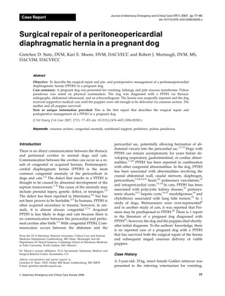
23.2 cmH2O (reference range 0–3 cmH2O). Thoracic rad-
iographs showed an enlarged cardiac silhouette with an
unusual irregular fluid-filled opacity along the ventral
portion of the thorax extending to the diaphragm (Fig-
ure 1). Differential diagnoses for the opacity included
pleural effusion, fat, or a PPDH. An abdominal ultra-
sound was performed and showed live feti within the
uterus, with an estimated pregnancy time of approxi-
mately 50 days. A round 6 3.6 cm structure surround-
ed by anechoic fluid was seen caudal to the heart
(Figure 2). The structure seemed to communicate with
the liver in the abdomen, but had a slightly different
echogenicity when compared with the liver. The he-
patic veins were considered enlarged, suggestive of
vascular compromise or possibly secondary to IV fluid
therapy. The presence of liver tissue in contact with the
heart in the left cranioventral thorax was suggestive of
a diaphragmatic hernia. Based on the appearance of the
liver tissue, necrosis secondary to vascular compromise
Figure 1: Right lateral thoracic radiograph showing an enlarged
cardiac silhouette.
Figure 2: Abdominal ultrasound image showing liver tissue in
contact with the heart.
Veterinary Emergency and Critical Care Society 2006, doi: 10.1111/j.1476-4431.2006.00200.x
78
G.D. Statz et al.](https://image.slidesharecdn.com/oski4-230511202039-ed253855/85/oski-4-pdf-2-320.jpg)


![circulation from the prior hepatocellular hypoxia and
ischemia associated with the liver tissue that was en-
trapped in the pericardium. Likewise, the alkaline
phosphatase elevation may have been related to
cholestasis during liver entrapment. Nutritional sup-
port after surgical repair of the PPDH was another
concern in this case. Fetal size increases rapidly during
the last 3–4 weeks of gestation, which causes a 15–25%
increase in energy requirements.51
A 20–50% increase
over maintenance caloric intake is required from the
fourth to seventh weeks to provide adequate energy for
fetal growth and to prepare for lactation.52
Increasing
the fat content in the diet provides increased caloric
density and should be balanced with a proportionate
increase in protein, vitamins, and minerals.52,53
The dog
in this case experienced postoperative vomiting and
regurgitation, which made total enteral nutrition diffi-
cult. An NG tube was placed to provide for partial en-
teral nutritional support and to maintain the health of
the enterocytes. Esbilac has been recommended by the
manufacturer as a nutritional supplement for pregnant
and lactating bitches to provide additional fat, protein,
and minerals and was administered via the NG tube.
The dog was also supplemented with parenteral
nutrition at approximately 2 times her resting energy
requirement taking into account illness energy require-
ments and those of late gestation. The resting energy
requirement was calculated using the standard formula
(30 BW[kg]170) and was multiplied by an illness
factor of 1.4, followed by a factor of 1.25 for late preg-
nancy requirements. Gastric reflux is common in preg-
nant humans because of the increased intra-abdominal
pressure during pregnancy and because of a hormone-
induced relaxation of the gastro-esophageal sphincter.44
Gastric contents become more acidic, and gastric emp-
tying is delayed during pregnancy.32
These changes
lead to important considerations for supportive care in
sick, hospitalized pregnant animals. The dog in this
case was treated with gastroprotectants, H2 receptor
blockers, and promotility drugs to help decrease the
acidity of the gastric contents, protect the lining of the
esophagus and stomach, increase lower gastroesopha-
geal sphincter tone, and stimulate motility of the upper
gastrointestinal tract. The NG tube was also used to
prevent esophageal reflux and potential aspiration
pneumonia by suctioning the gastric contents.
It is important to monitor and treat for hypoxemia in
critically ill and pregnant animals. The dog in this case
had poor oxygenation following surgical intervention
to correct the PPDH. Oxygen desaturation occurs easily
during pregnancy.54
Pregnant animals have a decreased
functional residual capacity and total lung volume.30
Atelectasis has been shown to occur more readily.55
Other possible causes for the decreased oxygen
saturation during the management of this case may
have included pulmonary parenchymal disease such as
pneumonia or pulmonary edema associated with fluid
overload, hypoalbuminemia, or re-expansion injury,
barotrauma because of mechanical ventilation during
anesthesia, hypoventilation, or vasculitis. A decision to
take the dog back to surgery for the cesarean section
was based on several factors including the decline in
body temperature and serum progesterone concentra-
tion. Body temperature declines 12–36 hours before
whelping22
and progesterone levels less than 2 ng/mL
have been consistently documented 36–48 hours before
whelping.22
The dog in this case was too weak to
deliver the puppies on its own, and the increased intra-
abdominal pressure from labor could have been detri-
mental, given the recent abdominal surgery with hernia
repair and the associated healing incisions.
Conclusion
This report describes a case of PPDH complicated by
pregnancy. It demonstrates the fact that PPDH may be
subclinical until a complicating factor like pregnancy
occurs. The standard surgical and post-surgical treat-
ment for PPDH had to be approached carefully in this
case. The bitch was provided with cardiovascular and
respiratory support before, during, and after PPDH
surgery to sustain adequate uterine perfusion. Nutri-
tional support between the staged surgeries in this case
was essential to allow continued growth of the puppies
and to prepare for lactation. This intensive monitoring
and aggressive supportive care for several days after
repair of the PPDH enabled a successful cesarean
delivery and survival of 7 healthy puppies and their
mother.
Footnotes
a
Bayer Corporation, West Haven, CT.
b
Baxter, Deerfield, IL.
c
Dinamap, Johnson Johnson, Tampa, FL.
d
Intracath, Becton Dickinson Vascular Access, Sandy, UT.
e
Fort Dodge Animal Health, Fort Dodge, IA.
f
Smith Kline Beecham Pharmaceuticals, Philadelphia, PA.
g
Fort Dodge Animal Health.
h
Elkins-Sinn Corporation, Cherry Hill, NJ.
i
Abbott Laboratories, North Chicago, IL.
j
Ethicon Inc., Somerville, NJ.
k
Feeding Tube and Urethral Catheter, Tyco Healthcare Group LP,
Mansfield, MA.
l
Hetastarch 6%, Abbott Laboratories.
m
Ben Venue Labs Inc., Bedford, OH.
n
Lidocaine 2%, Abbott Laboratories.
o
Potassium chloride, Abbott Laboratories.
p
Warrick Pharmaceuticals, Corp., Reno, NV.
q
Abbott Laboratories.
r
Faulding Pharmaceutical Co., Paramus, NJ.
s
Pedi-Tube Nasogastric Feeding Tube, Kendall Health Care,
Mansfield, MA.
t
Nestle, Glendale, CA.
Veterinary Emergency and Critical Care Society 2006, doi: 10.1111/j.1476-4431.2006.00200.x 83
PPDH in a pregnant dog](https://image.slidesharecdn.com/oski4-230511202039-ed253855/85/oski-4-pdf-7-320.jpg)


![10[1].pdf](https://cdn.slidesharecdn.com/ss_thumbnails/101-230511232659-e6af98b0-thumbnail.jpg?width=640&height=640&fit=bounds)


