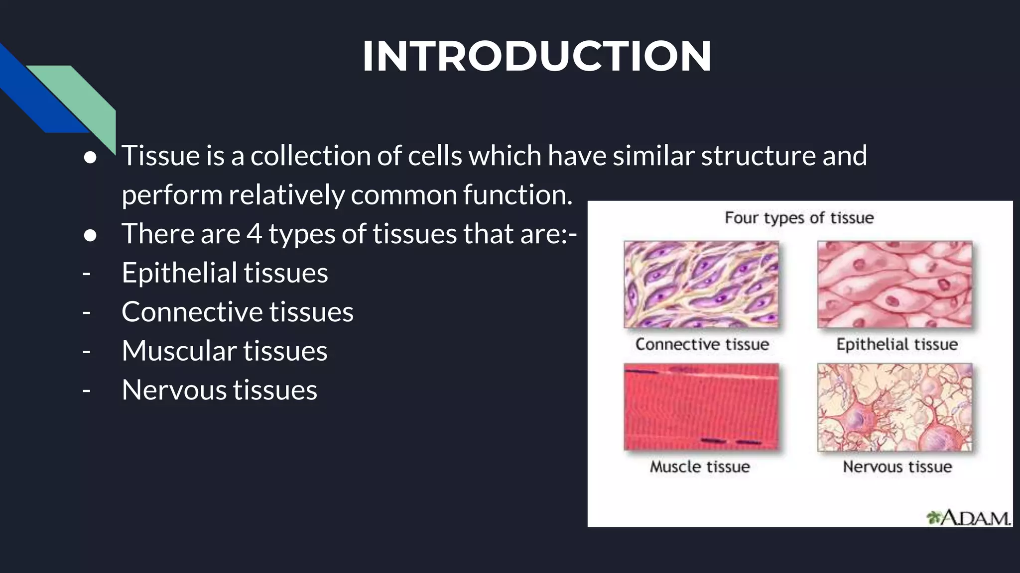
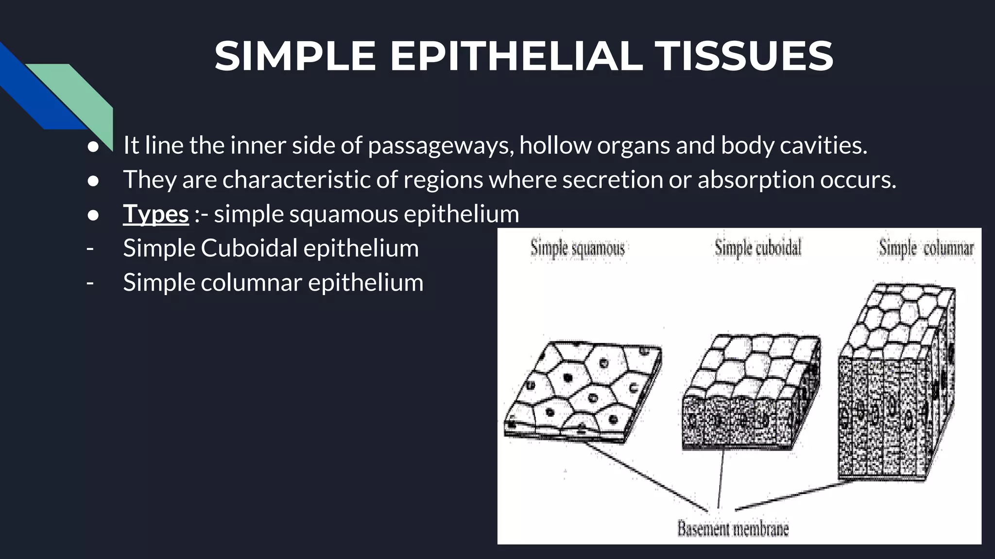
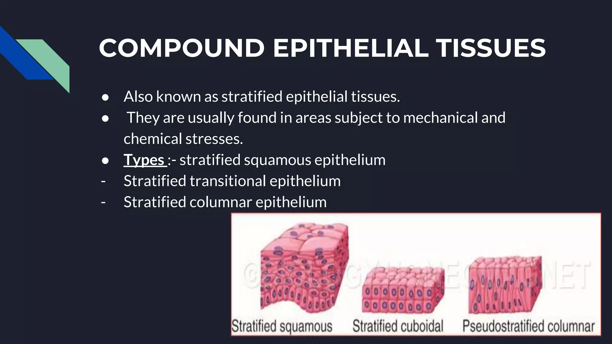
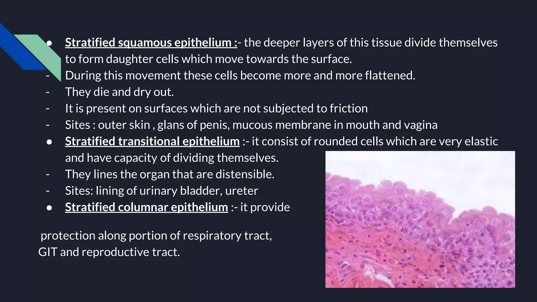
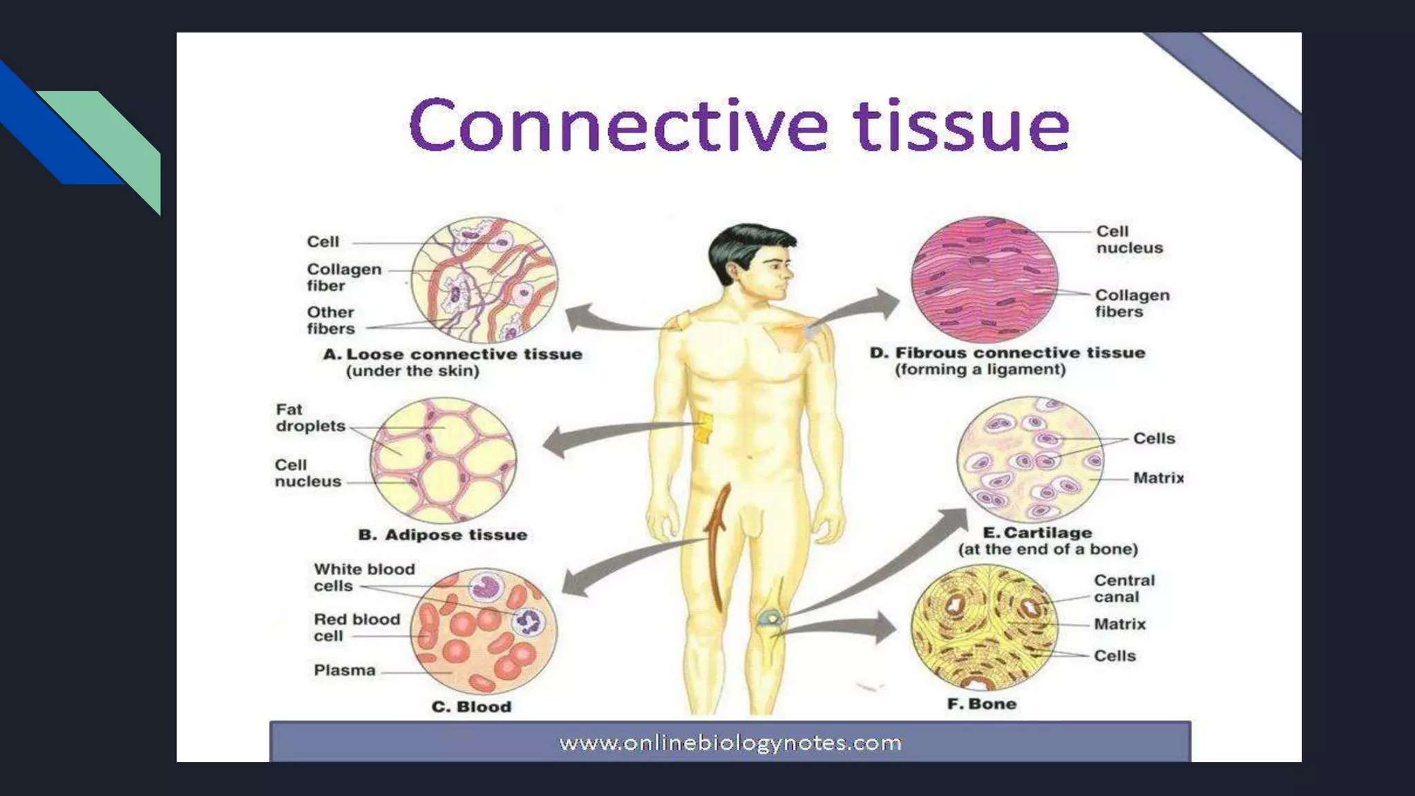
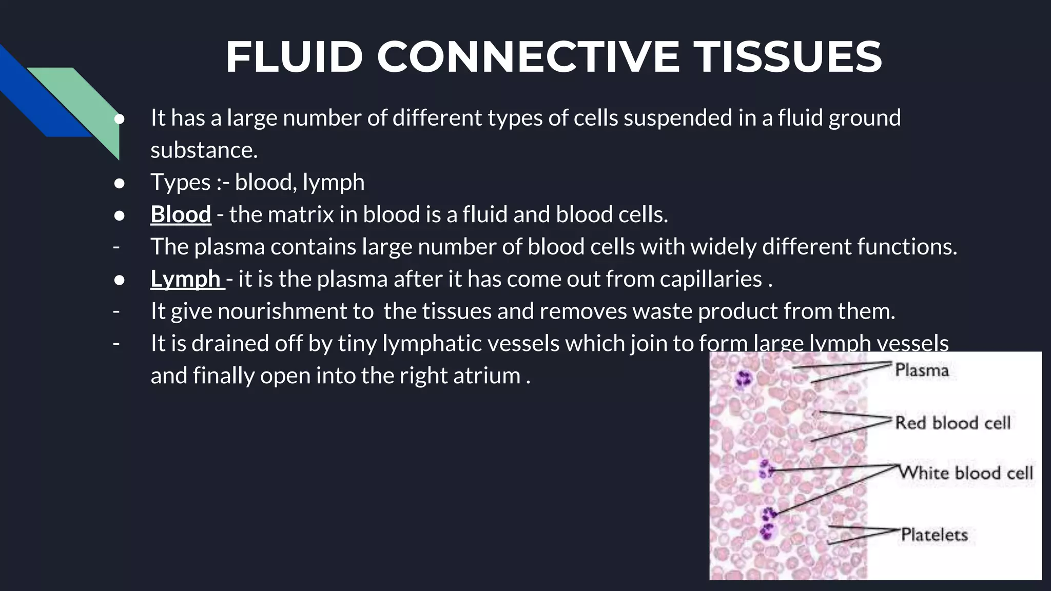
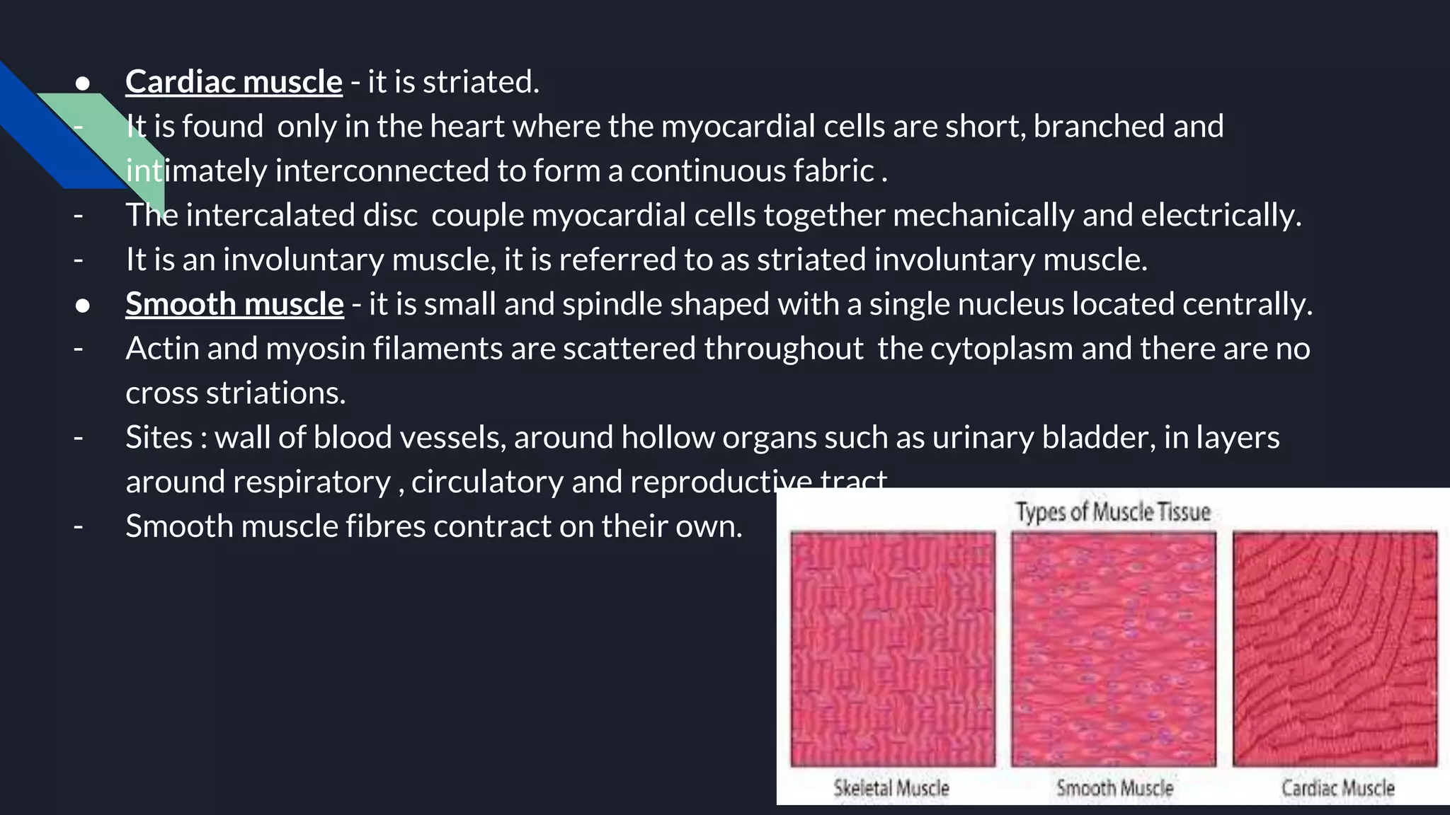
ORGANISATION OF THE
HUMAN BODY AT THE
TISSUE LEVEL
BY - MUSKAN KAPOOR
INTRODUCTION
● Tissue is a collection of cells which have similar structure and
perform relatively common function.
● There are 4 types of tissues that are:-
- Epithelial tissues
- Connective tissues
- Muscular tissues
- Nervous tissues
EPITHELIAL
TISSUES
● In this cell are closely packed without any
intracellular spaces.
● Location :- from the outer and inner lining
of most of the organs such as
gastrointestinal tract(GIT), urinary tract,
blood vessels, heart chambers etc.
- Found on the entire exposed surface of the
body such as skin.
● Functions :- play important role of defence
and protect body organs.
- Show different function in different organs
such as :- secrete gastric juice in stomach,
absorb digested food in intestine, remove
waste as sweat in skin.
● Types :- simple epithelial tissues
- Compound epithelial tissues
SIMPLE EPITHELIAL TISSUES
● It line the inner side of passageways, hollow organs and body cavities.
● They are characteristic of regions where secretion or absorption occurs.
● Types :- simple squamous epithelium
- Simple Cuboidal epithelium
- Simple columnar epithelium
● Simple squamous epithelium :- they are extremely thin and flat.
- The width of such cells is much greater than the height.
- They are found in areas where exchange takes place.
- Site :- lung alveoli, bowman’s capsule in kidney, blood vessels.
● Simple cuboidal epithelium :- they are cube shaped and on surface
views cell look either polygonal or hexagonal.
- They provide limited protection to an organ.
- Sites :- pancreas, salivary glands, kidney tubules, duct that transport
exocrine secretion.
● Simple columnar epithelium :- they are tall, slender and rectangular in shape and are
placed in shape and are placed side by side like columns.
- They occur in regions where absorption or secretion occurs.
- Sites :- most of GIT lining and excretory glands.
- Types :- ciliated columnar epithelium : it consist of modified columnar cells.
: it has motile hair like projection called cilia which push the
substances in a definite direction
: site - oviducts and renal tubules.
- Pseudostratified columnar epithelium :- pseudo means false and stratified means
multilayered.
:it consist of columnar cells of unequal heights.
: sites - trachea lining and parts of male reproductive tract.
COMPOUND EPITHELIAL TISSUES
● Also known as stratified epithelial tissues.
● They are usually found in areas subject to mechanical and
chemical stresses.
● Types :- stratified squamous epithelium
- Stratified transitional epithelium
- Stratified columnar epithelium
● Stratified squamous epithelium :- the deeper layers of this tissue divide themselves
to form daughter cells which move towards the surface.
- During this movement these cells become more and more flattened.
- They die and dry out.
- It is present on surfaces which are not subjected to friction
- Sites : outer skin , glans of penis, mucous membrane in mouth and vagina
● Stratified transitional epithelium :- it consist of rounded cells which are very elastic
and have capacity of dividing themselves.
- They lines the organ that are distensible.
- Sites: lining of urinary bladder, ureter
● Stratified columnar epithelium :- it provide
protection along portion of respiratory tract,
GIT and reproductive tract.
CONNECTIVE TISSUE
● It connect one tissue or organ with other tissues or organs.
● They are made up of cells which are found individually or in groups of two or more.
● Space between the cells is called matrix which is filled with different substances.
● Functions :- provide support and protect internal delicate organ of the body as bone
and cartilage do.
- Transport material from one part of the body to other as blood does.
- Store energy sources such as fats, in large quantities.
- Defend body from invasion by microorganisms.
● Types :- connective tissue proper
- Supporting connective tissue or skeleton tissue
- Fluid connective tissue
CONNECTIVE TISSUE PROPER
● Connective tissue fibres :- it is of 3 types - collagen fibres
- Elastic fibres
- Reticular fibres
➔ Collagen fibres :- they are strong and almost inflexible.
- They consist of protein collagen and are arranged in
bundles.
➔ Elastic fibres :- they are flexible and branched fibres
which consist of protein elastin.
- Site - in walls of arteries and lining of nerve cells.
➔ Reticular fibres :- they are much thinner than other two types of fibres.
- The substance reticulin forms very finely branched out fibres which cannot be
stretched.
- Site - spleen and liver
CONNECTIVE TISSUE PROPER
● It is of 2 types :- loose connective tissue
- Dense connective tissue
● LOOSE CONNECTIVE TISSUE :- it is of 3 types - areolar tissue
- Adipose tissue
- Reticular tissue
➔ AREOLAR TISSUE -it contain all 3 fibres types
- Site : beneath epithelium of skin, GIT, respiratory and urinary tract.
➔ ADIPOSE TISSUE - It is made up of rounded cells filled with fat globule in their cytoplasm
- Site : fat cells, deeper layer of skin, buttocks, breast.
- Function : serve as reserve store of fuel
: act as heat insulator
➔ Reticular tissue : it is basic framework for many organs that have complex tissue
- Site : liver, spleen , lymph nodes
- Function: provide supporting framework
DEEP
CONNECTIVE
TISSUE
● It is occupied by densely packed collagen
fibres.
● Example - tendons, ligaments and fasciae.
● Tendons :- they are mainly white tightly
packed fibres of protein collagen that run
parallel to form cord like structures.
- They are strong, flexible and resistant to
pull
- They connect muscles with bones and
facilitate in movement of limbs.
● Ligaments :- they are primarily made up of
densely packed yellow fibres of protein
elastin that run parallel to form cord like
structures.
- They can tolerate a moderate amount of
stretching.
● Fasciae :- they are sheets of connective tissues which contain
collagen fibres.
- It surround organs such as skeletal muscles, attach skeletal
muscles to bone and distribute the forces of muscular
contraction over a large area.
SUPPORTING TISSUE PROPER
● It provide strong framework that support and give definite shape to the body.
● They also protect internal delicate organ of the body.
● Example :- cartilage and bone
● Cartilage - it is relatively soft and flexible
- They are surrounded by smooth and clear walls called lacunae.
- Surface of cartilage is formed by tough connective tissues fibres i.e. perichondrium.
- Functions - it reduces friction between bony surfaces .
- It provide stiff but somewhat flexible support to body surfaces.
● Bone - they are relatively harder and stronger than cartilage because matrix consist of
yellow elastic fibres combine with phosphate and carbonates of calcium and magnesium
salts.
- All long bones of the body have a yellow central cavity which is filled with cells called bone
marrow.
FLUID CONNECTIVE TISSUES
● It has a large number of different types of cells suspended in a fluid ground
substance.
● Types :- blood, lymph
● Blood - the matrix in blood is a fluid and blood cells.
- The plasma contains large number of blood cells with widely different functions.
● Lymph - it is the plasma after it has come out from capillaries .
- It give nourishment to the tissues and removes waste product from them.
- It is drained off by tiny lymphatic vessels which join to form large lymph vessels
and finally open into the right atrium .
MUSCULAR TISSUE
● It is specialised for contraction .
● It is of 3 types :- skeletal muscle
- Cardiac muscle
- Smooth muscle
● Skeletal muscle :- they are generally attached to bones at both ends by means of tendon,
hence contraction produces movement of the skeleton.
- The tongue, superior portion of oesophagus, and sphincter and diaphragm are also
connected to skeletal muscle but they don’t cause movement of skeleton.
- Each muscle fibre is made up of many fibrils called myofibrils, they lie parallel to each
other and are striated.
- In between successive myofibrils there is a thin layer of cytoplasm called sacroplasm .
● Cardiac muscle - it is striated.
- It is found only in the heart where the myocardial cells are short, branched and
intimately interconnected to form a continuous fabric .
- The intercalated disc couple myocardial cells together mechanically and electrically.
- It is an involuntary muscle, it is referred to as striated involuntary muscle.
● Smooth muscle - it is small and spindle shaped with a single nucleus located centrally.
- Actin and myosin filaments are scattered throughout the cytoplasm and there are no
cross striations.
- Sites : wall of blood vessels, around hollow organs such as urinary bladder, in layers
around respiratory , circulatory and reproductive tract.
- Smooth muscle fibres contract on their own.
NERVOUS TISSUE
● It is specialised for conduction of electrical impulses from one region of
body to another.
● It contains 2 type of cells: conducting cells or neuron cells and
supporting cells or glial cells.
● Neuron : - it is structural and functional unit of
the nervous system.
- It consist of cell body or nerve cell with several
cytoplasmic extensions.
- It has 5-7 short processes called dendrites,
they transmit impulses towards the cell body.
- A single long process is called axon, it conveys
impulses away from the cell body.
- In addition it contains Nissl’s granules i.e.
membrane bound cavities which consist of
ribonucleoproteins.
- Neurons are specially designed to receive
stimuli from the environment.
- The axon is coated with a thick layer called
myelin sheath.
- Myelin facilitates conduction of nerve impulse,
it confines the nerve impulse to individual
● Neuroglia - also known as glial
cell.
- It support the nerve cells.
- There are about ten times as
many glial cells as neurons.
- Functions - isolate and protect
the cell membrane of neurons.
- Provide a supporting framework
for neural tissue .
- Regulate the composition of
intestinal fluid.
- Act as phagocytes that defend
neural tissue from pathogens
THANK YOU

organisation of human body at tissue level

  • 1.
    ORGANISATION OF THE HUMANBODY AT THE TISSUE LEVEL BY - MUSKAN KAPOOR
  • 2.
    INTRODUCTION ● Tissue isa collection of cells which have similar structure and perform relatively common function. ● There are 4 types of tissues that are:- - Epithelial tissues - Connective tissues - Muscular tissues - Nervous tissues
  • 3.
    EPITHELIAL TISSUES ● In thiscell are closely packed without any intracellular spaces. ● Location :- from the outer and inner lining of most of the organs such as gastrointestinal tract(GIT), urinary tract, blood vessels, heart chambers etc. - Found on the entire exposed surface of the body such as skin. ● Functions :- play important role of defence and protect body organs. - Show different function in different organs such as :- secrete gastric juice in stomach, absorb digested food in intestine, remove waste as sweat in skin. ● Types :- simple epithelial tissues - Compound epithelial tissues
  • 4.
    SIMPLE EPITHELIAL TISSUES ●It line the inner side of passageways, hollow organs and body cavities. ● They are characteristic of regions where secretion or absorption occurs. ● Types :- simple squamous epithelium - Simple Cuboidal epithelium - Simple columnar epithelium
  • 5.
    ● Simple squamousepithelium :- they are extremely thin and flat. - The width of such cells is much greater than the height. - They are found in areas where exchange takes place. - Site :- lung alveoli, bowman’s capsule in kidney, blood vessels. ● Simple cuboidal epithelium :- they are cube shaped and on surface views cell look either polygonal or hexagonal. - They provide limited protection to an organ. - Sites :- pancreas, salivary glands, kidney tubules, duct that transport exocrine secretion.
  • 6.
    ● Simple columnarepithelium :- they are tall, slender and rectangular in shape and are placed in shape and are placed side by side like columns. - They occur in regions where absorption or secretion occurs. - Sites :- most of GIT lining and excretory glands. - Types :- ciliated columnar epithelium : it consist of modified columnar cells. : it has motile hair like projection called cilia which push the substances in a definite direction : site - oviducts and renal tubules. - Pseudostratified columnar epithelium :- pseudo means false and stratified means multilayered. :it consist of columnar cells of unequal heights. : sites - trachea lining and parts of male reproductive tract.
  • 7.
    COMPOUND EPITHELIAL TISSUES ●Also known as stratified epithelial tissues. ● They are usually found in areas subject to mechanical and chemical stresses. ● Types :- stratified squamous epithelium - Stratified transitional epithelium - Stratified columnar epithelium
  • 8.
    ● Stratified squamousepithelium :- the deeper layers of this tissue divide themselves to form daughter cells which move towards the surface. - During this movement these cells become more and more flattened. - They die and dry out. - It is present on surfaces which are not subjected to friction - Sites : outer skin , glans of penis, mucous membrane in mouth and vagina ● Stratified transitional epithelium :- it consist of rounded cells which are very elastic and have capacity of dividing themselves. - They lines the organ that are distensible. - Sites: lining of urinary bladder, ureter ● Stratified columnar epithelium :- it provide protection along portion of respiratory tract, GIT and reproductive tract.
  • 9.
    CONNECTIVE TISSUE ● Itconnect one tissue or organ with other tissues or organs. ● They are made up of cells which are found individually or in groups of two or more. ● Space between the cells is called matrix which is filled with different substances. ● Functions :- provide support and protect internal delicate organ of the body as bone and cartilage do. - Transport material from one part of the body to other as blood does. - Store energy sources such as fats, in large quantities. - Defend body from invasion by microorganisms. ● Types :- connective tissue proper - Supporting connective tissue or skeleton tissue - Fluid connective tissue
  • 11.
    CONNECTIVE TISSUE PROPER ●Connective tissue fibres :- it is of 3 types - collagen fibres - Elastic fibres - Reticular fibres ➔ Collagen fibres :- they are strong and almost inflexible. - They consist of protein collagen and are arranged in bundles. ➔ Elastic fibres :- they are flexible and branched fibres which consist of protein elastin. - Site - in walls of arteries and lining of nerve cells. ➔ Reticular fibres :- they are much thinner than other two types of fibres. - The substance reticulin forms very finely branched out fibres which cannot be stretched. - Site - spleen and liver
  • 12.
    CONNECTIVE TISSUE PROPER ●It is of 2 types :- loose connective tissue - Dense connective tissue ● LOOSE CONNECTIVE TISSUE :- it is of 3 types - areolar tissue - Adipose tissue - Reticular tissue ➔ AREOLAR TISSUE -it contain all 3 fibres types - Site : beneath epithelium of skin, GIT, respiratory and urinary tract. ➔ ADIPOSE TISSUE - It is made up of rounded cells filled with fat globule in their cytoplasm - Site : fat cells, deeper layer of skin, buttocks, breast. - Function : serve as reserve store of fuel : act as heat insulator ➔ Reticular tissue : it is basic framework for many organs that have complex tissue - Site : liver, spleen , lymph nodes - Function: provide supporting framework
  • 13.
    DEEP CONNECTIVE TISSUE ● It isoccupied by densely packed collagen fibres. ● Example - tendons, ligaments and fasciae. ● Tendons :- they are mainly white tightly packed fibres of protein collagen that run parallel to form cord like structures. - They are strong, flexible and resistant to pull - They connect muscles with bones and facilitate in movement of limbs. ● Ligaments :- they are primarily made up of densely packed yellow fibres of protein elastin that run parallel to form cord like structures. - They can tolerate a moderate amount of stretching.
  • 14.
    ● Fasciae :-they are sheets of connective tissues which contain collagen fibres. - It surround organs such as skeletal muscles, attach skeletal muscles to bone and distribute the forces of muscular contraction over a large area.
  • 15.
    SUPPORTING TISSUE PROPER ●It provide strong framework that support and give definite shape to the body. ● They also protect internal delicate organ of the body. ● Example :- cartilage and bone ● Cartilage - it is relatively soft and flexible - They are surrounded by smooth and clear walls called lacunae. - Surface of cartilage is formed by tough connective tissues fibres i.e. perichondrium. - Functions - it reduces friction between bony surfaces . - It provide stiff but somewhat flexible support to body surfaces. ● Bone - they are relatively harder and stronger than cartilage because matrix consist of yellow elastic fibres combine with phosphate and carbonates of calcium and magnesium salts. - All long bones of the body have a yellow central cavity which is filled with cells called bone marrow.
  • 17.
    FLUID CONNECTIVE TISSUES ●It has a large number of different types of cells suspended in a fluid ground substance. ● Types :- blood, lymph ● Blood - the matrix in blood is a fluid and blood cells. - The plasma contains large number of blood cells with widely different functions. ● Lymph - it is the plasma after it has come out from capillaries . - It give nourishment to the tissues and removes waste product from them. - It is drained off by tiny lymphatic vessels which join to form large lymph vessels and finally open into the right atrium .
  • 18.
    MUSCULAR TISSUE ● Itis specialised for contraction . ● It is of 3 types :- skeletal muscle - Cardiac muscle - Smooth muscle ● Skeletal muscle :- they are generally attached to bones at both ends by means of tendon, hence contraction produces movement of the skeleton. - The tongue, superior portion of oesophagus, and sphincter and diaphragm are also connected to skeletal muscle but they don’t cause movement of skeleton. - Each muscle fibre is made up of many fibrils called myofibrils, they lie parallel to each other and are striated. - In between successive myofibrils there is a thin layer of cytoplasm called sacroplasm .
  • 19.
    ● Cardiac muscle- it is striated. - It is found only in the heart where the myocardial cells are short, branched and intimately interconnected to form a continuous fabric . - The intercalated disc couple myocardial cells together mechanically and electrically. - It is an involuntary muscle, it is referred to as striated involuntary muscle. ● Smooth muscle - it is small and spindle shaped with a single nucleus located centrally. - Actin and myosin filaments are scattered throughout the cytoplasm and there are no cross striations. - Sites : wall of blood vessels, around hollow organs such as urinary bladder, in layers around respiratory , circulatory and reproductive tract. - Smooth muscle fibres contract on their own.
  • 20.
    NERVOUS TISSUE ● Itis specialised for conduction of electrical impulses from one region of body to another. ● It contains 2 type of cells: conducting cells or neuron cells and supporting cells or glial cells.
  • 21.
    ● Neuron :- it is structural and functional unit of the nervous system. - It consist of cell body or nerve cell with several cytoplasmic extensions. - It has 5-7 short processes called dendrites, they transmit impulses towards the cell body. - A single long process is called axon, it conveys impulses away from the cell body. - In addition it contains Nissl’s granules i.e. membrane bound cavities which consist of ribonucleoproteins. - Neurons are specially designed to receive stimuli from the environment. - The axon is coated with a thick layer called myelin sheath. - Myelin facilitates conduction of nerve impulse, it confines the nerve impulse to individual
  • 22.
    ● Neuroglia -also known as glial cell. - It support the nerve cells. - There are about ten times as many glial cells as neurons. - Functions - isolate and protect the cell membrane of neurons. - Provide a supporting framework for neural tissue . - Regulate the composition of intestinal fluid. - Act as phagocytes that defend neural tissue from pathogens
  • 23.