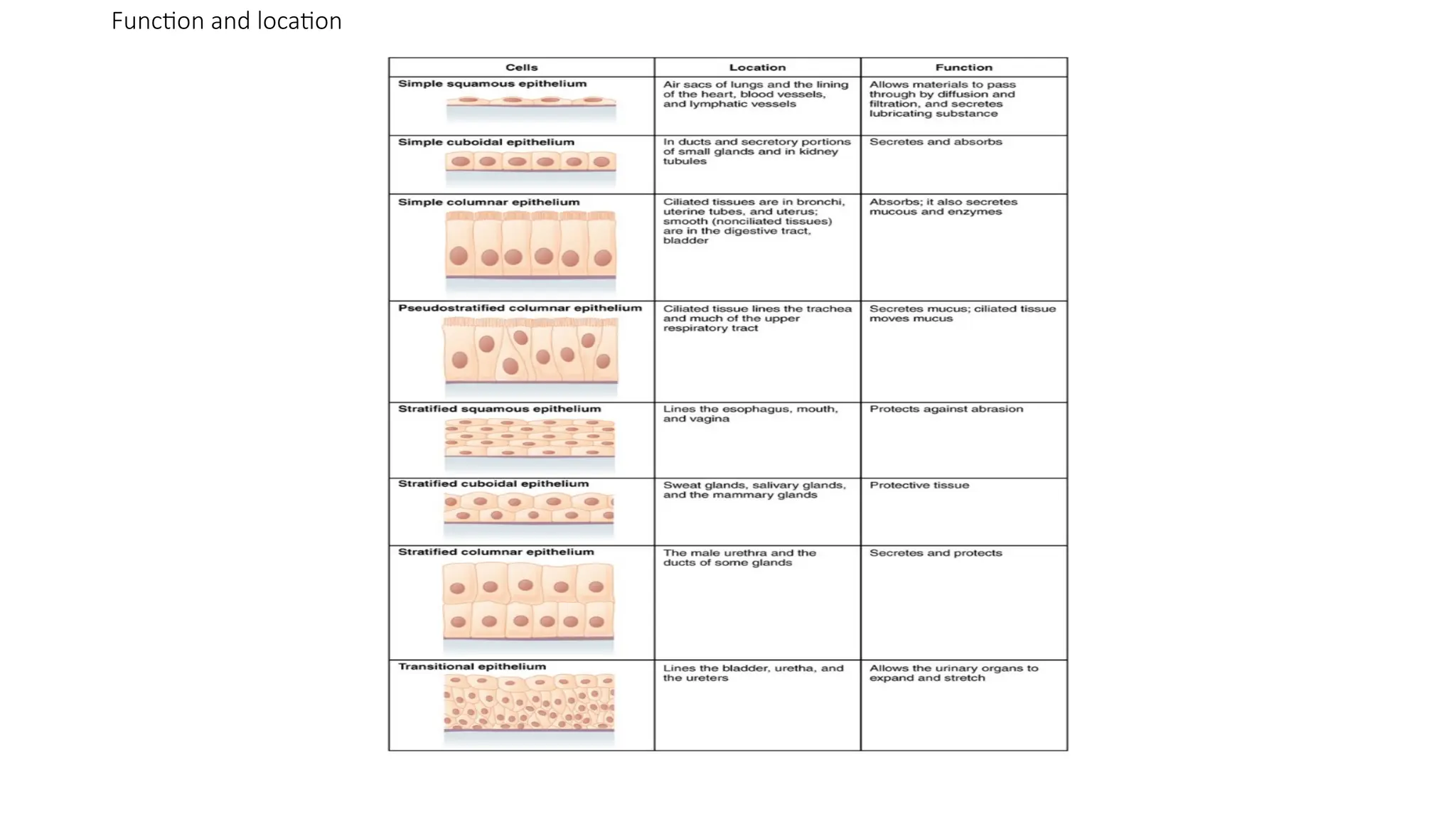
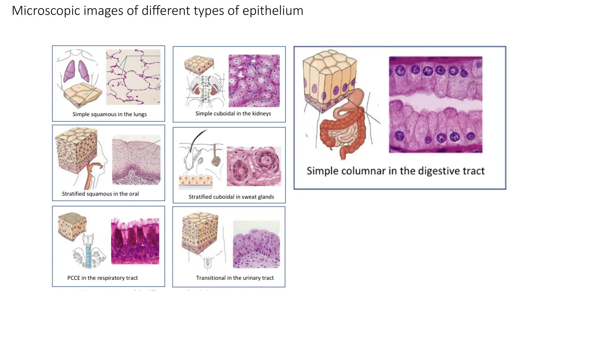
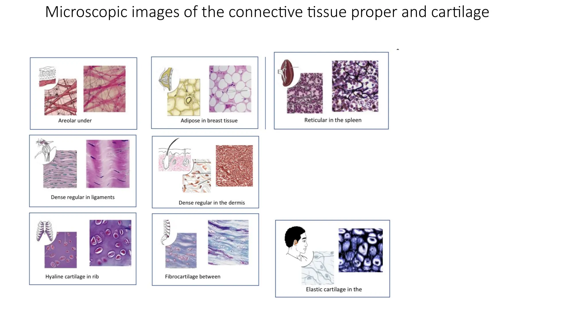
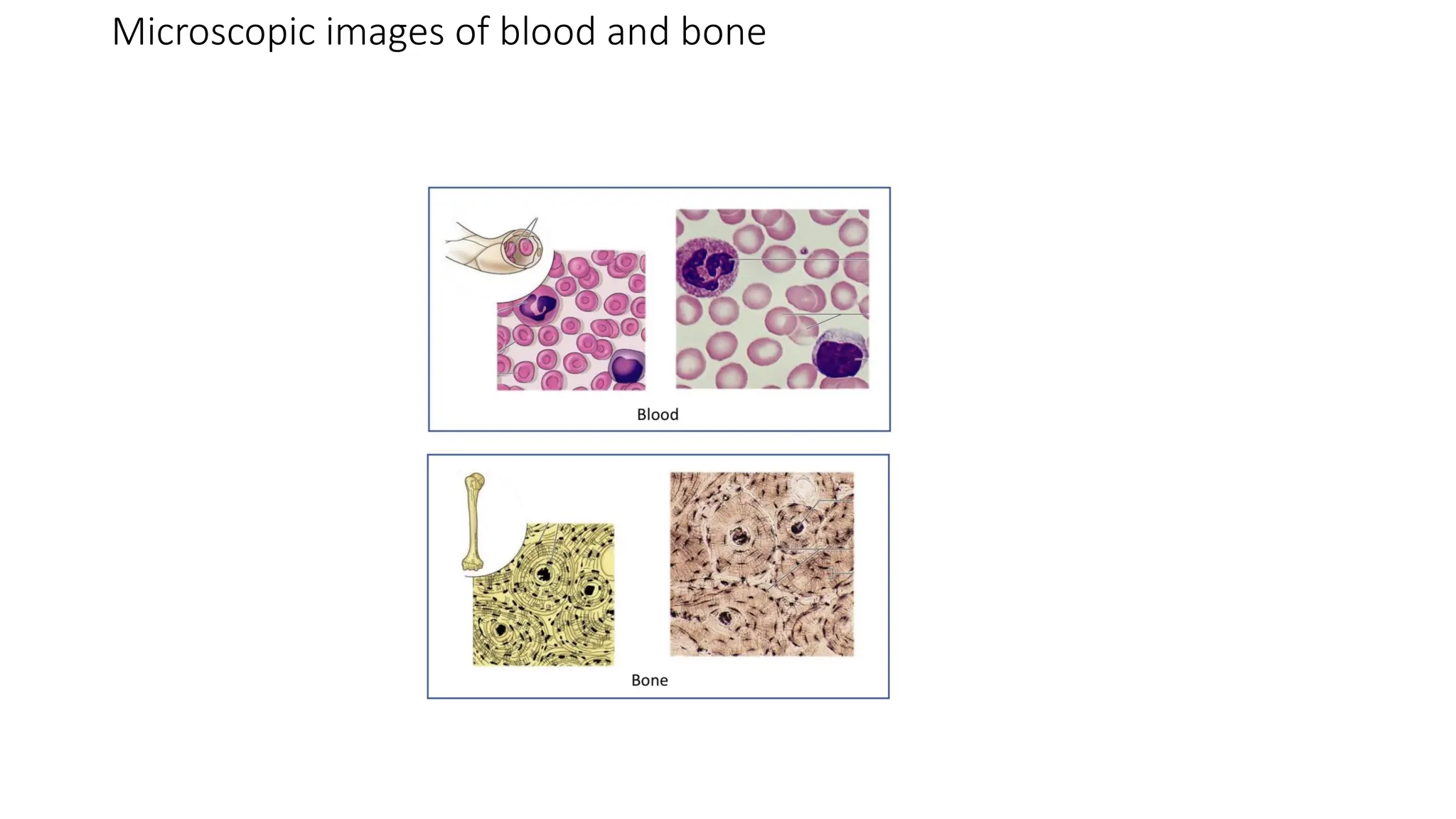
INTRODUCTION TO ANATOMY PRACTICAL
• Anatomical terms
• Anterior: Front
• Posterior: Back
• Medial: Towards the midline
• Lateral: Away from the midline
• Intermediate: Between and in the middle
• Proximal: Towards the beginning of a structure
• Distal: Towards the end of a structure
• Deep: Internal
• Superficial: External
• Ventral: Belly or front
• Dorsal: Back
• Cephalic: Head
• Caudal: Tail
• Sagittal plane: separates the body into right and left
• Transverse plane: separates the body into superior and inferior
UPPER LIMBS
• BONES:
• Shoulder: Clavical, Scapular,
• Arm: Humerus
• Forearm: Ulna(medial), Radius(lateral)
• Hand: Carpals, metacarpals and phalanges
HISTOLOGY
• Histology is the study of tissues
• Four basic tissues: Epithelium, connective, muscular and nervous
• Epithelium: Epi(above) and thelium(tissue), epithelium is a group of tissues
located above (covering or lining) all other tissue of the body
• Connective: this binds (connects) tissues to one another
• Muscular: movement
• Nervous: communication
• WHERE THEY ARE FOUND IN THE BODY
• Epithelial tissue: lining of GI tract and skin surface (epidermis)
• Connective tissue: bone, tendon
• Muscle tissue: cardiac muscle, smooth muscle, skeletal muscle
• Nervous tissue: brain, spinal cord, nerves
EPITHELIUM
• They are above other tissues and are constantly exposed to the external environment.
They are avascular and have a high rate of regeneration to replace damaged and lost cells.
• Epithelium has two surfaces:
• Apical surface: this is exposed to the external environment
• Basal surface: this is bound to the tissue below
• Five characteristics of the epithelia
• A high rate of regeneration
• A free and a bounded surface (polarity)
• Tightly packed cells
• Avascularity with innervation
• Basement membrane which is between the epithelium above and connective tissue below
Function of the epithelium
• It provides linings for the external and internal surfaces that face the harsh
environments. The outer layer of the skin is epithelial tissue, the innermost
layer of the digestive tract, the blood vessels.
• It forms glands that secrete materials onto epithelial surfaces or into the
blood. Examples of glands made of epithelial tissues are sweat glands,
salivary glands, mammary glands, and pituitary glands.
Function and location
Classification of epithelial tissue
Epithelial tissues are often classified according to the number of layers of
cells present and by shape of the cells.
Layers of cells:
• Single layer of cells, where all the cells touch the basement membrane is
known as a simple epithelium.
• Two or more layers of cells, which indicates the tissue has strata(layers), are
called stratified epithelium.
• Pseudostratified epithelium: is a specialized form of a simple epithelium, at
first glance it appears to be more than one layer of epithelial cells, but a
closer inspection reveals that each cell in the layer extends to the
basolateral surface of the epithelium.
Classification of epithelial tissue
Shape of cells:
• Squamous epithelial cells: under the microscope it looks flat and like scales
of fish
• Cuboidal epithelial cells: looks close to a square
• Columnar epithelial cells: looks like a column or a tall rectangle
• Transitional epithelial cells: they are few and are specialized to change
shape if they are stretched laterally. They can transition from columnar or
cuboidal looking shapes in their un-stretched state to more squamous
looking shapes in their stretched state.
Microscopic images of different types of epithelium
Connective tissue
This is found throughout the body and it is usually in association with other tissues.
It often serve to connect different tissues together.
Characteristics of connective tissues
• Dispersed cells: connective tissues generally have cells that are not tightly connected
to one another except the adipose tissue(fat).
• The tissue has more extracellular material than cells. Most connective tissues are
solid because all the volume between the dispersed cells is filled with an extracellular
matrix of viscous ground substance and protein fibers.
• An extensive network of protein fibers is found in the extracellular matrix. Protein
fibers are complexes of millions of individual proteins threaded into long fibrous
structure that provide strength and elasticity to the tissue as a whole.
Classification of connective tissue
• Connective tissue proper: It is further subdivided into
Dense connective tissue proper, which have dense arrangement of
extracellular protein fibers that give the tissue strength and toughness.
Tendon connecting muscles to bone and ligaments connecting bone and
bone are examples of dense connective tissue proper.
Loose connective tissue proper have fewer extracellular protein fibers and
more ground substance( the extracellular material surrounding the protein
fibers), making the tissues spongier but more fragile. Areolar tissue, found in
the hypodermis of the skin and below the epithelial layers of the digestive,
respiratory and urinary tracts, is a loose connective tissue proper and adipose
tissue(fat).
Types of connective tissue proper
Classification of connective tissue
• Supporting connective tissue: their extracellular matrix is tougher, denser and are
more solid than the various types of connective tissue proper. Bones and cartilage
are the two types of connective tissue in this sub category.
• Fluid connective tissue: blood and lymph are the two types of connective tissue in
this sub category. Both are fluid and both lack the network of cellular protein
fibers found in the other types of connective tissue.
Microscopic images of the connective tissue proper and cartilage
Microscopic images of blood and bone
Muscular Tissue
• This is subdivided into three categories: skeletal muscle, cardiac muscle and smooth muscle.
The three types of muscle tissue can be distinguished on the basis of morphologic and
functional characteristics.
Properties of Muscular Tissue
• Contractibility
• Extensibility
• Elasticity
• Excitability
• Highly vascularized
• Striated or smooth appearance
Function of muscular tissue
• Responsible for movement of the body and internal organs of an organism.
• Skeletal muscles are responsible for maintaining the posture of the body and
supporting the weight.
•
• Production of heat as a byproduct of metabolic processes. The heat helps to
maintain body temperature.
• Protection of internal organs
Skeletal Muscle
• They are found attached to bones. They are also called striated muscles because of their
striped appearance.
• They consist of long multinucleate fibers. The fibers run the entire length of the muscle
they come from and are usually too long to have their ends viewed under the
microscope. The fibers are long, wide but unbranched.
• Blood vessels are present in skeletal muscles
• Organism movement is done via skeletal muscles.
Cardiac Muscle
• They are only found in the heart. The heart pumps blood due to the contraction of cardiac
muscle.
• They are involuntary muscles and are striated like skeletal muscle.
• Cardiac muscle fibers are mononucleate, with only one nucleus per fiber and they can
sometimes be branched.
• Unlike skeletal muscle fibers, cardiac muscle fiber have distinct end to them called
intercalated discs.
• Intercalated discs are dark lines that run from one side of the fiber to the other. They are
darker and thicker than striation.
Smooth Muscle
• They are found in the walls of internal organs such as organs of the digestive tracts, blood
vessels etc.
• Smooth muscles are non striated because the fibers(actin and myosin) are extremely thin and
arranged randomly.
• Are involuntary muscles, they are controlled via autonomic nervous system.
• It consists of mononucleate fibers.
• It animates the contractility of the stomach, urinary tracts etc.
Nervous Tissue
• This comprises of neurons, the cells specialized for propagation of electrochemical signals,
and neuroglia, the supporting cells of nervous tissue.
Neuron
This is the structural and fundamental unit of the nervous system. It has the following features
• A large cell body (soma) in which the nucleus and other major organelles are found
• Dendrites, which are usually represented as numerous small projections extending from the
cell body.
• A single axon, which are like dendrites but much longer.
• Multiple axon terminal that branch off at the end of the axon.
• Cell body where most of the cellular processes of the neuron take place such as protein
synthesis, metabolism etc.
• Synapse, this is a junction between two neurons where impulses pass.
Nervous tissue
Neuroglia (Glial cells)
They provide support and protection to neurons. There are four types of cells:
• Astrocytes: provides physical and metabolic support
• Oligodendrocytes: form myelin sheaths around multiple axons
• Microglial: phagocytic cells
• Ependymal cells: lines the ventricle of the brain and the central canal of the spinal cord.
Some glial cells are associated with the pheripheral nervous system. They includes:
• Schwann cells: isolates axons from the surrounding extracellular compartment. Can be further
classified into myelinating and non-myelinating.
• Satellite cells: small cuboidal cells that surround the nerve cells in ganglia.
• Specialized glial cells: are associated with specific tissues or organs.

Introduction to anatomy practical and terms in anatomy

  • 1.
    INTRODUCTION TO ANATOMYPRACTICAL • Anatomical terms • Anterior: Front • Posterior: Back • Medial: Towards the midline • Lateral: Away from the midline • Intermediate: Between and in the middle • Proximal: Towards the beginning of a structure • Distal: Towards the end of a structure • Deep: Internal • Superficial: External • Ventral: Belly or front • Dorsal: Back • Cephalic: Head • Caudal: Tail • Sagittal plane: separates the body into right and left • Transverse plane: separates the body into superior and inferior
  • 2.
    UPPER LIMBS • BONES: •Shoulder: Clavical, Scapular, • Arm: Humerus • Forearm: Ulna(medial), Radius(lateral) • Hand: Carpals, metacarpals and phalanges
  • 3.
    HISTOLOGY • Histology isthe study of tissues • Four basic tissues: Epithelium, connective, muscular and nervous • Epithelium: Epi(above) and thelium(tissue), epithelium is a group of tissues located above (covering or lining) all other tissue of the body • Connective: this binds (connects) tissues to one another • Muscular: movement • Nervous: communication • WHERE THEY ARE FOUND IN THE BODY • Epithelial tissue: lining of GI tract and skin surface (epidermis) • Connective tissue: bone, tendon • Muscle tissue: cardiac muscle, smooth muscle, skeletal muscle • Nervous tissue: brain, spinal cord, nerves
  • 4.
    EPITHELIUM • They areabove other tissues and are constantly exposed to the external environment. They are avascular and have a high rate of regeneration to replace damaged and lost cells. • Epithelium has two surfaces: • Apical surface: this is exposed to the external environment • Basal surface: this is bound to the tissue below • Five characteristics of the epithelia • A high rate of regeneration • A free and a bounded surface (polarity) • Tightly packed cells • Avascularity with innervation • Basement membrane which is between the epithelium above and connective tissue below
  • 5.
    Function of theepithelium • It provides linings for the external and internal surfaces that face the harsh environments. The outer layer of the skin is epithelial tissue, the innermost layer of the digestive tract, the blood vessels. • It forms glands that secrete materials onto epithelial surfaces or into the blood. Examples of glands made of epithelial tissues are sweat glands, salivary glands, mammary glands, and pituitary glands.
  • 6.
  • 7.
    Classification of epithelialtissue Epithelial tissues are often classified according to the number of layers of cells present and by shape of the cells. Layers of cells: • Single layer of cells, where all the cells touch the basement membrane is known as a simple epithelium. • Two or more layers of cells, which indicates the tissue has strata(layers), are called stratified epithelium. • Pseudostratified epithelium: is a specialized form of a simple epithelium, at first glance it appears to be more than one layer of epithelial cells, but a closer inspection reveals that each cell in the layer extends to the basolateral surface of the epithelium.
  • 8.
    Classification of epithelialtissue Shape of cells: • Squamous epithelial cells: under the microscope it looks flat and like scales of fish • Cuboidal epithelial cells: looks close to a square • Columnar epithelial cells: looks like a column or a tall rectangle • Transitional epithelial cells: they are few and are specialized to change shape if they are stretched laterally. They can transition from columnar or cuboidal looking shapes in their un-stretched state to more squamous looking shapes in their stretched state.
  • 9.
    Microscopic images ofdifferent types of epithelium
  • 10.
    Connective tissue This isfound throughout the body and it is usually in association with other tissues. It often serve to connect different tissues together. Characteristics of connective tissues • Dispersed cells: connective tissues generally have cells that are not tightly connected to one another except the adipose tissue(fat). • The tissue has more extracellular material than cells. Most connective tissues are solid because all the volume between the dispersed cells is filled with an extracellular matrix of viscous ground substance and protein fibers. • An extensive network of protein fibers is found in the extracellular matrix. Protein fibers are complexes of millions of individual proteins threaded into long fibrous structure that provide strength and elasticity to the tissue as a whole.
  • 11.
    Classification of connectivetissue • Connective tissue proper: It is further subdivided into Dense connective tissue proper, which have dense arrangement of extracellular protein fibers that give the tissue strength and toughness. Tendon connecting muscles to bone and ligaments connecting bone and bone are examples of dense connective tissue proper. Loose connective tissue proper have fewer extracellular protein fibers and more ground substance( the extracellular material surrounding the protein fibers), making the tissues spongier but more fragile. Areolar tissue, found in the hypodermis of the skin and below the epithelial layers of the digestive, respiratory and urinary tracts, is a loose connective tissue proper and adipose tissue(fat).
  • 12.
    Types of connectivetissue proper
  • 13.
    Classification of connectivetissue • Supporting connective tissue: their extracellular matrix is tougher, denser and are more solid than the various types of connective tissue proper. Bones and cartilage are the two types of connective tissue in this sub category. • Fluid connective tissue: blood and lymph are the two types of connective tissue in this sub category. Both are fluid and both lack the network of cellular protein fibers found in the other types of connective tissue.
  • 14.
    Microscopic images ofthe connective tissue proper and cartilage
  • 15.
    Microscopic images ofblood and bone
  • 16.
    Muscular Tissue • Thisis subdivided into three categories: skeletal muscle, cardiac muscle and smooth muscle. The three types of muscle tissue can be distinguished on the basis of morphologic and functional characteristics. Properties of Muscular Tissue • Contractibility • Extensibility • Elasticity • Excitability • Highly vascularized • Striated or smooth appearance
  • 17.
    Function of musculartissue • Responsible for movement of the body and internal organs of an organism. • Skeletal muscles are responsible for maintaining the posture of the body and supporting the weight. • • Production of heat as a byproduct of metabolic processes. The heat helps to maintain body temperature. • Protection of internal organs
  • 18.
    Skeletal Muscle • Theyare found attached to bones. They are also called striated muscles because of their striped appearance. • They consist of long multinucleate fibers. The fibers run the entire length of the muscle they come from and are usually too long to have their ends viewed under the microscope. The fibers are long, wide but unbranched. • Blood vessels are present in skeletal muscles • Organism movement is done via skeletal muscles.
  • 19.
    Cardiac Muscle • Theyare only found in the heart. The heart pumps blood due to the contraction of cardiac muscle. • They are involuntary muscles and are striated like skeletal muscle. • Cardiac muscle fibers are mononucleate, with only one nucleus per fiber and they can sometimes be branched. • Unlike skeletal muscle fibers, cardiac muscle fiber have distinct end to them called intercalated discs. • Intercalated discs are dark lines that run from one side of the fiber to the other. They are darker and thicker than striation.
  • 20.
    Smooth Muscle • Theyare found in the walls of internal organs such as organs of the digestive tracts, blood vessels etc. • Smooth muscles are non striated because the fibers(actin and myosin) are extremely thin and arranged randomly. • Are involuntary muscles, they are controlled via autonomic nervous system. • It consists of mononucleate fibers. • It animates the contractility of the stomach, urinary tracts etc.
  • 21.
    Nervous Tissue • Thiscomprises of neurons, the cells specialized for propagation of electrochemical signals, and neuroglia, the supporting cells of nervous tissue. Neuron This is the structural and fundamental unit of the nervous system. It has the following features • A large cell body (soma) in which the nucleus and other major organelles are found • Dendrites, which are usually represented as numerous small projections extending from the cell body. • A single axon, which are like dendrites but much longer. • Multiple axon terminal that branch off at the end of the axon. • Cell body where most of the cellular processes of the neuron take place such as protein synthesis, metabolism etc. • Synapse, this is a junction between two neurons where impulses pass.
  • 22.
    Nervous tissue Neuroglia (Glialcells) They provide support and protection to neurons. There are four types of cells: • Astrocytes: provides physical and metabolic support • Oligodendrocytes: form myelin sheaths around multiple axons • Microglial: phagocytic cells • Ependymal cells: lines the ventricle of the brain and the central canal of the spinal cord. Some glial cells are associated with the pheripheral nervous system. They includes: • Schwann cells: isolates axons from the surrounding extracellular compartment. Can be further classified into myelinating and non-myelinating. • Satellite cells: small cuboidal cells that surround the nerve cells in ganglia. • Specialized glial cells: are associated with specific tissues or organs.