
Recommended
More Related Content
Similar to openings allow microbial colonization of the central nervous system
Similar to openings allow microbial colonization of the central nervous system (20)
Recently uploaded
Recently uploaded (20)
openings allow microbial colonization of the central nervous system
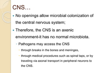
- 1. CNS… No openings allow microbial colonization of the central nervous system; Therefore, the CNS is an axenic environment-it has no normal microbiota. ◦ Pathogens may access the CNS through breaks in the bones and meninges, through medical procedures such as spinal taps, or by traveling via axonal transport in peripheral neurons to the CNS.
- 2. CNS… Microbes carried in the blood or lymph may penetrate the blood-brain barrier by infecting and killing cells of the meninges, causing meningitis. Some pathogens gain access to the CNS when localized inflammation distorts the cells of the blood-brain barrier, changing its permeability; such change is more likely during chronic infection by many pathogens. Circulation of cerebrospinal fluid can carry infective microbes throughout the cranial cavity and spinal column.
- 3. Bacterial Diseases of the Nervous System Not only can bacteria infect cells of the nervous system, But toxins released by bacteria growing elsewhere in the body can also affect neurons. In the following sections, we consider diseases of both types-leprosy, which is a disease of cells of the PNS, botulism tetanus, which involve toxins. However, the most common bacterial infection of the nervous system is bacterial meningitis, which we consider next
- 4. CNS… Bacterial meningitis involves inflammatory bacterial infection of the meninges, commonly the pia mater and arachnoid mater and more rarely the dura mater.
- 5. CNS… 1. Neisseria meningitidis Neisseria meningitidis is one of only two species of Gram-negative cocci that regularly causes disease in humans. 13 antigenic strains; strains A, B, C, and W135 cause most cases of disease in humans. The cells of all strains of Neisseria are nonmotile and are typically arranged as diplococci (pairs) with their common sides flattened in a manner reminiscent of coffee beans
- 6. Neisseria meningitidis The bacterium is known as the meningococcus and its disease as meningococcal meningitis. • Pathogenesis and clinical findings – Aspiration of infective bacteria – Attach to epithelial cells of nasopharyngeal and oropharyngeal mucosa – Cross the mucosal barrier & enter the bloodstream – May enter the CNS and cause meningitis. – Meningococcal LPS is responsible for the toxic effects found in meningococcal disease – Also antiphagocytic polysaccharide capsule play a role in the pathogenesis
- 7. Neisseria meningitidis • Manifestations of meningococcal infections • Fulminant Meningococcemia (Purpura Fulminans) – The most severe form of meningococcemia is the life- threatening Waterhouse-Friderichsen Syndrome. – Characterized by high fever, shock, wide spread purpura, disseminated intravascular coagulation, and adrenal insufficiency
- 8. Neisseria meningitidis – Meningococci shed LOS which stimulate • monocytes, neutrophils, and endothelial cells, release cytokines and other mediators • In addition, meningococci invade the vascular endothelium, produces molecules that can be procoagulant as well as adhesive for leukocytes.
- 9. Neisseria meningitidis • Fulminant meningococcemia: – Occurs in 5 to 15% of patients – begins abruptly with sudden high fever, chills, myalgias, weakness, nausea, vomiting, and headache. – typically, no signs of meningitis – Pulmonary insufficiency develops within a few hours – and patients many die within 24 hours even with appropriate antibiotic therapy and intensive care.
- 10. Neisseria meningitidis Petechial rash and neck extension characteristic of meningococcal meningitis.
- 11. Neisseria meningitidis • Meningococcal meningitis – the meninges are inflamed, with thrombosis of blood vessels – after an Ip of 1-3 days, • the onset of meningococcal meningitis is sudden with a sore throat, headache, & drowsiness • and signs of meningitis which include fever, irritability, stiff neck , photophobia and an increased level of PMNs in spinal fluid
- 12. Neisseria meningitidis • Diagnostic Laboratory Tests – Specimens: blood for culture, and CSF for smear and culture – Culture: CSF specimens are plated on • "chocolate" agar and incubated at 37 °C in an atmosphere of 5% CO2 • A modified Thayer-Martin medium with antibiotics (vancomycin, colistin, amphotericin) – oxidase +ve: a key test for identifying Neisseriae – Serology: antibodies to polysaccharides can be measured by latex agglutination
- 13. • Epidemiology, Prevention, & Control Humans only natural hosts Person-to-person transmission by aerosolization of respiratory tract secretions in crowded conditions Close contact with infectious person (e.g., family members, day care centers, military barracks, prisons, and other institutional settings) Highest incidence in children younger than 5 years and particularly those younger than 1 year of age 5% to 30% Commonly colonize nasopharynx of healthy individuals Vaccine for groups A, C, Y, and W-135 are the capsular polysaccharides
- 14. • Treatment –Penicillin G is the drug of choice –Either chloramphenicol or a third- generation cephalosporin is used in persons allergic to penicillins
- 15. 2. Streptococcus pneumoniae Streptococcus pneumoniae Louis Pasteur discovered Streptococcus pneumoniae “Diplococcus.” Ninety-two different strains of S. pneumoniae, collectively called pneumococci, are known to infect humans as normal members of the microbiota of the throat that opportunistically grow in the lungs, sinuses, and middle ear and from those locations move into the meninges via the blood. Streptococcus pneumoniae is the leading cause of meningitis in adults.
- 16. Streptococcus pneumoniae About 40 – 70% of healthy adults are carriers Infections usually arise from normal flora (endogenous)
- 17. Virulence factors Capsule – Major determinant of virulence – Anti-phagocytosis by preventing C3b complement deposition on the surface Cell Wall – Stimulate leukocytes for production of cytokines – Teichoic acid enhances inflammatory activity
- 18. Pneumococcal surface protein A, PspA (Choline binding protein A, CBpA) • Surface – exposed protein – Adherence to host tissue, colonization • prevent the deposition of C3b on the cell surface • binds to lactoferrin, may be involved in iron acquisition
- 19. • Autolysin. breaks the peptide cross-linking of the cell wall peptidoglycan, leading to lysis of the bacteria. • Autolysis enables the release of pneumolysin and large amounts of cell wall fragments. • The massive inflammatory response to these peptidoglycan fragments is an important component of the pathogenesis of pneumococcal pneumonia and meningitis.
- 20. Pneumolysin: • Intracellular membrane-damaging toxin released by autolysis. • Pneumolysin inhibits: • neutrophil chemotaxis • phagocytosis • lymphocyte proliferation and immunoglobulin synthesis.
- 21. • IgA1 protease: • cleaves human IgA1 in the hinge region. • Enables these pathogens to evade the protective functions of the principal immunoglobulin isotype of the upper respiratory tract • Hyaluronate lyase – Surface protein • Facilitates tissue invasion by breaking down the extracellular matrix components.
- 22. • One of the three leading causes of bacterial meningitis • Signs and symptoms are similar to those produced by other bacteria • Acute purulent meningitis may follow pneumococcal pneumonia • May also develop after trauma involving the skull • Mortality and frequency of sequelae are slightly higher than with other forms of pyogenic meningitis
- 23. • Common causes of sinusitis and otitis media – Otitis frequently occurs in children in association with viral infection • Chronic infection of the respiratory sinus sometimes extends to the subarachnoid space to cause meningitis • May cause endocarditis, arthritis, and peritonitis, usually in association with bacteremia • Pneumococci do not cause pharyngitis or tonsillitis
- 24. Laboratory Diagnosis • Detection of the pathogen in appropriate test samples – Smear: CSF, Sputum and others – Culture: Blood, CSF, Sputum and others • Stained Smears – Gram-stained film of rusty-red sputum shows typical organisms, many PMNs, and many red cells – Capsule Swelling Tests • Fresh emulsified sputum mixed with antiserum causes capsule swelling (quellung reaction)
- 25. • Culture: – grows well overnight on blood agar – α – hemolytic colonies – Colonies are bile soluble – Inhibited by optochin (ethylhydrocupreine) • Detection of capsular Ag in body fluids is possible
- 26. • Immunity – antibodies to capsular polysaccharide (type-specific) • Treatment – Penicillin G is the drug of choice • Resistant strains due to modified penicillin-binding proteins reported (not due to penicillinase) • Resistance rate ~ 10% – Penicillin resistant strains can be treated with: • Erythromycin, vancomycin or quinolones • High dose of third-generation cephalosporins • Pneumococci remain susceptible to vancomycin
- 27. Vaccination • Type-specific polysaccharides – Probably provide 90% protection against bacteremic pneumonia – Pneumovax vaccin available: • Contains 25 mg of the purified capsul polysaccharides of each of the 23 most frequent serovars • protection of high-risk individuals older than two years. • 80 to 90 % of all isolated pneumococci have antigens contained in this vaccine • hepta-valent conjugate vaccine (PCV7): – recommended for all children aged six weeks to five years of age, to help prevent ear infections
- 28. • Heterogeneous collection of α-hemolytic and non- hemolytic • Have the basic features of streptococci but lack specific Ags; toxins, & virulence factors of the other group • Optochin resistant and bile insoluble
- 29. • Genus Haemophilus – Small, pleomorphic, Gm-negative bacilli or coccobacilli – Facultative anaerobes or fermentative. – Require enriched media, blood or its derivative. • X factor (i.e., hemin) and/or V factor (NAD or NADP). – Haemophilus influenzae type b is an important human pathogen – other species are NF of mucous membranes and only occasionally cause disease.
- 30. • Haemophilus influenzae – grow both aerobically and anaerobically. – Its growth requires: hemin (X factor) and NAD (V factor). • six major serotypes identified(a–f) based on the capsular polysaccharide antigen. – some strains lack capsule and are referred to as nontypable strains. – subtype b (Hib) cause over 90% of all invasive infections
- 31. – Unencapsulated strains may cause: • Sinusitis, otitis media, and bronchitis • may also cause pneumonia in children in developing regions or in adults with immune defects. – Hib strains cause: • meningitis, bacteremia, epiglottitis, septic arthritis, pericarditis, as well as placentitis and peripartum septicemia in mothers. • A small proportion of invasive disease is caused by other capsular types, notably, a and f.
- 32. • About 50% of all reported invasive infections caused by Hib are meningitis • case-fatality rates may reach 40% in developing countries. • complications include: – subdural effusions and CNS sequelae occur in 11% to 40% of survivors of Hib meningitis, with deafness being a common finding.
- 33. • EPIDEMIOLOGY – H. influenzae is the NF of nasopharynx – spreads by airborne or direct contact person to person. – Nonencapsulated strains are frequently found in the URT. • up to 3/4 of healthy adults – Carriage of Hib can occur up to 30% of children in developing countries
- 34. • Diagnostic Laboratory Tests • Specimens – nasopharyngeal swabs, pus, blood, and spinal fluid for smears and cultures. • Direct Identification – immunologic detection of H .influenzae antigens in spinal fluid. • Culture – grown on chocolate agar typically (small, flat, colorless/transparent) colonies appear. – differentiated from related gram-negative bacilli by its requirements for X and V factors – and no hemolysis on blood agar
- 35. • Immunity – maternal antibodies for infants under age 3 months • Prevention – Polysaccharide polyribose phosphate capsule vaccines • Treatment – many strains of Hib are susceptible to ampicillin, but up to 25% produce -lactamase – all strains are susceptible to the third- generation cephalosporins.
- 36. Streptococcus agalactiae (group B Streptococci) Gm +ve cocci, short chains & diplococcal pairs Larger colonies β-hemolytic The only species that carries the Lancefield group B antigen Nine different capsular polysaccharide have been identified (Ia, Ib, and II-VIII). Serotype III most frequently associated with neonatal infections , adult infections are distributed over the different serotypes. Resident in the GIT Members of the NF of 10 to 30% female genital organ
- 37. May gain access to the amnoitic fluid or colonize the newborn as it passes through the birth canal A major cause of sepsis and meningitis in neonates Risk is much higher when factors are present that: ◦ decreased infant’s innate resistance (prematurity) or ◦ increased chances of transmission (ruptured amniotic membranes)
- 38. Immunocompromised individuals: ◦ Infections of the skin & connective tissues, Sepsis, UTI, Pneumonia & Peritonitis Virulence factor Capsular polysaccharide Haemolysins C5a peptidase hyaluronidase (not all strains)
- 39. Clinical diseases defined by the age of the patient at presentation Neonatal infection i) Early-onset neonatal disease Occur within the first week of life, with a median age of 20 hrs at the onset of illness Infection acquired during or shortly before birth from organisms colonizing the maternal genital tract
- 40. results from ascending spread of Str. agalactiae from the vagina into the amniotic fluid Then aspirated by the infant, and results in septicaemia of infant or mother or both. ~ 50% of infants delivered vaginally by carrier mothers become colonized ◦ only 1 to 2% develop clinically evident infection
- 41. Purulent Meningitis most common manifestation but septic arthritis, osteomyelitis, conjunctivitis, sinusitis, otitis media, endocarditis and peritonitis also occur Findings: fever, lethargy or irritability, poor feeding, and seizures
- 42. Laboratory diagnosis Culture: blood, CSF, or other appropriate specimen Definitive identification: reaction to specific antiserum to the group B carbohydrate antigen Biochemical tests (presumptive identification): ◦ hydrolysis of sodium hippurate (99% +ve) ◦ hydrolysis of bile esculin agar (99 to 100% -ve) ◦ bacitracin susceptibility (92% resistant)
- 43. Immunity Abs to the capsular polysaccharide afford protection Treatment Penicillin is the treatment of choice (less susceptible than GAS) Neonatal infections often initially treated with combinations of penicillin (or ampicillin) and an aminoglycoside Prophylactic administration of ampicillin or penicillin during delivery reduce the risk of infection
- 44. Focused on reducing contact of the infant with the organism In colonized women: ◦ attempts to eradicate the carrier state not successful ◦ intrapartum antimicrobial prophylaxis with penicillin or ampicillin reduce transmission and disease in high-risk populations